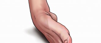
Dislocations, sprains, bruises and ruptures (ligaments, for example) - injuries with damage to muscles and joints.
Such injuries are common companions for people who lead an active lifestyle and play various sports. Often the cause of these injuries is a person’s unpreparedness for a given physical activity - which is why it is recommended to specially prepare for each sports achievement.
1
Treatment of dislocations, sprains, ligament tears
2 Treatment of dislocations, sprains, ligament tears
3 Treatment of dislocations, sprains, ligament tears
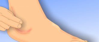
However, dislocations, bruises, sprains and ligament tears can also be caused at home. Dislocations of the shoulder and knee joints are most often recorded. A dislocation of the ankle joint, for example, is much less common (usually this is an injury to lovers of high-heeled shoes).
Let's try to figure out how to properly treat dislocations and other injuries.
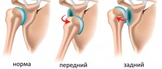
Symptoms of dislocation
Depending on the type of injury, be it, for example, a dislocated hip, a dislocated leg, or a dislocated shoulder, the manifestations of the disease may differ from each other. So, with a similar problem with the hip joint, received at birth, the gait is often disturbed, the posture is bent, and one leg may even become shorter than the other.
However, there are still general symptoms of dislocation, based on which one can assume a dislocation of a certain part of the body:
- redness of the skin in the affected area;
- intense pain that intensifies when moving the dislocated limb;
- external change in the shape of the joint, an increase in its size;
- increased swelling in the injured area;
- loss of sensation in the limbs (this symptom is possible due to injury to nerve endings);
- difficulty in motor activity;
- increase in body temperature, high fever, chills.
Sprains
Sprains occur in a person if he performs sudden movements with a force exceeding the permissible load on the joint. When a ligament is sprained, there is partial damage or incomplete rupture of the capsule of the ligaments that strengthen the joint.
The ligaments most often affected by sprains are the ankle ligaments (especially when the foot rolls in) and the wrist joint. Somewhat less commonly, this injury affects the ligaments of the knee joint. When wearing high-heeled shoes, a woman often gets her legs turned inward and the Achilles tendon stretched.
Degrees of sprain
The following degrees of ligament damage can be distinguished:
- 1st degree , preservation of the mechanical integrity of the ligament with rupture of individual fibers. The presence of slight edema without hemorrhage is characteristic. The patient may feel moderate pain, with limited movement and support;
- Grade 2 , accompanied by partial damage to the articular capsule of the ligaments, the appearance of multiple fiber ruptures, frequent blood loss and moderate swelling. Difficulty in support, movements are quite painful and limited. Some instability of the joint is revealed.
- Grade 3 , complete rupture of the ligaments, which is marked by severe pain, significant swelling and bruising. When moving, joint instability is felt.
For grade 1 and 2 sprains, conservative treatment may often be sufficient.
For complete ligament ruptures, surgical treatment is used.
Causes
The main cause of dislocation, whether it is a dislocation of the shoulder joint or jaw, is indirect trauma. So, for example, if a person accidentally falls directly on his hand, stretching it forward, then, most likely, it is not the hand itself that will suffer, but his shoulder joint.
Often, damage occurs along with a number of severe bone diseases, as additional complications. These include:
- tuberculosis;
- arthritis;
- osteomyelitis;
- arthrosis and others.
Ligament tears
Ligament rupture is a fairly common type of injury for people whose work involves prolonged physical activity. Most often, ligament rupture accompanies a dislocation or fracture, but it can also be an independent injury.
In accordance with the reasons that caused the injuries, traumatic and degenerative ligament ruptures are distinguished. If the first type of damage can be obtained as a result of injuries, then the second occurs during the aging process of the body.
Symptoms of ligament rupture:
- pain and limitation of joint function (impossibility to straighten and lift the injured arm or leg);
- the appearance of edema, hematoma;
- change in the external contours of the damaged joint (joint instability);
- the occurrence of numbness and tingling in the affected limb.
A special place is occupied by ligament ruptures such as meniscus damage.
When should you see a doctor?
If you suspect you have a dislocated hip or dislocated hip joint, you should immediately seek qualified medical help. Otherwise, there is a risk of aggravating the condition of an already damaged joint.
It is necessary to provide first aid for a dislocation. The joint should be immediately immobilized and a cooling compress applied to it to reduce swelling. If the injured person feels unbearable pain, it is necessary to give him an anesthetic drug. Then you need to transport the victim to the hospital to provide him with proper medical care by an experienced trauma surgeon.
Specialists with many years of qualifications and enormous work experience are ready to accept patients at JSC “Medicine” (clinic of Academician Roitberg) at any time. The multifunctional medical institution is located in the center of Moscow, at 2nd Tverskoy-Yamskaya Lane, 10, close to the Mayakovskaya, Belorusskaya, Tverskaya, Novoslobodskaya and Chekhovskaya metro stations.
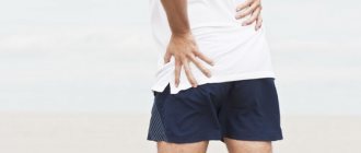
Causes of hip dislocation in adults
It is important to know! Doctors are shocked: “An effective and affordable remedy for joint pain exists. " Read more.
In adults, dislocations of the hip joint are rare. This is explained by the fact that the head of the femur is firmly fixed in the acetabulum, and the joint itself is strengthened by ligaments and muscles. In order for the femur to move from its place, a high-velocity traumatic factor is needed.
- car crashes;
- natural disasters;
- landslides;
- falling from a height;
- disasters.
Dislocation occurs due to the indirect action of an external force placed directly on the femur. The latter acts as a lever. The impact of a traumatic factor on the hip joint itself usually leads to fractures of the femoral neck or pelvic bones. In some cases, dislocation may be accompanied by pinching of the obturator nerves, compression of the femoral vessels, contusion of the sciatic nerve and fractures of the bones that form the acetabulum. If not treated in a timely manner, these complications can lead to the death of the soft tissues of the limb or even complete paralysis of the leg.
Hip joint dislocations usually occur in men and women under 50 years of age. Elderly people suffer from osteoporosis. The disease makes bones very fragile, making them easy to break. Therefore, at older ages, fractures of the femoral neck occur much more often.
| View | Description | Development mechanism |
| Suprapubular | The femoral head is located anterior to the ilium. The patient's leg is extended and turned outward. A convex formation is visible in the groin area, and the buttock on the side of the injured limb looks flattened | A sharp fall from a height onto an abducted, bent and turned outward leg |
| Obturator | The head of the femur is located next to the pubis. The lower limb is strongly turned outward, bent at the knee and hip joints | Similar to previous |
| Iliac | The femur is located behind the ilium. The leg is bent and turned inward. There is a protrusion in the buttock area, and a recess in the groin. On examination, shortening of the leg is noticeable. | Sudden bending or rotation of the leg at the knee and hip joint, which usually occurs in an accident |
| Sciatic | The head of the femur is located near the ischium. The deformity is more pronounced than with iliac dislocation | Similar to previous |
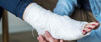
Treatment of injury
It includes mandatory repositioning of the displaced joint and returning it to its previous position. The manipulation can be performed under both general and local anesthesia. Finally, a plaster cast is applied to the damaged area.
In addition, there is often a need for additional procedures:
- therapeutic massage;
- recreational gymnastics;
- acupuncture sessions.
In particularly difficult situations, surgery may be indicated. If the cause of the dislocation is a bone disease, you should definitely give it due attention and carefully treat it, otherwise the injuries will recur again and again.
Treatment of joint dislocations and reduction
Treatment of traumatic dislocations consists of reduction (relocation) and fixation of the articular elements. This procedure should be carried out immediately if the patient is in satisfactory condition.
If the dislocation is not corrected as soon as possible, this can lead to a number of complications:
- development of contracture due to contraction of muscle fibers;
- the formation of scar tissue in the joint cavity - usually at the site of a dense blood clot (fibrin).
There are two options for joint realignment:
- open;
- closed.
The open method is used if the closed method is contraindicated for medical reasons. Its essence lies in joint arthroscopy. Through surgery, the doctor removes from the joint cavity coagulated blood and articular elements destroyed due to injury. He then returns the displaced parts to their original position using lever-like movements. This is a painful procedure, so it is performed under general anesthesia. Anesthesia allows not only to achieve pain relief, but also has a relaxing effect on muscle fibers, facilitating the operation.
The closed method involves returning the articular heads to their place without surgical intervention. After reduction, the joint must be immobilized. To do this, a plaster fixing bandage is applied to it for 14–20 days. After reduction, a control x-ray is taken, which shows whether the displaced structures are in the correct position.
Rehabilitation after injury
After the dislocation has been treated at the proper level and the patient has gone home with the doctor’s recommendations, the recovery period begins.
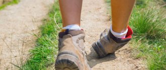
In order to strengthen muscles and bones and quickly return them to their previous condition, it is recommended:
- take a course of restorative massage;
- physical therapy course;
- take up regular swimming;
- do not avoid long walks in the fresh air;
- balance your own diet by consuming enough proteins, fats and carbohydrates required by the body.
Prevention of dislocation of the shoulder joint, jaw or other part of the body consists, first of all, of taking good care of your own health, proper nutrition and regular exercise.
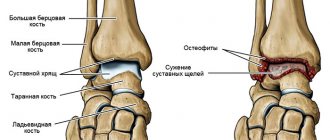
Meniscal tears
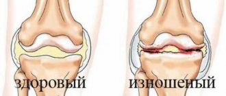
The menisci of the knee (internal and external) are a kind of cartilage pads between the bones that protect the articular cartilage and ensure smooth movements when walking.
A meniscus tear is a damage to the cartilage formation, which is accompanied by injury to the structures of the knee joint. Injuries such as cruciate ligament injury and rupture of the collateral ligaments (the lateral ligaments of the knee) often occur simultaneously with a meniscus tear. Meniscal tears are common among athletes, especially representatives of contact sports.
The second reason for damage to the meniscus of the knee joint is degenerative disorders in cartilaginous formations. If the meniscus is damaged, the function of the entire joint deteriorates, pain, swelling, cracking and popping appear when bending the knee.
1 Treatment of dislocations, sprains, ligament tears
2 Treatment of dislocations, sprains, ligament tears
3 Treatment of dislocations, sprains, ligament tears
Main features
Pathological displacement can be one- or two-sided, this affects the clinical picture. Bilateral posterior dislocation is characterized by the following symptoms:
- normal mouth closure is difficult;
- the lower teeth are behind, their position is unnatural;
- severe pain is observed below the ears, after some time reporting develops in this area;
- there is copious secretion of saliva, speech becomes slurred;
- in a horizontal position, suffocation is experienced; the patient can only stand or sit.
Unilateral posterior pathology manifests itself in the same way, but there is pain and swelling only on the affected side, asymmetry of the jaw, and curvature of the mouth are observed.
With lateral bilateral pathology, the following signs are observed:
- swelling, severe pain in the area below the ears;
- slurred, impaired speech;
- the presence of increased salivation;
- the lower jaw is strongly displaced, this is not only visible from the outside, but is also strongly felt by the patient himself.
For bilateral, the following clinical symptoms are observed:
- the jaws do not close, the mouth is constantly open;
- slurred speech;
- Below the ear area there is severe swelling and pain;
- hypersalivation (saliva secretion) appears.
A unilateral anterior dislocation has the same symptoms, but the mouth is skewed to only one side. A person cannot swallow saliva; jaw movements are possible, but they are limited and accompanied by pain.
Features of treatment
| Click to sign up for a FREE consultation |
Treatment of TMJ must begin with reduction, for which the doctor sits the patient down and stands directly in front of him. Next, he takes hold of the lower jaw on both sides, his thumbs should rest against the teeth, and the rest completely cover the jaw. Then with your thumbs you should press on the jaw, lowering it, and at the same time lifting its front part up. The fact that the joint is in place is indicated by a click and a feeling of sinking of the lower jaw. Then a sling-shaped bandage is applied to the joint area, the duration of its wearing is up to 5-7 days. At this time, the patient will be able to take only pureed and liquid food to consolidate the result. The same diet is followed after the treatment of the dislocation is completed.
Before realigning the jaw, the doctor must make sure that there is no jaw fracture, otherwise serious harm may occur. In addition, the reduction itself must be performed strictly according to established rules; a mandatory examination is required before starting treatment. In some cases, surgery will be required.
joint
joint
- unsuccessful attempts at treatment make further reduction difficult, so surgery may be required;
- you can further damage the joint by significantly damaging the soft tissue, making the dislocation irreducible;
- there are no opportunities for adequate pain relief at home;
- there are no diagnostic devices at home to assess the degree of damage to intra-articular structures;
- the lack of adequate treatment leads to an old dislocation, which cannot be reduced in a closed way, but is treated with surgery.
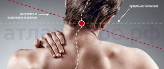
Types and causes of Atlanta injuries
Displacement or skew of the atlas most often occurs in newborns due to the actions of obstetricians during the birth process or due to incorrect presentation of the fetus. Subluxation is sometimes characterized by limited neck mobility or torticollis.
During childbirth, the baby experiences an incredible external mechanical load for its small size. The cervical spine suffers the most as a result of high pressure, especially if the birth process is forced to be stimulated using obstetric forceps and other methods.
At one time in the Soviet Union there was a practice of “preserving the perineum of a woman in labor.” The so-called “humane” method was aimed at reducing the rate at which the fetus comes out, which was supposed to reduce the recovery period of the mother and help avoid injury to the perineum. In fact, the midwife simply pressed the baby’s head in the opposite direction with her palm, increasing the already high load on the baby’s neck.
The difference between the Swiss method and traditional methods of treatment
Since the craniovertebral (upper cervical) zone of the spine is a “risk zone” - the most dangerous for exposure, doctors and osteopaths usually try to use conservative treatment: gentle traction and a system of physical exercises to strengthen the muscles, as well as manual therapy, and so on. However, these osteopathic methods in the vast majority of cases do not provide a complete return of C1 to its natural position.
Correction of C1 using the Swiss technique is based on a discovery in the field of physiology that allows you to overcome muscle memory. If the effect is carried out not with the help of a device that vibrates for a sufficiently long time, but with the help of manual therapy, then the result from realigning the vertebra will not last long at best: sooner or later instability of the first cervical vertebra will appear, and the atlas will again take an incorrect position in the atlantoaxial joint In the worst case, there will be no result at all, or even greater harm will be caused to the musculoskeletal system and the entire body.
If you want to learn more about how the reduction of the atlas vertebra is carried out and what the effect of the method is, then you can read and watch detailed reviews and video reviews about it.
Diagnostics.
After an accident, the victim must be immediately taken to the traumatology department, and he must not make any leg movements (active or passive). Upon admission of the patient, the doctor performs a detailed examination of the injured limb and takes a complete anamnesis. The main method for diagnosing foot dislocations remains radiography. Only with the help of X-rays can a traumatologist make an accurate diagnosis (determine the type of dislocation) and begin appropriate treatment. In the case of complicated dislocations with fractures, surgical intervention may be required.
Symptoms and consequences of atlas subluxation in children
Even minor trauma to the cervical spine and displacement of the cervical vertebrae lead to a narrowing of cerebral blood flow and compression of the vertebral artery and important blood vessels. The consequences of this are increased intracranial pressure, headaches, migraines, impaired growth and the development of diseases of the musculoskeletal system, and delayed motor development. Such problems are observed even in children 6, 8, 10 years old. Attempts to treat concomitant diseases and prescribing exercise therapy (physical therapy) do not eliminate the problem.
Children who have suffered a birth injury to the neck often experience poor posture and scoliosis. Due to poor blood supply to the brain, the central nervous system suffers, and this affects the character, psyche of the child, and his physical development.
Not all consequences of birth trauma go away with age; some remain for life and only get worse over time.
How do they adjust it?
An orthopedic doctor always knows how to correct a dislocation of a particular joint. This is easiest to do in the first 3 hours after the injury. A dislocation in the first three days is considered fresh. During this period, it can be reduced in almost all patients. Certain difficulties appear from 3 days to 3 weeks. After 3 weeks, dislocations are considered old. They often have to be realigned during surgery.
Large joints often have to be realigned using the physical strength of several people at once. To facilitate this process, medications that eliminate swelling and relax muscles (muscle relaxants) may be used. Joint dislocations are always painful. Therefore, they are adjusted using local or general anesthesia.
After reduction of a closed dislocation, the limb is immobilized. The duration of immobilization is determined by the severity of the injury, the nature and location of the dislocation. The patient wears the bandage for a long time (several weeks). In this case, early loading on the injured limb is not recommended. As a result of anatomical damage, repeated dislocations are possible.
How to diagnose subluxation of the upper vertebra?
Diagnosis of C1 displacement at the Atlas-Standard clinic is made on the basis of:
- Kinesiological tests (muscle tone and the relationship between their tension and the functioning of internal organs are assessed). Using this method, you can detect whether there are malfunctions in the craniovertebral zone (cervical spine), and most importantly, the tests help to see the consequences of these malfunctions.
- X-rays, CT (computed tomography) and MRI (magnetic resonance imaging) demonstrate statistical changes in the vertebra. They are done to control the condition of the spinal column.
MRI picture: subluxation of the first cervical vertebra (atlas).
What specific methods will be selected for correcting rotational subluxation will be clarified during a consultation with a specialist. Note that hardware research methods can display the structure of the joint, but cannot demonstrate the consequences of displacement. An example would be a paralyzed limb where the cause of paralysis is a subluxation of C1. This is why special kinesiological tests are needed.
The same tests are performed after correction of the C1 misalignment to ensure that there is no misalignment and the position of the vertebrae allows the body to function normally.