Fractures of the bones of the hand are injuries in which the integrity of the bones of the wrist, metacarpus or phalanges of the fingers is disrupted. These are common injuries, accounting for about a third of all bone fractures. This trend is associated both with the relative fragility of the hand, adapted for performing fine manipulations, and with its high activity. A fracture can occur as a result of a fall on a bent hand, a blow from a fist, the edge of the palm or fingers, or a direct blow to the hand.
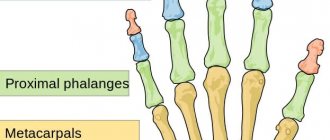
The bone apparatus of the hand consists of 27 bones of three sections - the wrist, metacarpus and fingers. The wrist consists of 8 spongy bones arranged in two rows - proximal, closer to the forearm, and distal, closer to the metacarpus. The first, proximal row, starting from the thumb, are the scaphoid, lunate and triquetral bones, which form an articulation with the radius bone of the forearm - the wrist joint. The fourth bone of the proximal row, the pisiform, does not participate in the formation of the wrist joint.
The second, distal row - the polygonal, trapezoid, capitate and hamate bones - connect to the five tubular bones of the metacarpus, radiating from the wrist. The distal ends of the metacarpal bones form five metacarpophalangeal joints - the connections of the metacarpus with the fingers. The first finger of the hand consists of two phalanges, the rest - of three. The phalanges of the fingers - short tubular bones - are connected to each other by interphalangeal joints.
The bones most susceptible to fractures are the phalanges and metacarpal bones. Wrist bones are broken quite rarely. The vast majority of injuries to the bones of the wrist occur as a result of a fracture of the scaphoid bone; the lunate and pisiform bones are less commonly affected. Fractures of the hamate and distal carpal bones are practically never encountered in clinical practice.
Fractures of the bones of the hand are accompanied by sharp pain and swelling in the area of damage; when bone fragments are displaced, deformation of the hand is possible. A hematoma may appear at the site of swelling. With some fractures, you can feel the displaced bone fragments under the skin or hear their crepitus. The diagnosis can be established by a traumatologist, who will clarify the complaints, ask in detail about the mechanism of injury, examine and palpate the fracture area, and check the preservation of movements in the joints. The diagnosis is confirmed by an X-ray examination of the hand, which can visualize the fracture line, assess the degree of displacement of fragments and, as a result, determine treatment tactics.
Treatment of hand fractures includes mandatory immobilization with a plaster cast for a period of 3 to 8 weeks. In case of displacement of fragments, closed reduction is performed; if it is ineffective, skeletal traction or osteosynthesis is performed. Careful comparison of fragments and consolidation of the fracture are important to preserve not only the aesthetics of the hand, but also its full function.
Classification of hand bone fractures
Depending on the presence or absence of skin damage over the fracture, the following are distinguished:
- closed fractures - the integrity of the skin is not compromised;
- open fractures - in the area of damage there is a wound in which bone fragments can be identified.
According to the position of bone fragments:
- without displacement - the broken bone maintains its position, the fragments exactly touch along the fracture line;
- with displacement - bone fragments diverge to the sides and, as a result, cannot heal along the fracture line without their comparison - reposition.
According to the involvement of articular structures in the fracture:
- extra-articular fractures - the fracture line passes outside the joint cavity;
- intra-articular fractures - the fracture line is located inside the joint cavity;
- fracture-dislocations - a violation of the integrity of the bone in combination with a dislocation in the adjacent joint.
According to the location of the fracture:
- wrist bone fractures;
- metacarpal bone fractures;
- fractures of the phalanges of the fingers.
You can also classify hand fractures depending on the number of fragments, degree of displacement, and infection. The etiology of the fracture is also important - whether it was traumatic or pathological - arising against the background of bone disease. All these factors influence the choice of treatment tactics for fractures and, ultimately, the possibility of completely restoring the function of the damaged hand.
What are the possible treatment options for brachymetacarpy and how does it work?
To lengthen the bones of the hand, there are three main options for operations: one-stage bone grafting, two-stage bone grafting and distraction osteosynthesis according to G.A. Ilizarov. The method of choice in the treatment of brachymetacarpy is the third option - distraction osteosynthesis or lengthening with a distraction device according to the method of G.A. Ilizarov. With brachymetacarpy, the shortening of the metacarpal bone always exceeds 1 cm, and one-stage bone grafting does not eliminate the shortening of the metacarpal bone by more than 1 cm at a time, so the choice of this technique for the treatment of brachymetacarpy is not the most competent decision. Two-stage bone grafting can be used to treat this pathology, however, lacking the advantages of one-stage bone grafting (only one operation and a relatively short recovery period), it has the same disadvantage - the need for bone grafting from another area. Lengthening with a distraction device allows one to avoid significant postoperative scars and does not require trauma to the donor area, despite the rather long period of wearing the device (about 3 months), and therefore this technique is the operation of choice for brachymetacarpy.
Wrist fractures
The bones of the wrist, due to their shape, structure and position, are broken quite rarely. The most susceptible bone to fracture is the scaphoid, the large bone at the base of the big toe. Injuries to the lunate and pisiform bones of the wrist also occur. The triquetral bone, as well as the bones of the distal row - polygonal, trapezoid, capitate and hamate - are subject to fractures extremely rarely; usually their fractures are combined with dislocations in the corresponding joints.
Scaphoid fractures
The cause is a fall on a bent hand, a blow with a fist, or a direct injury to the wrist. The following options are possible:
- intra-articular fracture of the scaphoid - the fracture line is located inside the cavity of the wrist joint;
- extra-articular fracture - separation of the tubercle of the scaphoid;
- de Quervain's fracture-dislocation - a simultaneous fracture of the scaphoid and dislocation of its proximal fragment and the lunate from the wrist joint.
Symptoms are pain and swelling at the base of the thumb, inability to move the hand at the wrist joint, or clench the hand into a fist. The diagnosis is established based on the patient’s complaints, data on the nature of the injury, examination and radiography of the hand bones. Sometimes, in the absence of displacement of fragments, the fracture line with all its signs is not determined. In this case, immobilization is still carried out with repeated radiography after 7-10 days, when, due to the activation of regenerative processes, the fracture line becomes clearly visible.
Treatment is immobilization with a plaster cast for a period of 4 weeks, followed by monitoring and prolongation of immobilization in case of insufficient consolidation of the fracture. In case of displacement of fragments and fracture dislocation, closed reduction is ineffective; fixation of fragments of the scaphoid bone with a wire is indicated. Fractures of the scaphoid are often complicated by the development of a false joint or lysis of bone fragments due to damage to the blood vessels supplying them during injury. Therefore, it is important to follow all the doctor’s recommendations and take control photographs in a timely manner to avoid complications and deterioration in the function of the wrist joint. After restoring the integrity of the scaphoid bone, physiotherapeutic treatment and exercise therapy are indicated to restore hand function.
Lunate fractures
The cause is a fall on a bent hand or direct injury, a blow to the wrist. It manifests itself as pain and swelling, intensifying with movements in the third, fourth and fifth fingers and with extension of the hand. The diagnosis is established taking into account complaints, the mechanism of injury, an objective examination of the area of damage and the results of an x-ray examination. To treat a lunate fracture, a plaster cast is applied for 4-8 weeks. Recovery usually proceeds without complications.
Fractures of the pisiform bone
The cause is a blow with the edge of the palm or direct injury. It manifests itself as pain and swelling of the wrist on the little finger side, increasing pain when it moves. The diagnosis is made taking into account complaints, anamnesis of injury, examination of the area of damage and radiography of the bones of the hand. For complete consolidation of a fracture of the pisiform bone, 4-5 weeks of immobilization are sufficient. The injury is rarely complicated.
Main functions
The main function that these bones perform is to ensure unhindered movement of the fingers. The metacarpal bones contribute to their flexibility, thanks to which the hand has the ability to bend and extend, which is very important for reflex movements.
The absence of these abilities greatly complicates human activity and limits his capabilities. A similar phenomenon can occur when an injury occurs, as a result of which a person is temporarily unable to fully use the fingers of the palm. In severe cases, if the victim is not provided with first aid, injuries can lead to disability.
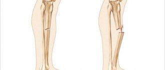
Metacarpal fractures
The long, thin metacarpal bones are often broken by a punch or direct trauma. Muscle traction and movements in the hand until the fracture is immobilized often lead to displacement of bone fragments. There are epiphyseal fractures, when the fracture line is localized in the area of the bone heads, and diaphyseal fractures, when the fracture line is located in the bone body.
First metacarpal fracture
The cause is a blow with a bent first finger, less often a direct blow to the first metacarpal bone.
Fracture of the base of the first metacarpal bone . A typical injury for boxers and MMA fighters. A Bennett fracture is a separation of the base of the first metacarpal bone, which is held by ligaments, with simultaneous dislocation of most of it in the carpometacarpal joint. Rolando's fracture is a comminuted fracture-dislocation of the first metacarpal bone. Both injuries present with pain, deformation and swelling in the “anatomical snuffbox” area - the area under the base of the first finger - with increased pain when moving or trying to make a fist. Diagnosis is carried out taking into account complaints, trauma history, examination of the area of injury and x-ray of the hand. Bennett and Rolando fractures are treated surgically using osteosynthesis - restoring bone integrity by fixing fragments with metal knitting needles, pins or plates.
Fracture of the middle part of the first metacarpal bone . More often it occurs due to a direct blow to the bone. It manifests itself as pain, swelling and deformation in the area of the first metacarpal bone. The diagnosis is established taking into account the patient's complaints, information about the mechanism of injury, examination of the area of the first metacarpal bone and x-ray examination of the bones of the hand. Treatment is plaster immobilization for a period of 4-5 weeks; if the fragments are displaced, preliminary closed reposition is required. If conservative reduction is ineffective, an operation - pin osteosynthesis - is performed to compare the fragments.
An example of Dr. Valeev’s operation to restore after a fracture of the first metacarpal bone:
Before surgery:
After operation:
Fracture of II, III, IV, V metacarpal bones
The cause is a blow with a fist or a fall on fingers clenched into a fist. They can be single, but more often several metacarpal bones are broken, usually the fourth and fifth. It manifests itself as pain, swelling and deformation of the hand, and a hematoma often occurs. Diagnosed on the basis of complaints, history of injury, objective examination and X-ray results of the bones of the hand. To treat a non-displaced fracture, immobilization is performed for a period of 4-5 weeks. If fragments are displaced, closed reduction is indicated, and if it is ineffective, skeletal traction or pin osteosynthesis is indicated.
Cervical injury
Damage to the metacarpal bones in the neck area occurs as a result of a sharp push. As a result, the head of the bone fits directly into its body without back cushioning. In this case, the bone itself does not move, but remains in place. Sometimes this condition may be considered normal, however, with a very strong blow, the bone can go deep into the cavity, which will noticeably shorten the finger.
If the fracture angle is slight, it can be treated without surgery, otherwise urgent surgery will be required. Leaving an injury untreated is not recommended, even if it is so small that it seems trivial. Over time, it will make itself felt: the consequences may be different, but restrictions on motor function and pain when trying to restore them are guaranteed.
Fracture of the phalanges of the fingers
The cause is a blow with the fingers, an injury while fixing the fingers, or a direct blow to the phalanges. Fractures of the phalanges of the fingers can be:
- intra-articular;
- extra-articular;
- single;
- multiple - within one finger or several;
- combined with dislocations in the metacarpophalangeal or interphalangeal joints.
Symptoms: pain, swelling, hematoma, deformity. The pain intensifies when you try to move your fingers. The diagnosis is established on the basis of complaints, trauma history, objective examination and X-ray results. To treat a fracture of the phalanges of the fingers without displacement, fixation is performed with a plaster cast for 3-4 weeks. In case of fracture-dislocations, joint reduction is performed; in case of displacement of fragments, closed reduction is performed. If it is not possible to compare the fragments using a closed method, skeletal traction or pin osteosynthesis is indicated.
How to identify an injury
The origin of fractures is very diverse, however, the signs indicating injury have similar properties. General signs of injury are expressed as follows:
- manifestation of acute pain in the affected area;
- the presence of a bluish or purple tint to the skin;
- significant swelling;
- with some injuries, internal bleeding occurs, forming a pronounced hematoma;
- if the bones of the foot are damaged, pain is felt when walking, which makes movement painful;
- There are cases when, with a strong blow, the bone undergoes severe deformation, and its head is difficult to feel.
First aid to the victim
Some cases require first aid. Such injuries include open injuries with bleeding. The first step is to take measures to stop the bleeding and call an ambulance. Further hospitalization of the victim is carried out under the supervision of a traumatologist or other specialist.
With closed lesions, the injured arm must be immobilized to ensure that its mobility is limited. This is done in order not to accidentally touch the sore spot, and to avoid post-traumatic shock or loss of consciousness when damaged joints are displaced. The arm can be tied with any fabric available at hand. The main thing is that the fingers of the broken hand are bent.
Preventive actions
To avoid injury, experts recommend following simple rules
- Observe safety precautions at work and at home.
- Be extremely careful when transporting heavy objects, playing sports, as well as under other circumstances associated with an increased risk of injury.
- Constantly monitor the condition of the musculoskeletal system, perform physical exercises that help strengthen bone tissue.
- Take vitamin complexes and minerals containing rich amounts of calcium.
Never perform any physical activities while intoxicated.
Diagnosis of injuries
To diagnose the condition of the injury, its characteristics and degree of complexity, specialists use the following methods.
- First of all, it is necessary to interview the patient, and then conduct a visual examination, take the necessary tests and determine the cause of the injury.
- Taking an x-ray. Photographs of the hand from both sides and from the side, depending on the location of the fracture.
- For multiple fractures with complications, computed tomography is necessary.
Typically, for experienced traumatologists, diagnosis is limited to a simple examination, and an x-ray is sufficient to confirm it.