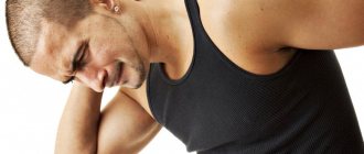
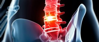
Pain and stiffness in the neck occur with improper load, as well as with certain pathological conditions of the cervical spine. Painful sensations in the neck are almost always accompanied by limited mobility - when trying to turn or tilt the head, a feeling of sharp or increasing pain occurs. Sometimes it is so strong that a person is forced to move the entire body instead of moving the neck. In this case, the pain can concern not only the neck, but also radiate to nearby areas - chest, arms, head, shoulders; accompanied by tinnitus, numbness of hands and fingers, dizziness.
In order to prescribe competent treatment, you need to determine the causes of neck pain - this will be the task of the specialist you contact. Let's figure out what can cause pain in the cervical spine.
Causes of neck pain
There are 2 main causes of pain and limitation of neck movements: muscle stiffness and diseases of the cervical spine.
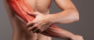
Neck muscle stiffness
Rigidity is a condition where muscles become hypertonic (unable to relax) and resist any attempts to move. Stiffness of the neck muscles is not an independent disease - it occurs either due to lack of physical activity, or accompanies certain diseases.
Due to a sedentary lifestyle - working at a computer, driving, lack of sports activity - the cervical vertebrae can become displaced, causing aching pain and spasms caused by improper blood circulation in the cervical area.
Stiff neck muscles can also be a symptom of:
- encephalitis, meningitis, cerebral hemorrhages. Other symptoms of these diseases: headache, weakness, nausea, fever, dizziness, rash.
- parkinsonism. It is accompanied by tremors throughout the body, impaired balance and automaticity of movements, and problems with speech.
- neck injuries. In such cases, pain always precedes some serious mechanical impact on the neck (fall, blow).
Diseases of the cervical spine
Pain and restrictions of movement in the cervical spine can occur due to pathologies of the spinal column:
- Hernia. Severe pain radiates to the shoulders and arms, the palms may tingle and go numb, and a “lumbago in the neck” occurs - a sharp pain that makes it difficult to move the entire upper body.
- Cervical osteochondrosis. Accompanied by headache, tinnitus, decreased visual acuity, flashing “spots” before the eyes, pain radiating to the arms and shoulders.
- Cervical arthrosis. Pain occurs not only in the neck, but also in the shoulder area. It bothers me most in the mornings and evenings.
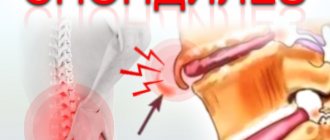
- Spondylosis. Accompanied by occipital pain, numbness and tingling of the neck, problems with hearing and vision, and dizziness may occur.
- Asymmetry of the cervical artery. It is characterized by tinnitus, pain in the head, darkening of the eyes, and increased blood pressure.
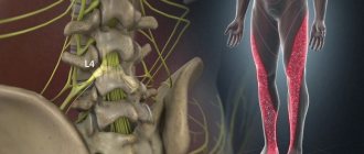
Stiffness of the leg muscles. Symptoms and treatment
Tension can be associated with prolonged stay in one position and poor circulation. The pathological condition caused by the disease is associated with excessive excitability of the neurons of the anterior horns of the spinal cord.
Types of voltage:
- Spastic. Causes the tone of individual muscles and spreads unevenly.
- Rigid. It affects all muscles at once and causes difficulty walking.
There are many serious illnesses in which rigidity is a symptom. These may include stroke, spinal injuries, inflammation in the brain, nerve disorders, as well as congenital cerebral palsy and phenylketonuria.
Stroke
A stroke causes severe damage to the central nervous system and disruption of communication between the muscles and the brain. As a result, painful, spastic contractions occur, making it difficult to perform normal daily activities. To restore damaged connections, you need to perform special exercises. Taking medications that stimulate the muscular and central nervous systems, as well as massage, contribute to successful recovery.
Hypoxia
Hypoxia – low oxygen content in tissues. The cause of muscle tissue hypoxia is the low activity of mitochondrial enzymes, which leads to impaired cellular respiration. The cause may be a lack of B vitamins, as well as poisoning with heavy metals and microbial toxins. One of the symptoms is parkinsonism, or shaking palsy. Treatment is carried out by taking drugs that perform the function of the missing enzymes.
Cerebral palsy
Cerebral palsy manifests itself in the form of delayed motor development, convulsions and epileptic seizures. The mild form is very difficult to identify, so observation by a neurologist is necessary at an early age.
The main causes of the pathology:
- violation of the development of the cerebral cortex;
- fetal hypoxia, ischemic brain damage during childbirth;
- jaundice caused by Rh conflict between mother and newborn;
- intrauterine infection, such as herpes virus;
- poisoning of a child during childbirth.
Phenylketonuria
Phenylketonuria is a hereditary disease associated with the inability to break down the amino acid phenylalanine. Since the liver lacks the enzyme phenylalanine 4-hydroxylase, it is converted not into tyrosine, but into byproducts that damage the brain. This leads to disruption of nerve impulse conduction, rigidity and developmental delays. A child can grow up healthy with timely diagnosis and exclusion of animal proteins from the diet.
Brain injuries
In case of brain injury, a person experiences:
- memory loss;
- Strong headache
; - spontaneous reflexes;
- muscle spasms.
This is caused by increased arousal and damage. To make a diagnosis, an MRI, X-ray, or, in case of severe traumatic brain injury, a lumbar puncture should be done. After a thorough examination and determination of the cause of muscle stiffness, the patient is prescribed a pastel regimen, as well as taking absorbent agents.
Spinal cord injuries
Spinal injuries can result in vertebral fragments being pushed into the spinal cord. Damaged cells do not receive enough oxygen and die. With significant damage, the process of apoptosis, that is, programmed cell death, is triggered, causing rupture of the spinal cord. After this, the ability to move is temporarily lost, since the body supports the functioning of only vital systems. To avoid complete atrophy of the lower extremities it is necessary:
- immediate surgical intervention;
- administration of drugs that stimulate damaged cells;
- supporting muscle function with electrical impulses.
Sclerosis
Sclerosis causes destruction of the myelin sheaths of neuron axons. As a result, the innervation of muscles, including the lower extremities, is disrupted, and rigidity occurs. In this case, there is poor coordination of movements and severe pain when straightening the leg. Tendon reflexes are especially strong. The patient cannot bend or straighten his legs normally, and his gait becomes wooden.
Nerve impulse conduction disorder
Occurs when nerves are injured. It can also cause muscle stiffness due to neuropraxia, which is a dysfunction of the nerve. Basically, there is a disorder of motor function. Timely diagnosis and surgery using microsurgical techniques help restore nerve function and get rid of nerve impulse conduction disorders.
Meningitis
The disease is associated with inflammation of the membranes of the spinal cord and brain. Penetrating infection causes the development of bacteria:
- meningococci;
- streptococci;
- staphylococci;
- Pseudomonas aeruginosa.
Rigidity manifests itself in the form of Kernig's sign, when a spasm of the knee joint occurs and the patient is unable to straighten the leg.
Encephalitis
Encephalitis is an inflammation of the brain. May be caused by viruses and bacteria. One of the symptoms is muscle rigidity of the lower extremities, manifested by parkinsonism, spasms and convulsions. Encephalitis is difficult to detect due to its asymptomatic early stages. Slight drowsiness and increased fatigue quickly turn into mental disorders and impaired muscle tone.
Treatment
Regardless of the cause, stiffness is treated in two stages:
- elimination of the disease;
- treatment of muscle spasms.
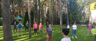
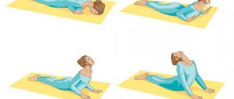
Daily physical exercise will be an excellent prevention and will help treatment.
Surgery is required for serious injuries to the brain and spinal cord, as well as when drug treatment fails. Author: K.M.N., Academician of the Russian Academy of Medical Sciences M.A. Bobyr
Diagnosis of the cause of neck pain
After an oral interview, the doctor examines the patient: palpates the surface of the neck, checks the sensitivity of the arms and legs, the state of reflexes, and determines the presence of muscle spasms and hypertonicity.
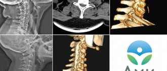
The following hardware techniques are used:
- radiography;
- MRI;
- CT scan;
- electromyography (EMG);
- Doppler ultrasound (USDG) of neck vessels.
Blood and urine tests can be taken to identify inflammatory processes in the body.
Postural instability
Violation of postural fixation can occur in the early stages, for example, patients cannot hold their arms extended forward. But these are not such serious deviations that both patients and doctors pay attention to them. But closer to the later stages of the disease, serious problems begin. For example, there are difficulties with starting movement and stopping it, no matter how difficult it is to overcome the inertia of rest and movement. During movement, the body tilts along the course of inertia; in particular, when moving forward, it tilts and, as it were, moves ahead of the legs. This disrupts the center of gravity and leads to frequent falls and injuries.
Methods for treating pain and stiffness in the cervical spine
Treatment for neck soreness may include:
- Physiotherapy sessions (magnetic therapy, electrophoresis, ultrasound therapy, shockwave therapy).
- Prescription of painkillers, sedatives, anti-inflammatory drugs, muscle relaxants.
- Methods of holistic medicine (acupuncture, massage, moxotherapy, hirudotherapy, stone therapy, vacuum therapy).
- Physical therapy, proper organization of the workplace, control of posture.
- Surgical intervention. Radical methods of surgical treatment are prescribed only if conservative methods are ineffective or for a more serious diagnosis that initially requires surgical intervention.
Muscular dystrophy, or myodystrophy
3987 21 September
IMPORTANT!
The information in this section cannot be used for self-diagnosis and self-treatment.
In case of pain or other exacerbation of the disease, diagnostic tests should be prescribed only by the attending physician. To make a diagnosis and properly prescribe treatment, you should contact your doctor. Muscular dystrophy: causes, symptoms, diagnosis and treatment methods.
Definition
Muscular dystrophies are a large group of inherited diseases that are characterized by progressive weakness and degeneration of skeletal muscles, that is, loss of muscle mass. The most common muscular dystrophies include Landouzy-Dejerine muscular dystrophy (scapulohumeral-facial myopathy), Duchenne muscular dystrophy and Becker dystrophy. This group also includes Emery-Dreyfus dystrophy, Erb-Roth muscular dystrophy and others.
Duchenne muscular dystrophy affects mainly boys, and the prevalence of this disease is 3.3 per 100,000 population; 1 in 3,500 newborn boys suffers from this pathology. This type of dystrophy is often classified into the same group with Becker muscular dystrophy (incidence rate 1 in 20,000 newborns). Duchenne and Becker muscular dystrophies are inherited in an X-linked recessive manner. This means that the damage is located on the X-sex chromosome and is transmitted from mother to son, and daughters are carriers and, as a rule, do not get sick themselves.
Landouzy-Dejerine myopathy (humeral-scapular-facial myopathy) occurs with a frequency of 0.9-2 per 100,000 population. The disease is inherited in an autosomal dominant, autosomal recessive (the rarest) or X-linked manner. For an autosomal dominant type of inheritance, one copy of the defective gene from one of the parents is sufficient.
The frequency of development of Emery-Dreyfus muscular dystrophy is not precisely known; 7 genetic forms have been described, but the frequency has been established for only one of them - the X-linked recessive form, it is 1 in 100,000.
The incidence of Erb-Roth muscular dystrophy ranges from 1.5 to 2.5 cases per 100 thousand population. Both boys and girls are susceptible to this type of muscular dystrophy. Erb-Roth muscular dystrophy is inherited in an autosomal recessive manner, that is, the pathology manifests itself if a child receives an abnormal gene from each parent. Each parent can be a carrier of the defective gene, but usually remains healthy. About 30% of gene mutations occur de novo
, that is, they are not inherited, but appear “out of nowhere” and can then be passed on to offspring.
Causes of muscular dystrophy
The cause of the development of various muscular dystrophies is pathologies in genes - about 25 genes are known to be responsible for the development of congenital muscular dystrophies.
In Duchenne muscular dystrophy, due to a mutation, the production of the protein dystrophin, which ensures the strength, stability and functionality of muscle fibers, is disrupted, and its deficiency leads to damage to the membranes of muscle cells (myocytes).
Classification of diseases
Muscular dystrophies can be classified depending on which protein has been mutated. In addition, they are divided according to the type of inheritance: autosomal dominant, autosomal recessive, X-linked.
Symptoms of muscular dystrophy
Duchenne muscular dystrophy usually manifests itself between 2 and 3 years of age. Pathological processes first occur in the leg muscles, children can walk on their toes, waddle, and there is an excessive forward arching of the spine - lordosis. It becomes difficult for children to run, jump, climb stairs, and get up from the floor. For different types of muscular dystrophies, Govers' symptom is characteristic - due to weakness of the muscles of the thighs and pelvic girdle, the patient, in order to rise from a squatting position, has to lean his hands on the floor, then rise, leaning his hands on his knees. Muscle weakness progresses, children develop scoliosis and flexion contractures - when the child cannot fully straighten the limb. Children often fall, so there is a high risk of broken arms or legs. Individual muscle groups may be replaced by fatty or fibrous tissue, resulting in muscle pseudohypertrophy, especially noticeable in the ankles. If the myocardium (heart muscle) suffers, then there is a predisposition to the development of rhythm and conduction disturbances of the heart, as well as dilated cardiomyopathy (a condition where the chambers of the heart are enlarged and the walls are thinned), leading to heart failure.
In 20-30% of cases with Duchenne muscular dystrophy, intellectual and memory impairments appear.
By age 12, most children are forced to use a wheelchair. At the age of 15-20 years, patients already require respiratory support; patients with Duchenne muscular dystrophy die from respiratory or cardiac complications at the age of 12-25 years.
Symptoms of Becker muscular dystrophy usually appear later, and the disease itself is a little easier.
Becker's muscular dystrophy debuts at 10-20 years of age and progresses slowly; the ability to walk independently is maintained for 15-20 years from the onset of the disease. The symptoms are similar to Duchenne muscular dystrophy, weakness extends to the muscles of the hips, pelvis, shoulders, patients walk on their toes or waddle, and hypertrophy of the leg muscles is also observed.
The development of the disease is very individual; some patients require a wheelchair by the age of 30, while others manage with a cane for a long time.
Patients with Becker muscular dystrophy also experience damage to the heart muscle with the development of heart failure.
The first signs of Landouzy-Dejerine muscular dystrophy appear mainly at the age of 10-20 years. First, atrophy and muscle weakness are observed in the shoulder girdle with damage to the muscles of the shoulder blades and shoulders, then spread to the face with characteristic asymmetry. The initial manifestations are difficulty raising the arms above the head, protruding “wing-shaped” shoulder blades and scoliosis. As the disease progresses, the facial muscles suffer, and the patient cannot close his eyes tightly and purse his lips. Later, facial expressions become poor and speech is unintelligible. Characteristic symptoms are a transverse smile (“Gioconda’s smile”), everted lips (“tapir lips”), and a “polished” forehead. Sometimes atrophy extends to the leg muscles. Other clinical signs of Landouzy-Dejerine muscular dystrophy may include retinal vascular abnormalities, retinal edema and detachment, and hearing loss.
Emery-Dreyfus muscular dystrophy is characterized by contractures of the elbow and ankle joints that occur in early childhood (shortening of the Achilles tendons leads to the fact that the child cannot sit on his heels), stiffness of the spine, slowly progressive weakness of the scapulohumeral and pelvic-peroneal muscles (hip muscles). and lower legs), as well as severe cardiomyopathy with rhythm and conduction disturbances. The severity of heart disease often determines the prognosis of the disease due to the high likelihood of sudden cardiac death or the development of progressive heart failure.
Erb-Roth muscular dystrophy is accompanied by weakness of the muscles of the lumbar region and limbs. The first signs appear at the age of 10-20 years: difficulties in running, fast walking, jumping, Govers' symptom is characteristic. Over time, posture and gait begin to change, and the tone of the muscles of the shoulder girdle decreases. As the disease progresses, the patient may completely lose the ability to walk. Total wasting of the trunk muscles leads to the fact that the patient’s shoulder blades begin to protrude, the waist becomes very thin, and the lumbar lordosis increases. A characteristic symptom of loose shoulder girdles is that when you try to lift the patient, holding his armpits, the patient’s shoulders move freely upward and the head seems to “fall” between them.
Diagnosis of muscular dystrophy
The doctor can make a preliminary diagnosis during the examination, observing the child’s attempts to run, jump, climb stairs, and get up from the floor.
To confirm the diagnosis, the following is carried out:
- Blood test for serum creatine kinase, AST, ALT.
Treatment of neck pain in medical
The high professionalism and impressive work experience of our doctors allows us to select a suitable treatment program for neck diseases for each patient. Our medical center is equipped with modern high-tech and reliable devices for laboratory and instrumental diagnostics, physiotherapy and treatment. And experienced massage therapists and chiropractors will help not only relieve neck pain, but also improve the overall health of the body.
If you are concerned about neck pain and limited mobility, make an appointment with us without expecting the condition to worsen.
Causes
Doctors at the Yusupov Hospital identify the cause of muscle rigidity and use innovative methods for treating diseases of the central and peripheral nervous system, extrapyramidal disorders, the manifestation of which is muscle rigidity. Muscle stiffness, or muscle tension, in particular is a symptom of Parkinson's disease.
How to communicate with rigid people?
Such an interlocutor is absolutely not interested in someone convincing him, teaching him something new, or opening up unknown horizons for him; he feels good as it is (perhaps it’s actually bad, but he won’t show it). Still, if the discussion touches on important issues related to work, family and other values, you can use simple instructions.
Get your interlocutor interested
“Do you want an orange?
- No. “What if I clean it?” A person driven by curiosity or laziness will be willing to give up his usual stubbornness and listen to you.
Use human-readable examples
He may not like smartphones, but he loves science fiction. This can be a starting point for building a dialogue.
End the conversation
If they argue with you for the sake of arguing.
In this case, you will achieve nothing. A little rigidity never hurts. Nevertheless, flexibility of thinking, according to psychologists, makes people much happier: they do not get stuck in negative emotions and show greater agility in conditions of uncertainty.
If you want to develop flexible thinking and learn to quickly adapt to changing circumstances, the Agile course will help you.
The Unobvious Benefits of Rigidity
The resulting definition does not paint the picture of the most pleasant person to talk to. He is persistent, stubborn, even narrow-minded in some ways. One would like to call such a person narrow-minded, but even brilliant scientists can afford to dislike Zoom conferences and other innovations.
And yet, rigid people are not necessarily bores, potential abusers and retrogrades. Rigidity provides people with such qualities as: perseverance, confidence in their ideas. This same quality allows you to maintain concentration when performing routine tasks at work or at home; it goes well with linear thinking and helps you act “on autopilot.”
Rigidity in work situations can be compensated by flexibility in communication; the reluctance to learn new technologies is quite balanced by ingenuity when it comes to outdated techniques (do you know how to repair a push-button telephone?).
Varieties of the syndrome
As noted earlier, muscular dystonia is deviations in the functioning of individual muscles or muscle groups, which are manifested by excessive stiffness, limitation of motor activity, and uneven tone. In this regard, there are two main forms of the disease: hypertonicity and hypotonicity. With hypertonicity, increased muscle tension is noted, and with hypotonicity, decreased muscle tension.
The disease is classified into several forms according to localization and extent of spread:
- Local. Covers a small area of the body, only one muscle or muscle group is involved. Another name is focal.
- Segmental - several parts of the body that are nearby are involved. Manifestations of dystonia are observed in several muscle groups.
- Multifocal. Multiple body parts that are not adjacent to each other are involved.
- Hemidystonia - occurs in the muscles of one half of the body.
- Generalized – the whole body is involved, symptoms can appear in any muscle group, despite the fact that there is usually a predominant one.
Focal dystonia is more common than other forms. There are also several types of them:
- Cranial. It manifests itself as blepharospasm - the so-called involuntary squinting of the eyes. Along with this, other symptoms are also present.
- Pharyngeal (another name is laryngeal).
- Cervical.
- Occupational dystonia, which occurs with severe muscle tension.
Focal dystonias most often occur in adults of working age. Symptoms of the disease reduce the quality of life, contribute to a decrease in performance, and increase the risk of social maladjustment and disability. The syndrome often leads to the formation of functional defects - for example, visual disturbances with blepharospasm up to functional blindness, difficulty holding the head in the cervical form of the disease. Therefore, treatment is aimed not only at eliminating symptoms, but also at restoring and maintaining body functions.