In accordance with the ICD, degenerative-dystrophic diseases of the spine (DDSD) in medicine are understood as a broad group of pathologies of osteochondral tissue, which often cause chronic pain and gradual destruction of the spinal column. Such changes, sometimes with disabling consequences, include: intervertebral hernias, protrusions, osteochondrosis, spondylolisthesis, long-term consequences of spinal fractures and injuries.
Degenerative diseases of the spine may not bother the patient until a certain point, so they are most often “incidental findings” on CT or MRI. However, over time, the situation worsens - stenosis leads to narrowing of the intervertebral foramina and spinal canal, osteoporosis - to increased fragility and degeneration of the vertebrae, osteophytes and hernias - to neuralgia.
Contrary to popular stereotypes, degenerative spinal diseases are not only common in older patients and adults (median age 40 years), but can also be detected in younger patients. Some common DDSDs, such as Schmorl's hernia, do not affect the patient's quality of life and do not require special treatment or surgery. However, their timely diagnosis will help the patient adjust their lifestyle (change motor habits, add exercise therapy, conservative treatment procedures) and thus prevent possible complications.
In this article we will tell you what other diseases belong to the group of DDSD, how they manifest themselves, and what to do if this diagnosis appears in the CT report?
What are the types of degenerative-dystrophic diseases of the spine?
A large group of degenerative-dystrophic diseases of the spine are united by at least three characteristics:
- DDSDs are potentially chronic diseases. In the absence of adequate therapeutic and conservative measures, the negative effects increase over time.
- Most degenerative diseases of the spine are associated with the natural aging of osteochondral tissue, but its destruction, thinning, and fragility can also be caused by other reasons (sports or household injury, physical inactivity, metabolic disorders, congenital connective tissue dysplasia).
- Some DDSDs, such as spinal stenosis, intervertebral hernias, and tumors lead to compression of the nerve endings or roots of the spinal cord. This causes a gross disruption of the biomechanics of the human body, muscle atrophy, weakness of the limbs, lameness, as well as intense pain, which can lead to depression.
Depending on the location, it is customary to distinguish degenerative-dystrophic diseases:
- Cervical spine;
- Thoracic spine;
- Lumbosacral spine.
The localization of DDZP influences treatment tactics.
Degenerative-dystrophic diseases of the spine include:
- Osteochondrosis;
- Intervertebral hernia;
- Protrusion;
- Spondylosis;
- Spondyloarthrosis;
- Spondylolisthesis;
- Spinal stenosis;
- Spinal cyst;
- Osteophytes;
- Spondylosis;
- Spinal osteoporosis;
- Sacroiliitis;
- Myofascial syndrome;
- Facet syndrome (facet joint syndrome).
Causes of degenerative spine diseases
Among the “triggers” for the development of degenerative-dystrophic diseases of the spine, the following should be mentioned:
- Aging of osteochondral tissue , loss of moisture and elasticity by the intervertebral discs (accordingly, a decrease in shock-absorbing properties, as well as the height and density of the disc, higher trauma);
- Connective tissue dysplasia (excessive elasticity, fragility, fibrousness);
- Physical inactivity or “sedentary” lifestyle: weak back muscles reduce the intensity of local blood circulation, as a result of which the nutrition of the vertebrae is disrupted;
- Overweight;
- Diabetes; ;
- Primary metabolic diseases (gout, mucopolysaccharidosis, pathologies of protein metabolism);
- Genetic predisposition to DDSD, congenital anomalies of the structure of the spine;
- Infectious and inflammatory processes;;
- Systematic overload , violation of biomechanics (work related to heavy lifting, heavy physical labor, professional sports);
- Injuries, vertebral displacement.
General information
Osteosclerosis is a pathological condition caused by an increase in bone density and thickening of bone trabeculae (beams), a decrease in the volume of bone marrow cells as a result of excessive formation of bone components, as well as compact substance.
It develops under conditions of imbalance in the functional viability of osteoclasts and osteoblasts - when synthesis processes prevail over destruction processes. Osteosclerosis can be physiological - it is noted during the development of skeletal structures in growth zones, but it is pathological osteosclerosis that is dangerous, since it leads to a decrease in the elasticity of bone formations.
Schematic representation of normal bone and osteosclerosis
Osteosclerosis may be accompanied by benign dysplasia ( melorheostosis ), inhomogeneity and spotty ossification ( osteopoikilosis ), myelofibrosis, increased fragility and failure of bone marrow tissue as in osteopetrosis . Moreover, narrowing of the bone marrow canal and its complete obliteration due to thickening of the cortical layer is possible.
Osteosclerosis occurs in the form of genetic diseases, including marble disease , which develop in childhood, as well as in the form of adult osteomyelosclerosis, which is characteristic mainly of older people.
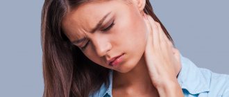
Pain in the spine (dorsopathies)
Dorsopathies in medicine refer to a variety of pain in the back (and limbs) caused by degenerative-dystrophic diseases of the spine.
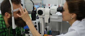
If the patient experiences pain for longer than 12 weeks, it is called chronic. The causes of dorsopathies are clarified based on the results of MRI of the spine (CT is most often only an auxiliary method). Treatment is carried out by neurologists, osteopaths, algologists or spinal neurosurgeons.
Degenerative-dystrophic diseases of the spine often lead to compression of the nerve endings and roots of the spinal cord, so the pain syndrome can be very intense and spread to other associated parts of the skeleton: arms, feet, chest, etc.
Thus, problems with the spine are indicated not only by dorsopathies localized in the back area, but also by other pains - with irradiation and neuralgic symptoms, for example:
- Symptom of intermittent claudication;
- “Shooting” pain in the leg;
- Numbness in the arms or legs;
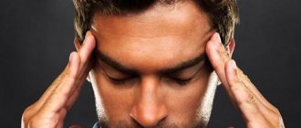
- Dizziness and headaches;
- Weakness of the limbs, marked decrease in muscle tone;
- Loss of sensation in limbs.
As a rule, pain becomes more intense after physical activity and goes away during rest, but it also happens the other way around, when it is at rest that the patient begins to experience pain.
DDZD of the cervical spine causes pain in the neck and forearm, paresis of the arms, headaches and dizziness, tinnitus and blurred vision.
DDSD of the thoracic spine can manifest as pain behind the sternum, which intensifies with breathing and coughing. The pain spreads along the ribs with radiation to the back. The symptoms are somewhat reminiscent of angina pectoris.
DDSD of the lumbosacral spine is associated with intense lower back pain radiating to the legs (numbness, weakness, lameness). It becomes difficult for the patient to play sports, walk, bend over, or remain in a static position for a long time.
Symptoms
The clinical picture is usually long-lasting and asymptomatic. Changes in the musculoskeletal system can cause the following symptoms:
- numbness of the limbs;
- sensation of a foreign body in the joint;
- a feeling of stiffness and a decrease in the range of motion of joints - flexion, extension, rotation, bending, etc.;
- discomfort and pain in the joints, especially during physical activity;
- neurological manifestations caused by pinched nerve endings: ringing in the ears, dizziness, headache, auditory and visual disturbances;
- it is difficult to inhale and exhale;
- with lesions of the hip joint, disorders affecting internal organs, for example, the genitourinary system, are possible.
Marble disease is manifested by anemia due to the excessive development of the number of bone osteophytes against the background of a sharply reduced volume of red bone marrow.
Osteoarthritis can lead to complete immobility, difficulty breathing and disability of the patient.
Spinal injuries
Spinal fractures can result from accidents, sports injuries, falls from a height, blows and bruises. Secondarily, they occur against the background of osteoporosis, aggressive growth of hemangiomas or other tumors. It should be noted that not all spinal fractures are visible on x-rays - due to the low resolution and two-dimensional nature of the visualization, the capabilities of this method in diagnosing fractures and injuries are limited. The “gold standard” is considered to be a CT scan of the spine - a high-precision volumetric slice-by-slice scan that allows you to obtain an authentic 3D model of the body area under study.
Spinal injuries are classified into three groups, depending on the severity of the injury:
- Vertebral fractures (compression wedge-shaped, comminuted) without distraction and axial torsion - the spinal cord and important neurovascular components are not affected.
- Fractures of the vertebral bodies with distraction (rupture) of the anterior and/or posterior segments, there are fracture-dislocations, local compression of the spinal cord is possible.
- Vertebral fractures with distraction, displacement (twisting) of the vertebrae, compression of nerve fibers and parts of the spinal cord.
At the moment, computed tomography is the only diagnostic method that provides comprehensive answers about the nature of the bone injury and a complete visual picture.
Diagnosis of cervical sclerosis
The most accurate diagnostic methods will be instrumental x-ray methods. The Yusupov Clinic has all the necessary equipment to determine this pathology. Our specialists have significant experience in diagnosing spinal diseases using various images.
Sclerosis of the endplates is a sign of the presence of a pathological process caused by the underlying disease. For its differential diagnosis and confirmation of diagnosis, the following instrumental examinations are used:
- radiography;
- ultrasonography;
- magnetic resonance imaging;
- computed tomography;
- osteodensitometry;
- Sometimes a genetic test is required to rule out a genetic disease.
These imaging methods make it possible to determine the degree of damage to the joint, changes in its structure and the amount of connective tissue growth. Dense structures will appear lighter than others. An extremely interesting method is densitometry, which can be used to assess bone mineral density. It can be ultrasonic, x-ray and photon. The essence of the method is to compare the patient’s indicators with special criteria, which are statistical data. Use Z and T-tests. The T-score corresponds to normal peak bone mass, that is, the average value for the age at which bone mineral density in a given area reaches its maximum. The Z-score is characterized by the average value for a given age. The norm for a T-score is −1 or higher. From -1 to -2.5 a decrease in mineral density is determined; less than -2.5 – osteoporosis. The Z-score is normally positive. Negative values indicate low bone density.
Osteochondrosis
Behind the term “osteochondrosis” is a large group of pathologies that lead to degenerative processes affecting the intervertebral discs. As a result, they become smaller, delaminate, lose elasticity and ultimately rupture, leading to the formation of a protrusion or intervertebral hernia.
Osteochondrosis is considered to be the starting point for the development of other DDDDs, but in itself it is not their cause. A predisposition to osteochondrosis can be hereditary, but more common causes include metabolic disorders, posture and physical inactivity (weak back muscles, poor circulation and, accordingly, nutrition of the intervertebral discs).
Osteophytes of the spine
Osteophytes are hard, abnormal growths on the bone tissue around a vertebra. Such growths have a jagged shape and can painfully compress nerve endings and narrow the lumen of the vertebral foramen. Sometimes osteophytes are formed from the tissues of dead ligaments. In most cases, they do not affect quality of life due to their small size. However, if osteophytes grow over time, they ultimately cause acute compression, damage to surrounding tissue, and inflammation. Symptoms of spinal osteophytes are dull pain in the back or neck, which intensifies when walking or standing (sometimes radiating to the leg or arm) and paresis. In this case, osteophytes require surgical removal. If they are found along with the operated hernia, they can complicate surgical tactics, so in this case they are also removed.
Diet for osteosclerosis
Diet for osteoarthritis of the knee joint
- Efficacy: no data
- Timing: constantly
- Cost of products: 1700-1800 rubles. in Week
The best diet for osteosclerosis is a diet that enhances metabolism, strengthens and makes the body strong, so the menu should include:
- proteins - optimal sources are dietary poultry, eggs, seafood, fish and vegetable protein from legumes and other vegetables;
- complex carbohydrates - cereals, crispbread, whole grain bread, vegetables, dried fruits, fruits and berries, which additionally contain many vitamins immune- boosting substances ;
- calcium – you need to consume dairy and fermented milk products every day;
- healthy fats – various unrefined vegetable oils, seeds and nuts.
Spinal stenosis
Stenosis is a pathological narrowing of the spinal canal caused by hyperplasia of bone tissue, tumor growth, and the entry of fragments of osteochondral fragments during injury into the space occupied by the roots of the spinal cord or nerve fibers, which leads to their compression. Spinal stenosis manifests itself as pain with neuralgic symptoms. The most common complaint is back pain, which increases with walking and decreases with sitting (flexing the spine). An accompanying symptom is most often numbness and weakness of the legs, pain of a “shooting” nature. The latter is typical for lesions of the lumbosacral segment.
Pathogenesis
The mechanism of osteosclerosis is based on the opposite condition to osteoporosis , reflecting reparative processes in the bone - in the form of an increase in the bone-forming ability of osteoblasts. This nonspecific reaction in the form of an increase in bone mass occurs due to periosteal and endosteal ossification in response to various diseases, injuries and processes in the body. To stimulate true osteosclerosis, pathological processes are sufficient, the substrate of which is located in the spaces between the bone beams.
In addition to pathological changes in the bone—thickening of the trabeculae and compact substance—cancellous bone tissue also undergoes changes, taking on the appearance of a narrow-loop structure or a compact mass.
Intervertebral hernia
Intervertebral hernia is a local displacement of disc material (nucleus, cartilaginous nodule, fragments of the fibrous ring) into the spinal canal and intervertebral foramen. Intervertebral hernias can also cause compression of nerve fibers and spinal cord roots.
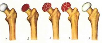
Depending on the stage of development of the disease, the following types of hernias are distinguished:
- Protrusion is a displacement of disc material (nucleus pulposus, annulus fibrosus) towards the spinal canal. Visualized as a small protrusion. The fibrous ring is stretched.
- Extrusion is a more pronounced displacement of disc material into the spinal canal. Often leads to damage to the fibrous ring.
- Sequestration is the prolapse of the nucleus pulposus and fragments of disc material into the spinal canal with rupture of the fibrous ring. Provokes severe pain.
Schmorl's hernias are cartilaginous nodules that invade the bodies of adjacent vertebrae and deform them. The depression forms at the border of the vertebra and the disc. Large Schmorl's hernias can provoke immune reactions in the body, accompanied by pain. However, the vast majority of such hernias do not affect the quality of life and do not require surgical treatment.
Prevention: useful tips
Like any other disease of the spine, osteosclerosis is better prevented. Since treatment can be very long and not always successful. This is especially true for patients at risk for developing this disease. Therefore, it is important to follow the advice and recommendations of specialists on the prevention of osteosclerotic changes in the bones of the spine.
- Hypervitaminosis of vitamin D. One of the main causes of increased bone density.
Therefore, persons living in southern regions with sufficient daylight hours do not need to take this vitamin additionally in the form of a solution. An exception may be the winter. Blood levels of vitamin D - To maintain normal bone metabolism, you should eat a balanced and regular diet. The diet should contain sufficient amounts of magnesium, calcium, phosphorus and iron.
- For blood diseases, it is important to undergo the necessary treatment in a timely manner to prevent hypochromic anemia.
- Compliance with recommendations for ensuring back health is of great importance in preventing osteosclerosis. Regular exercise, long walks, and sleeping on an orthopedic mattress are necessary.
In some cases, wearing an orthopedic corset may be a preventive measure. The orthopedic doctor who issued the prescription or referral should tell you how to choose and use it correctly.
Video - Bone Mineral Density
Spinal osteosclerosis is a serious disease of the musculoskeletal system. If detected early, it can be quite successfully regressed. Therefore, if you experience any back pain, you should immediately consult a doctor. To identify pathology in the early stages, when the pain syndrome is mild or completely absent, people at risk are recommended to undergo an annual X-ray examination of the spine. Treatment of osteosclerosis is carried out mainly by surgical methods. The question of the advisability of transplantation is decided individually, taking into account the overall picture of the disease.
Spinal neoplasms
Spinal tumors can be benign or malignant. The prevalence of the latter is relatively low, and most often it is not bone cancer that is diagnosed, but spinal cord cancer, which paralyzes the entire human body.
As benign tumors grow, they can also have disabling consequences. Neoplasms can compress nerve fibers and roots of the spinal cord, causing pain, neuralgia, and muscle atrophy.
Spinal injuries, bone diseases, and heredity increase the risk of spinal tumors.
Benign tumors of the spine include:
- Osteoma - develops from bone cells in the pedicles of the posterior part of the vertebra. It manifests itself as back pain, which bothers you at night. They are poorly visualized on X-ray; the only accurate method of visualizing them is CT.
- Bone cyst - a tumor forms on the back of the vertebra or affects it completely. Most often found in the cervical spine. If there are compressions, they are removed surgically.
- Giant cell tumors are formed primarily from bone cells and represent a deviation from their normal development. They reach large sizes and therefore cause pain. They are removed surgically and require pre- and postoperative monitoring, as they can degenerate into a malignant tumor of the spine.
- Granuloma is a spinal tumor, usually small in size. Accompanying damage to the vertebra, namely the thinning of its body. Vertebral granuloma can be an independent disease, or it can indicate malfunctions and damage to other organs. Treatment is carried out using surgical or radiation methods.
Malignant neoplasms of the spine include:
- Metastases are secondary foci of oncology, formed from malignant cells that migrated from other organs, bones and joints through the circulatory system or lymph flow. As a rule, the primary focus is located close, and depending on the segment of the spine, it can be the lungs, abdominal cavity, mammary gland, prostate gland, etc.
- Osteogenic sarcoma is a malignant tumor of bone tissue. Common among children (adolescents) and elderly patients.
- Myeloma (or multiple myeloma) is a malignant tumor of bone tissue. Multiple myeloma often affects not only the spine, but also other osteochondral structures. The highest prevalence of the disease is among patients over 40 years of age.
Treatment of osteosclerosis of the knee joint
Treatment for osteosclerosis of the knee joint should be started as early as possible. It is necessary to reconsider your diet, to eliminate the deficiency of calcium, magnesium, phosphorus, and some other vitamins and minerals. It is also necessary to conduct an examination of the large intestine, since if its function is impaired, there is a high probability of developing secondary osteoporosis even at a young age.
Treatment can be carried out using manual therapy methods. For example, our clinic uses the following methods:
- reflexology (acupuncture) allows you to influence biologically active points on the human body and trigger the processes of regeneration of affected tissues due to the hidden reserves of the body;
- laser exposure to sclerotic lesions stimulates rapid restoration of the physiological structure of the tissue;
- therapeutic exercises and kinesiotherapy restore the natural tone of the muscle fiber, enhance the process of diffuse nutrition of the cartilage tissue of the joint;
- osteopathy and massage improve microcirculation of blood and lymphatic fluid in the affected area, accelerate the process of restoring the elasticity of soft tissues;
- physiotherapy accelerates metabolic processes at the cellular level.
In the process of treating osteosclerosis of the knee joint, an integrated approach is important. The doctor gives the patient individual recommendations that eliminate the effect of negative risk factors. If rheumatoid or autoimmune pathology is detected, it is important to first normalize the patient’s condition and stop the inflammatory process. Then it is necessary to develop an individual rehabilitation course, during which the functionality of the knee joint will be completely restored.
If you are looking for safe and effective treatment for osteosclerosis of the knee, schedule a free initial appointment with a podiatrist at our manual therapy clinic today. Fill out the doctor appointment form located further down the page.
Spondylolisthesis of the vertebral segment
Spondylolisthesis is an abnormal displacement or slipping of a vertebra. As a result, the spinal segment loses stability. The consequences can be traumatic and dangerous - sometimes the displacement of one vertebra in relation to another (upper to lower) reaches 75%.
Spondylolisthesis of the spinal segment can be a consequence of injury, surgery, improper bone development, systematic improper loads, and diseases of the osteochondral tissue. Causes pain (passes after rest) with neuralgic symptoms, dysfunction of the pelvic organs.
To stabilize the spinal segment, the patient may be indicated for neurosurgical surgery with the installation of an implant, metal structure, or interbody cage.
A CT scan revealed a degenerative disease of the spine - what to do?
If back pain does not bother you, but the “accidental finding” on a CT or MRI turned out to be “degenerative-dystrophic disease of the spine,” you should clarify what kind of disease it is and assess the possible health risks in the future.
Timely consultation with a neurologist or osteopath will help develop measures for effective treatment of DDSD and prevention of complications. The worst option would be to ignore the problem and “let go” of the situation to the point of traumatic consequences and the need for surgical intervention.
If there is severe pain associated with an intervertebral hernia, stenosis or other type of DDSD, the patient should consult a neurologist, algologist or neurosurgeon. The pain must be relieved, as it makes it difficult to move and provokes the development of depression, thus aggravating the situation. The patient will also be prescribed therapy (NSAIDs, therapeutic blockades, exercise therapy, radiofrequency ablation, physiotherapy, etc.) or surgery will be recommended.
Treatment of cervical sclerosis
There is no etiotropic treatment, since this pathological process is a consequence of the underlying disease. After a thorough diagnosis and identification of the disease that caused osteosclerosis of the cervical spine, specialists at the Yusupov Hospital prescribe complex therapy specifically aimed at it. It should be borne in mind that therapy is extremely effective only in the initial stages, the symptoms of which do not bother the patient so much. Because of this, treatment is carried out later and the goal is not a cure, but to stop the progression. The changes that occur in the joint are irreversible and the sooner you see a doctor, the greater the chance of a complete recovery.
Treatment is multicomponent, it consists of drug therapy, physiotherapy, and in severe cases, surgical methods are added.
The following groups of drugs are used:
- non-hormonal anti-inflammatory drugs. Their goal is to eliminate the inflammatory process and reduce pain;
- analgesics - relieve pain;
- chondroprotectors - improve cartilage metabolism, slow down or stop its destruction, have a slight anti-inflammatory effect;
- muscle relaxants - help relieve muscle spasm that occurs as a protective reaction to reduce movement in the damaged joint;
- B vitamins, phosphorus, calcium are important for tissue regeneration.
You can also use various warming ointments to relieve pain. But this is a symptomatic treatment that only alleviates the condition.
Physiotherapy has proven itself extremely positively, since its use improves blood supply to the affected area, reduces inflammation and accelerates tissue regeneration, inhibiting further sclerosis.
Among the physiotherapeutic procedures that have proven themselves extremely positively:
- electrophoresis - improves blood flow in the affected area. If carried out together with the drug, it ensures its fastest penetration;
- ultra-high frequency therapy;
- mud and hydrogen sulfide baths.
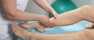
During an exacerbation, physical therapy and massage are prohibited. Their appointment is determined by the attending physician. Exercises should only be performed together with an instructor in a physical therapy room to prevent possible injuries. With the restoration of mobility, the strength of the back and neck muscles will increase. A muscle corset will stabilize the spine. Massage increases blood flow in the massaged area and reduces muscle spasm. In classic massage there are three main movements: stroking, rubbing and kneading.
If the problem is treated late, when osteophytes have arisen, surgical treatment is required, since they cannot be removed by any medications or physical therapy. But you can completely cure the joint.