Diseases of the cardiovascular system are in first place in mortality statistics in the world. Among the main factors in their development are heredity, obesity, physical inactivity, unhealthy diet, smoking and alcohol abuse. The risk of disease also increases with age. But most pathologies can be prevented by leading a healthy lifestyle and undergoing preventive examinations. If this fails, early diagnosis comes to the aid of patients.
Hardware research methods are not numerous, but one of the most common is ultrasound of the heart, which shows the size of its chambers, wall thickness, condition of the valves and the nature of contractions of the heart muscle.
When is it prescribed?
Ultrasound examination (echocardiography or EchoCG) is an informative and easy-to-use method, without which it is difficult to imagine diagnostics in modern cardiology. With its help, a sonologist (better known as an “uzist” or “ultrasound specialist”) can assess in real time the condition and functions of the organ and its surrounding tissues. Non-invasive diagnostics determines structural abnormalities and changes that can appear in various pathologies and developmental defects.
Thanks to its informativeness and high safety, cardiac ultrasound has become widely used in the diagnosis of cardiac diseases in adults and children. With its help, diseases of the myocardium, heart valves, pericardium, large vessels, including the aorta and pulmonary trunk, as well as other structural units of the cardiovascular and related systems are identified. Other benefits of such research include:
- lack of difficult-to-implement training recommendations;
- the ability to monitor current processes over time;
- affordable cost and high speed of execution;
- availability of mobile equipment for diagnosing urgent cases.
A referral for echocardiography is given to adult patients by a general practitioner or cardiologist, and to children by a pediatrician or pediatric cardiologist. The examination can be carried out for preventive purposes, in the presence of risk factors for the development of cardiac pathologies, for direct indications in the presence of obvious symptoms or diagnosed disorders.
Indications for use
The main advantage of echocardiography is the ability to diagnose pathologies even before the appearance of their first symptoms. But in general, indications for cardiac ultrasound include:
- complaints of shortness of breath, weakness, dizziness, swelling;
- cases of loss of consciousness, rapid heartbeat;
- difficulty breathing, blue fingertips and lips during physical activity;
- severe arrhythmia, so-called chest pain;
- detection of noises during auscultation;
- diseases with a risk of damage to the cardiovascular system, for example, rheumatism;
- often high blood pressure, diagnosed hypertension;
- the presence of changes in the electrocardiogram, for example, myocardial hypertrophy;
- suspicion of congenital defects in children, etc.
In some cases, echocardiography is performed once to confirm or refute existing suspicions, in others repeatedly at certain intervals to reliably track changes.
In addition, thanks to the active development of medical equipment and the expansion of its functionality for cardiac ultrasound, the indications for ultrasound may also be much wider. Thus, many models of modern devices are capable of combining classical ultrasound with Doppler ultrasound. This allows the specialist, during diagnosis, to track the direction and speed of blood flow in the chambers, which is important for a comprehensive assessment of the functioning of the organ and determination of disorders in its valve apparatus.
Contraindications
Along with safety and ease of implementation, another advantageous difference of ultrasound examination is the absence of absolute contraindications. In practice, specialists note only the relative difficulties they encounter when conducting diagnostics. Usually this:
- violations of the integrity of the skin in the area where the sensor is located (wounds, external manifestations of dermatological disorders, etc.);
- pronounced hair growth in the chest area;
- open injuries and deformations of the chest;
- extreme obesity, significant accumulation of fatty deposits in the examined area;
- performing ultrasound of the heart in newborns, etc.
There are no side effects or complications when performing ultrasound diagnostics. The safety of the method allows it to be used several times a week. There are no contraindications for it during pregnancy, since there is no threat to the health of the expectant mother and fetus.
But it is worth considering that some patients, in extremely rare cases, report an allergic reaction to the gel used for smooth sliding and better contact of the sensor with the body. In general, gels for ultrasound diagnostics are hypoallergenic, but exceptions to the rules may still exist. Therefore, when visiting a sonologist, it is better to immediately warn about a possible reaction. You can avoid it by replacing the gel, washing the examined area with soap immediately after diagnosis, or pre-applying baby cream to the skin, which will prevent the gel composition from penetrating the pores.
Use among pregnant women
During pregnancy, women are susceptible to many diseases. Due to changes occurring in the body, the load on the heart also increases.
Echocardiography is often performed on pregnant women
Indications for echocardiography:
- diabetes;
- hereditary predisposition to heart disease;
- if the patient fell ill with rubella while carrying a baby, or a high concentration of bodies for this disease was detected in the plasma;
- if in the first trimester the woman took any strong medications;
- in the presence of miscarriages in the medical history.
Ultrasounds are often performed on an unborn baby in the womb. The procedure is performed to detect heart defects in the fetus at an early stage and is performed at 18–22 weeks.
Preparing for the study
There are no special recommendations for preparing for an ultrasound examination. But in order for the results to be most reliable during ultrasound of the heart, it is necessary:
- exclude physical activity the day before;
- a day before, stop drinking coffee, alcohol, and strong tea;
- Avoid eating spicy and fatty foods before eating;
- do not eat for a couple of hours so that the stomach is not full and does not put pressure on the diaphragm;
- do not smoke 2-3 hours before;
- before entering the office, rest a little and calm down in the corridor;
- if there is thick hair on the chest, shave it off if possible;
- Before making an appointment, tell your doctor what medications you are taking and, if necessary, stop taking them for a while;
- lying on the couch, do not worry and remain as calm as possible;
- strictly follow all recommendations of a specialist.
In case of urgent need, echocardiography can be performed without any preparation. When planning a visit to a specialist, you do not need to take anything with you, in particular towels. Everything you need for a cardiac ultrasound is in the treatment room.
Progress of the procedure
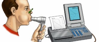
How is echocardiography done? The session takes no more than 30–40 minutes, and the patient does not experience discomfort or pain. No anesthesia is required. During the examination, the patient must remove clothes to the waist and lie down on the couch. The sensor is applied to several areas. This is the area of the jugular fossa, the area of the intercostal space to the left of the chest, the place where the sternum ends.
To ensure good contact of the sensor with the skin, the medical worker treats it with a special gel, which must be wiped with a napkin after the procedure.
How is ultrasound diagnostics of the heart done?
Echocardiography is a diagnostic method based on the action of ultrasonic waves that emanate from a sensor, penetrate the patient’s body and are reflected in a certain way from tissues of different densities. The received signals are converted into electrical impulses, and after processing by a computer they are displayed on the monitor in the form of specific two- or three-dimensional pictures. On the monitor, the sonologist or cardiologist receives a real-time image of the organ.
In addition to the standard equipment of the device, if necessary, equipment can be connected to create the Doppler effect. Doppler ultrasound of the heart provides the doctor with the opportunity to evaluate the color picture of blood flow in the chambers and determine the direction and speed of blood movement.
Ultrasound examination in its traditional sense is carried out in the supine or lateral position. Having taken into account all the recommendations for preparation, the person simply comes to the office, gives the direction and then does what he is told. The procedure algorithm looks like this:
- the person undresses to the waist and sits on the couch;
- the doctor applies a small amount of gel to the skin and distributes it over the area being examined;
- he moves a special sensor along the sternum and, if necessary, asks the patient to change position;
- During the procedure, the specialist adjusts the equipment parameters and takes appropriate measurements;
- assessing the picture displayed on the screen, the ultrasound specialist writes down the data;
- then within 5-20 minutes he fills out a form with the results and gives them to the visitor.
The ultrasound specialist does not interpret the results. The exception is when an ultrasound of the heart is performed directly by a cardiologist.
In addition to the traditional version of the diagnostic procedure, transesophageal echocardiography and special stress tests are also distinguished. In transesophageal diagnostics, the equipment sensor is inserted directly through the esophagus. In preparation for this procedure:
- It is recommended not to eat for about 4-6 hours and limit drinking;
- To reduce the gag reflex, local anesthesia of the pharynx and root of the tongue is used;
- if necessary, intravenous administration of certain drugs can be used for better tolerability;
- Patients need to remove dentures, if any.
When prescribing a cardiac ultrasound, the attending physician determines whether a transesophageal examination is worth doing. The procedure is very unpleasant, but it allows you to clearly see those parts that are not visible or are much less visible with traditional echocardiography. Indications for examination through the esophagus are severe cardiac pathologies and the consequences of unsuccessful surgical intervention. Contraindications include diseases of the esophagus, osteochondrosis of the cervical spine, diaphragmatic hernia and some other pathologies not related to the cardiovascular system. Also, this method is not recommended for examining children and pregnant women.
A stress test is in fact the same as an ultrasound of the heart, which shows the functioning and condition of the organ in real time. But, if standard echocardiography is performed at rest, then when performing stress tests on the body, on the contrary, a huge load is placed on the body. During the procedure, a person must run or walk quickly on a treadmill or exercise on an exercise bike. If he is unable to exercise, for example due to injury, to perform stress tests, special drugs are introduced into the body to artificially stimulate the active functioning of the organ being studied. In the rooms for conducting stress tests, it is necessary to have resuscitation equipment, in particular a defibrillator, an Ambu bag, etc. A similar study is relevant for ischemic heart disease. It is most informative for determining the early stages of myocardial diseases.
Description of the method
Many patients who have been referred for electrocardiography are interested in what it is and what the essence of the method is. Echo CG is performed in a hospital setting or at home using special equipment. For this, an ultrasound-emitting device, a special sensor and a transducer are used that transmits an image of the area under study to the screen.
Passing through the heart, ultrasound waves are absorbed and reflected by its tissues. Thanks to this, the device displays an image on the screen, from which a specialist can make a conclusion about the main parameters of the organ’s functioning.
Cardiac echocardiography is considered one of the most informative and health-safe methods for identifying pathologies.
What does an ultrasound of the heart show?
The results of an ultrasound of the heart and the indicators of its studied parts can only be interpreted by a doctor. The procedure itself helps:
- determine the size and parameters of the heart chambers;
- assess the condition of valves, arteries, blood flow, mobility of the heart muscle;
- measure the thickness of the myocardium and septa;
- determine the presence of fluid or neoplasms;
- detect congenital or acquired defects;
- diagnose coronary artery disease, hypertrophy, pericardial diseases;
- check for irregularities in the operation of the valve apparatus;
- identify chamber expansion, the presence of intracardiac thrombi, aneurysms, etc.
Together with other diagnostic methods in cardiology, such as electrocardiography, computed tomography or magnetic resonance imaging, echocardiography provides a comprehensive assessment of the state of the cardiovascular system. By assessing the results of a heart ultrasound, what the study shows and what changes are visible on the monitor, the doctor can make a reliable diagnosis, choose or adjust the most effective treatment. Or prescribe additional examinations if necessary.
Decoding EchoCG
After the examination, the doctor draws up a conclusion. First, a visual picture with the presumed diagnosis is described. The second part of the study protocol indicates the patient’s individual indicators and their compliance with standards.
Decoding the data obtained is not a final diagnosis, since the study can be done not by a cardiologist, but by an ultrasound diagnostic specialist.
It is the cardiologist, based on the collected medical history, examination results, interpretation of tests and data from all prescribed studies, who can draw accurate conclusions about your condition and prescribe the necessary treatment!
What indicators are considered normal?
Having received the results in hand, the cardiologist can interpret them. In this case, the specialist takes into account anamnesis, medical history, the effect of medications taken and many other related factors. To evaluate the results of cardiac ultrasound, the norm of indicators is prescribed in its standard version. Deviations from the norm do not always indicate the presence of serious pathologies. If you consult a doctor in a timely manner and start a correctly selected treatment, there is every chance of bringing the abnormal indicators back to normal.
| Indicator name | Women | Men |
| Left ventricular myocardial mass | From 95 to 142 g | From 135 to 180 g |
| Left ventricular mass index | From 71 to 88 g/m2 | From 71 to 92 g/m2 |
| Left ventricular end-systolic size | From 31 to 42 mm | |
| End diastolic size | From 45 to 58 mm | From 46 to 58 mm |
| Left ventricular wall thickness in diastole | Max 10mm | Max 11mm |
| Volume of blood ejection during left ventricular systole | From 60 to 100 ml | |
| Right ventricular wall thickness | 4.8 to 5 mm | 5 mm |
| Left atrium size | From 17.5 to 33 mm | From 18.5 to 33 mm |
| Left atrial end-diastolic volume | From 38 to 57 ml | From 50 to 82 ml |
| Right atrial end-diastolic volume | From 20 to 100 ml | |
| Thickness of the interventricular septum in systole | 5 to 9.0 mm | From 5 to 9.5 mm |
| Thickness of the interventricular septum in diastole | From 7.5 to 11 mm | |
| Aortic opening area | From 20 to 35 mm2 | |
| Thickness of the outer membrane of the pericardium | 1.2 to 1.7 mm | |
| Volume of fluid in the pericardial cavity | From 10 to 30 ml | |
Ultrasound parameters of the heart, normal for children from birth to 14 years
| Index | 0-1 m | 1-3 m | 3-6 m | 6-12 m | 1-3 g | 3-6 years | 6-10 years | 11-14 years old |
| LV EDC | 13-23 | 16-26 | 19-29 | 20-32 | 23-34 | 25-36 | 29-44 | 34-51 |
| TZS LV | 2-5 | 2-5 | 3-6 | 3-6 | 3-7 | 3-8 | 4-8 | 5-9 |
| LV ESD | 8-16 | 9-18 | 11-20 | 12-22 | 13-22 | 14-25 | 15-29 | 21-35 |
| Diameter AO | 7-13 | 9-15 | 10-16 | 10-17 | 11-18 | 13-21 | 13-26 | 15-30 |
| TM ZhPd | 2-6 | 2-6 | 2-6 | 2-6 | 2-6 | 3-7 | 4-8 | 5-8 |
| LA diameter | 9-17 | 10-19 | 12-21 | 14-24 | 14-26 | 15-27 | 16-31 | 19-32 |
| TSS PZhd | 1-3 | 1-3 | 1-3 | 1-4 | 1-4 | 1-4 | 1-4 | 1-4 |
| Pancreas diameter | 2-13 | 2-13 | 2-14 | 3-14 | 3-14 | 4-15 | 5-16 | 7-18 |
What is the difference between ECHO and ultrasound of the heart?
Not everyone knows how to do an ultrasound of the heart, or how ECHO differs from ultrasound. In essence, echocardiography and ultrasound are the same thing. Ultrasound emanating from the equipment's sensors is reflected from the tissue and captured by the device for subsequent registration and translation into an image. The name echocardiography was not chosen by chance, because these signals are formed according to the echo principle, reflecting differently from tissues with different acoustic densities.
| Interpretation of ultrasound (EchoCG) of the heart | To the list of articles |