- Incidence of soft tissue sarcomas
- Risk factors for soft tissue sarcomas
- Diagnosis of soft tissue sarcomas
- Treatment of soft tissue sarcomas
- What happens after treatment is completed?
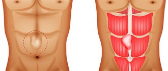
Soft tissue sarcomas are malignant tumors that develop from fat, muscle, nervous tissue, blood and lymph vessels, and joint tissue. They can occur in any part of the body. 50% of them are detected on the upper and lower extremities, the rest - on the trunk, in the head and neck, in the internal organs and retroperitoneum (posterior abdomen).
There are many types of soft tissue tumors, and not all of them are malignant.
TUMORS FROM ADITY TISSUE
Lipomas are the most common benign tumors of adipose tissue. Most of them are located under the skin. Lipoblastomas and hibernomas are also benign tumors of adipose tissue.
Liposarcomas are malignant tumors of adipose tissue. They are most often found on the thigh and in the retroperitoneal space in people aged 50-65 years. Some liposarcomas grow very slowly, while others grow rapidly.
TUMORS FROM MUSCLE TISSUE
There are two types of muscles: smooth and striated. Smooth muscles are located in the internal organs (stomach, intestines, blood vessels, uterus) and contract involuntarily, regardless of our desire and we cannot control their activity. Striated (skeletal) muscles allow movement of the arms, legs and other parts of the body. These movements depend on our desire.
Leiomyomas are benign smooth muscle tumors and are most often found in women in the uterus.
Leiomyosarcomas are malignant smooth muscle tumors that can develop in any part of the body, but are most often localized in the retroperitoneum and internal organs. They are rarely found in the muscles of the arms and legs.
Rhabdomyomas are benign tumors of striated muscles.
Rhabdomyosarcomas are malignant tumors of striated muscles. Most often they develop on the upper and lower extremities, but can also be detected in the head and neck, bladder and vagina. The disease is most often diagnosed in children.
Neurofibromas, schwannomas, and neuromas are benign tumors of the nerves.
Malignant schwannomas and neurofibrosarcomas (neurogenic sarcomas) are malignant forms of the disease.
Ewing group tumors include osseous Ewing sarcoma, extraosseous Ewing sarcoma, and primitive neuroectodermal tumor (PNET) . All of these tumors share some common features of neural tissue. They are common in children and rare in adults.
Joints are surrounded by synovial tissue, which produces a special fluid that reduces friction when the articular surfaces move. The synovium can be a source of tumors.
Nodular tenosynovitis (tenosynovitis) is a benign tumor of the joint tissue. It most often occurs on the hands, usually in women.
Synovial sarcoma is a malignant tumor that most often develops in the knee joint in young adults.
Hemangiomas are benign tumors of blood vessels. They are quite common and are often discovered at birth. They can be located both on the skin and in internal organs. Sometimes they go away on their own without treatment.
Lymphangiomas are benign tumors of the lymphatic vessels.
Angiosarcomas can develop from both blood (hemangiosarcoma) and lymphatic (lymphangiosarcoma) vessels. Sometimes these tumors occur in parts of the body that have previously been treated with radiation. Kaposi's sarcoma develops from cells that resemble the epithelium (lining cells) of blood and lymph vessels. Most often, this tumor occurs when the immune system is severely suppressed, for example, in patients with AIDS or in people who have undergone an organ transplant.
Hemangioendothelioma is a low-grade malignant tumor of blood vessels. It is less aggressive than hemangiosarcoma, but can destroy nearby tissues and metastasize (spread) to distant organs (liver, lungs).
Glomus tumors usually arise under the skin of the fingers and are benign.
Hemangiopericytoma is a malignant tumor that most often develops in adults on the lower extremities, pelvis and retroperitoneum.
Tendons and ligaments belong to fibrous tissue and can become sources of various tumors.
Fibromas, elastofibromas, superficial fibromatosis, and fibrous histiocytomas are benign tumors of fibrous tissue.
Fibrosarcoma is a malignant tumor that is most often detected at the age of 30-55 years on the upper and lower extremities and trunk.
Desmoid tumor (aggressive fibromatosis) has features of a benign and malignant tumor. It does not spread to distant organs, but can spread locally, leading to death.
Dermatofibrosarcoma is a low-grade tumor that occurs under the skin of the extremities and trunk. It affects tissues close to the tumor, but rarely metastasizes.
Malignant fibrous histiocytoma is the most common soft tissue tumor of the extremities in older adults. Less commonly, it is detected in the retroperitoneal space.
Myxoma is a benign tumor that usually occurs in the muscles, but does not develop from muscle cells. Myxoma cells produce a mucus-like substance.
Malignant mesenchymoma is a rare tumor that has features of several types of sarcomas.
Alveolar soft tissue sarcoma is rare in young adults and is usually located on the lower extremities.
Epithelioid sarcoma most often develops under the skin of the upper and lower extremities in adolescents and young adults.
Clear cell sarcoma is a rare tumor that occurs in the tendon area and is somewhat similar to melanoma (a malignant pigmented tumor).
Desmoplastic small cell tumor is a rare variant of sarcoma in adolescents and young adults and is usually found in the abdomen.
Inflammation and trauma can lead to the formation of nodes under the skin and muscles that resemble a true tumor. These conditions include nodular fasciitis and myositis ossificans.
FIBROHISTIOCYTIC TUMORS AND TUMOR-LIKE LESIONS
Xanthoma
A rare disease, most often localized in the skin. Occurs in people with impaired lipid metabolism, usually multiple. Also localized in tendons. Presented by small nodules, partly of the xanthelasma type.
Juvenile xanthogranuloma
A small nodule in the dermis or subcutaneous tissue. Disappears spontaneously.
Fibrous histiocytoma
It is more common in middle age and is localized mainly on the lower extremities. Usually has the shape of a dense knot up to 10 cm, grows slowly. After surgical removal, recurrences are rare.
Survival prognosis
Survival depends primarily on the stage of the tumor. The relative five-year survival rate for sarcomas is about 50% (including Kaposi's sarcoma):
- For localized sarcoma - 83%.
- When the tumor spreads to regional lymph nodes - 54%.
- If there are distant metastases - 16%.
Observed five-year survival rate for soft tissue sarcomas of the arms and legs:
- At stage I: 90%.
- At stage II: 81%.
- At stage III: 56%.
- At stage IV: no data.
If the tumor is not in the extremities, the prognosis is worse. Thus, with sarcoma of the retroperitoneal space, the survival rate is 40–60%.
The ten-year survival rate is approaching five years. If a patient is still alive five years after he was diagnosed with the disease, this often gives reason to believe that recovery has occurred.
BENIGN TUMORS OF ADIPOSE TISSUE
Lipoma
One of the most common benign tumors (30-40%). It can occur anywhere there is fatty tissue. When localized in the dermis, it is usually encapsulated, in other parts of the body it is poorly demarcated. Tumors localized in the retroperitoneal space can become malignant; other localizations practically do not become malignant. Lipomas are often multiple and sometimes develop symmetrically. Their growth is not related to the general condition of the body. The tumor has the shape of a lobular node. With long-term existence in the lipoma, dystrophic changes, calcification, and ossification can develop.
There are numerous variants of mature fatty tumors, which differ from the classic lipoma both in clinical manifestations and in some morphological features.
Myelolipoma
A rare tumor, most often found in the retroperitoneum, pelvic tissue, and adrenal glands. Does not become malignant.
Subcutaneous angiolipoma
Numerous painful nodes. It occurs more often in young men on the front wall of the abdomen, on the forearm.
Spindle cell lipoma
It is observed more often in adult men (90%). The node is round in shape, dense, slowly growing, most often localized in the area of the shoulder joint and back. Recurrence and metastasis after excision have not been described, despite the fact that the tumor can infiltrate surrounding tissues.
In chondro- and osteolipomas, metaplastic areas of bone and cartilage tissue are detected.
Benign lipoblastomatosis
It is divided into nodular (kind lipoblastoma) and diffuse (kind lipoblastomatosis) forms. Boys under 7 years of age (88%) get sick more often. The tumor is localized on the lower limb, in the buttocks and on the upper limb - the shoulder girdle and hand. Lesions of the neck, mediastinum, and trunk have also been described. The tumor node is encapsulated, lobulated, spherical in shape, and can reach 14 cm. After surgical treatment, relapses are possible, sometimes repeated. Metastases have not been described.
Gebernoma (fetal lipoma)
Lipoma from lipoblasts, pseudolipoma is an extremely rare tumor, localized in places where there is brown fat (neck, axillary region, sinus, mediastinum). Presented as a lobular node, usually small in size. Does not recur and does not metastasize.
Treatment
The main method of treating muscle cancer is surgery. Excision of the pathological focus should be carried out together with the surrounding tissues. This way you can reduce the likelihood of relapses.
Self-medication is dangerous with complications!
Attention
Despite the fact that our articles are based on trusted sources and have been tested by practicing doctors, the same symptoms can be signs of different diseases, and the disease may not proceed according to the textbook.
Pros of seeing a doctor:
- Only a specialist will prescribe suitable medications.
- Recovery will be easier and faster.
- The doctor will monitor the course of the disease and help avoid complications.
find a doctor
Do not try to treat yourself - consult a specialist.
Radiation therapy is prescribed in cases where for some reason it is impossible to carry out radical removal of the tumor. It is also necessary to stop the growth of pathological cells some time before the planned surgical operation.
Chemotherapy involves treating cancer tumors with cytotoxic drugs. This treatment method is applicable in situations where multiple metastases have spread throughout the body.
After the tumor is removed, the patient is prescribed medications that help strengthen the immune system.
BENIGN TUMORS OF MUSCLE TISSUE
Tumors of muscle tissue are divided into smooth muscle tumors - leiomyomas, and striated muscle tumors - rhabdomyomas. Tumors are quite rare.
Leiomyoma
Mature benign tumor. Occurs at any age in both sexes. It is often multiple. The tumor can become malignant. Treatment is surgical.
Leiomyoma, developing from the muscular wall of small vessels - small, often multiple, poorly demarcated and slowly growing nodes, often with ulcerated skin, is clinically very similar to Kaposi's sarcoma.
Genital leiomyoma is formed from the muscular lining of the scrotum, labia majora, perineum, and nipples of the mammary gland. May be multiple. Cellular polymorphism is often observed in the tumor. Hormone dependent. Treatment is surgical.
Angioleiomyoma from the trailing arteries
Clinically, a sharply painful tumor that can change size under external influences or emotions. The sizes are usually small, more often found in older people, on the limbs, near the joints. It is characterized by slow growth and a benign course.
Rhabdomyoma
A rare mature benign tumor based on striated muscle tissue. Affects the heart and soft tissues. It is a moderately dense node with clear boundaries, encapsulated. Metastases of rhabdomyoma have not been described. Relapses are extremely rare. Microscopically, 3 subtypes are distinguished - myxoid, fetal cell and adult. Rhabdomyoma of the female genitalia is also distinguished. The adult type mainly recurs.
Causes
To date, medicine cannot accurately determine the cause of the formation of a malignant tumor in muscle tissue. However, experts identify a number of factors that can trigger the development of myosarcoma.
Tissue damage
As a result of injuries or medical operations, active tissue restoration and cell division occur. Under such circumstances, the immune system cannot fully identify and destroy atypical cells, which subsequently cause cancer.
The formation of scars and scars can lead to the development of a tumor 1 to 3 years after the wound has healed.
Chemical substances
Exposure to carcinogens negatively affects the cell nucleus, which promotes DNA mutation. This will lead to changes in the structure of cells and their abnormal development in the future.
Ionizing radiation
Radiation therapy, working in hazardous industries, and living in areas with high levels of radiation have a negative effect on muscle tissue.
Precancerous diseases
It is believed that some types of benign neoplasms (rhabdomyomas, leiomyomas) have a tendency to malignancy.
Hereditary predisposition
Often, muscle tumors are congenital and are diagnosed in close relatives. In every third patient, myosarcoma is combined with dysfunction of the cardiovascular, digestive or central nervous systems.
Active cell growth also occurs in childhood and adolescence. Some of them remain immature and subsequently cause pathological processes.
BENIGN TUMORS OF BLOOD AND LYMPHATIC VESSELS
These lesions include various processes, a significant number of them are considered in dermatology. Some of them relate to malformations of the vascular system of a tumor-like nature, some to true tumors.
Capillary angioma
True neoplasm with proliferation of endothelial cells.
Benign hemangioendothelioma
Congenital pathology, occurs in newborns and infants, more often in girls, with localization in the head area.
Capillary hemangioma
After lipoma, the most common tumor of soft tissues, often multiple, reaches its maximum size by 6 months of age; with multiple lesions, localization in internal organs is possible
Cavernous hemangioma
A formation consisting of bizarre sinusoid-type cavities of various sizes. Localized in the skin, muscles, internal organs. Has a benign course.
Senile hemangioma
A true tumor is characterized by proliferation of capillaries followed by their cavernization with secondary changes.
Hemangioma
A mature benign tumor of vascular origin, common. It most often affects middle-aged people and is localized on the mucous membrane of the nose, lips, skin of the face, extremities, and in the mammary gland. It is a clearly demarcated grayish-pink node 2-3 cm in size. The tumor can often become malignant and develop into angiosarcoma.
Arterial angioma
A conglomerate of malformed vessels, no signs of a tumor.
Glomangioma (glomus tumor, Barre-Masson tumor)
It occurs as an isolated tumor or as multiple disseminated familial glomusangioma. The tumor is benign and occurs in older people, in the hands and feet, most often in the nail bed area. May affect the skin of the lower leg, thigh, face, and torso. In isolated observations, it was observed in the kidneys, vagina, and bones. When localized in the skin, the tumor is sharply painful. Does not recur and does not metastasize.
Hemangiopericytoma
It is rare and can occur at any age. Localized in the skin, less often in the thickness of soft tissues. It looks like a delimited dense red node. The tumor can become malignant - giving relapses and metastases, it is considered a potentially malignant process. Malignancy in up to 20% of cases has been described in adults. The process in children is benign.
Lymphangioma
It is observed more often in children as a malformation of the lymphatic vessels, but can occur at any age. Most often localized on the neck, oral mucosa.
Possible complications
The consequence of cancerous muscle tumors is often relapses. Almost every second patient experiences repeated cell mutation over time.
In addition, this pathology has the ability to spread throughout the body through the circulatory and lymphatic systems, affecting neighboring tissues, lymph nodes, bone structures and internal organs. Most often the disease affects the lungs and liver.
If a tumor develops on the head, complications may include paralysis of the facial muscles, as well as complete or partial loss of vision.
In advanced stages of the disease, the patient's death is very likely.
BENIGN TUMORS AND TUMOR-LIKE DISEASES OF SYNOVIAL TISSUE (JOINTS)
Benign synovioma without giant cells
The existence of benign synoviomas is debated. Most authors are inclined to believe that all synoviomas are malignant, regardless of the degree of maturity. The tumor mainly affects the knee joint, in the form of small dense nodes. Treatment is surgical, but patients should be observed for 5-9 years. The disease can cause relapses and metastases.
Benign giant cell synovioma (nodular tenosynovitis)
Pseudotumor process occurs quite often. In 15% the process occurs in the area of the synovial membrane of the joints, in 80% in the tendon sheaths, in 5% in the mucous bursae. It is a nodular formation, most often localized on the fingers, less often on the feet, and even more rarely in the area of large joints. Favorite localization is the interphalangeal joints. It is more common in women 30-60 years old. If it persists for a long time, it can cause atrophy of surrounding tissues, including bones. The process often recurs, most of the relapses are associated with incomplete removal. Does not give metastases.
Pigmented villonodular synovitis
It is located inside the shell of the joints, most often in the area of the knee, elbow and shoulder joints. Occurs in middle age. The etiology is not clear.
Diagnostics
The examination begins with an external examination of the patient and palpation of the problem area, studying the medical history and clarifying existing complaints. If muscle cancer is suspected, additional diagnostic methods are prescribed.
Using ultrasound, the location of the problem area, its depth and its effect on neighboring tissues are determined.
Thanks to CT and MRI, they obtain an image of a cancer tumor in three-dimensional space and detect metastases.
The use of radiography is necessary if pathological cells affect bone tissue.
To determine the type of myosarcoma, a puncture is done, and the resulting biomaterial is sent for research to the laboratory.
BENIGN TUMORS OF PERIPHERAL NERVES
Traumatic or amputation neuroma
Arises as a result of post-traumatic hyperregeneration of the nerve. It is a small painful node.
Neurofibroma
A single, slowly growing benign tumor of the mesenchymal sheath of the nerve trunk of any location, but most often develops on the sciatic nerve and intercostal nerves. Occurs in people of any age. Clinically defined as a small, densely elastic consistency with a smooth surface of the tumor node, upon palpation of which the pain radiates along the nerve. Some tumors can reach large sizes. Tumor growth can occur both to the periphery of the nerve and in the thickness of the nerve trunk, which is revealed during its morphological examination.
Treatment is surgical. The prognosis is good. A special disease is multiple neurofibromatosis (Recklinghausen's disease), which belongs to the group of dysplastic processes. Cases of malignancy of one of the multiple neurofibromas in this disease have been described.
Chemotherapy for sarcomas
Depending on the type and stage of sarcoma, chemotherapy can be used in combination with surgery or alone. Various chemotherapy drugs are prescribed: ifosfamide, doxorubicin, docetaxel, dacarbazine, cisplatin, oxaliplatin, methotrexate, gemcitabine, vincristine, paclitaxel, eribulin, vinorelbine.
A combination of drugs called MAID is used: Adriamycin, ifosfamide, dacarbazine and mesna, a drug that helps protect the urinary system from the toxicity of chemotherapy drugs.