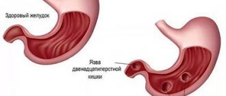
One of the manifestations of urolithiasis is the formation of stones in the bladder. This pathology is accompanied by pain, impaired urination, and blood in the urine. To diagnose the disease, the patient is prescribed an ultrasound examination of the urinary tract, a general urinalysis, cystoscopy, and cystography. Therapeutic measures consist of fragmentation and removal of stones. The contact method (or lithotripsy) or surgical intervention - open cystolithotomy - can be used.
general information
The formation of stones in the bladder is called cystolithiasis. Just like stones in the kidneys, ureter, urethra, this is a manifestation of urolithiasis. Stones are formed due to impaired physicochemical properties of urine (organic and inorganic compounds dissolve in it), the presence of physiological factors (congenital or acquired metabolic disorder - metabolic, inflammatory, medicinal, etc.).
Considering the place and mechanism of formation, stones come in different sizes, quantities, shapes, and chemical compositions. They also differ in their consistency and surface.
In most cases, the disease is diagnosed in men whose age exceeds 50-55 years. However, stones can also form in children.
Prevention of stones
The main direction in the prevention of urolithiasis is the normalization of metabolism.
If metabolic processes are not normalized, then relapse of the disease is inevitable.
The following preventive measures are recommended:
- daily physical activity;
- giving up alcohol;
- maintain normal weight;
- drink about 2 liters of liquid per day;
- reduce salt intake;
- When determining the type of stones, follow dietary recommendations.
- promptly treat inflammatory diseases of the urinary organs.
- be regularly examined by a urologist or nephrologist.
Why do they appear?
The most common reason that contributes to the formation of bladder stones is the presence of bladder outlet obstruction. This is not the name of the disease, but rather a whole list of possible pathologies. Such pathologies sooner or later lead to a violation of the removal of the full volume of urine from the body. As a result, stagnation occurs and urine accumulates. Subsequently, salt crystals form. They are the basis of a future bladder stone.
In addition, there are a number of other reasons that contribute to the formation of stones:
- Diseases of the gastrointestinal tract.
- Trauma, pathology of the skeletal system.
- Inflammatory process in the bladder.
- The connection between the bladder and the central nervous system is damaged. This is called a neurogenic bladder. Occurs with spinal cord injury.
- Constricted urethra. This is also the reason for the excretion of not the full volume of urine.
- The presence of a foreign body in the bladder. For example, there was suture material, a stand, and a catheter left.
- The inner muscular layer is damaged, the mucous membrane protrudes.
- If a woman’s bladder has dropped along with the walls of the vagina. This is called a cystocele.
- Removal of a stone that was located in the kidney through the ureter into the bladder.
- Carrying out surgery to eliminate urinary incontinence. This operation involves tissue transfer.
- If there is a sufficient amount of vitamins and ultraviolet radiation in the body.
- There is an infection that causes the body to lose water.
The formation of stones in the bladder can occur due to impaired metabolism. There are a number of factors that contribute to this. What factors include:
- A person regularly drinks water that contains a lot of salt. Hard water provokes rapid stone formation. People who live in an area with hard tap water must use various methods to soften the water. This could be a filter or a folk method for softening water. Thanks to this, there is a significant reduction in the risk of stone formation in the bladder.
- Poor nutrition. If a person regularly and in large quantities consumes sour, salty, spicy, fried foods, the acidity of the urine increases, resulting in the formation of stones. In addition, it is necessary to eliminate salt from the diet as much as possible and drink enough liquid throughout the day. It is necessary to remember that excluding harmful foods from the daily diet helps to minimize the risk of developing both urolithiasis and any other disease.
- The body does not receive enough fluid. A person drinks little water, urine becomes more concentrated. It liquefies due to a sufficient volume of fluid in the body. Blood viscosity also decreases. In this situation, the body not only gets rid of harmful substances more intensively, but also reduces the likelihood of stone formation. The generally accepted norm for daily fluid intake is two to three liters. This volume may vary depending on the presence of other pathologies of the urinary and cardiovascular systems. Only a qualified specialist can determine the amount of water that needs to be consumed every day.
- Failure to maintain an active lifestyle. Physical activity promotes normal blood circulation in the body. With a sedentary lifestyle, circulation is disrupted, so vital organs and systems receive insufficient amounts of oxygen and other nutrients. Urolithiasis often occurs in bedridden patients. Lack of sufficient physical activity causes disorders in the musculoskeletal system. There is a rapid leaching of calcium from bone tissue. As a result, there is an increase in calcium in the blood, which is excreted by the body in the urine. This means that the formation of stones begins.
- Climate. A hot climate contributes to dehydration. Especially if you don't drink enough liquid. There is an increase in the concentration of salt in the urine, as a result of which stones are formed.
- The kidneys and urinary tract do not work properly. Any injury to this organ leads to urine stagnation. This also causes narrowing of the ureter and upper urinary tract. An infection may also occur. Such a violation should be identified and treated promptly.
- Chronic disease of the digestive and genitourinary system. This happens with pyelonephritis, cystitis, and stomach ulcers. Basically, the presence of any infection can cause metabolic disorders. This means that any inflammation should be treated promptly.
- Bone disease, previous trauma. Osteomyelitis, osteoporosis and other diseases of the skeletal system can cause stones in the bladder. It is necessary to ensure that the musculoskeletal system functions normally.
- Hereditary factors. If a close relative has been diagnosed with urolithiasis, the likelihood of its occurrence increases. The presence of a hereditary predisposition does not guarantee that the disease will appear in any case. If a person follows a healthy diet and leads an active lifestyle, the likelihood of developing bladder stones will be significantly reduced.
- Vitamin deficiency and hypovitaminosis. The formation of stones can occur if vitamin C is present in large quantities in the body. The disease can also develop if there is not enough vitamin A and D.
- If a person suffers from alcoholism or uses excessive amounts of diuretics. Diuretics cause rapid loss of fluid from the body. As a result, the concentration of urine increases and stones form. It must be remembered that the dosage and duration of use of a diuretic drug is determined exclusively by the attending and qualified physician. Self-medication is strictly prohibited.
- Medications. Stones can be caused by ascorbic acid, sulfonamides and other drugs that disrupt metabolic processes.
Treatment of bladder stones
If conglomerate deposits have not become widespread, conservative treatment may be prescribed.
Having found out what the stone consists of, the doctor can prescribe a combination of drug therapy with the nutritional system. The diet prescribed for men and women with the formation of bladder stones depends on the chemical composition of the stone - phosphate, oxalate, urate or mixed type. To eliminate symptoms, stop pain and prevent recurrent stone formation, effective and safe drug therapy has been developed.
Preparations are used based on the perennial herb Goldenrod - Goldenrod (Solidágo virgáurea). The herbaceous plant of the Asteraceae family received its Latin name in the scientific classification from the word solidus - healthy, strong. It helps remove stones and prevents the formation of new conglomerates. If concomitant infectious diseases of the genitourinary system are detected, antibiotics are prescribed. Pain and colic are eliminated with the help of antispasmodics.
The following surgical methods are used in surgical treatment:
- stones are crushed and removed endoscopically;
- carry out cystoscopy inside the bladder, crushing the blockages;
- An operation is performed on the open abdominal cavity, removing stone formations with a scalpel.
Experts advise choosing one of the most gentle methods, called “shock wave lithotripsy.” The operation to crush conglomerates is carried out under UHF control using X-ray examination. When the procedure is completed, the crushed stones will leave the bladder on their own and come out painlessly.
How are stones classified?
The stones differ from each other, given their chemical composition.
- Oxalates. This stone contains calcium salts of oxalic acid. They are distinguished by their dense structure, dark color, and spiky, uneven surface.
- Phosphates. Such stones consist of calcium salts of phosphoric acid. They have a soft and crumbly consistency. Their surface is smooth and slightly rough, the color is white-gray. They are characterized by intensive growth, especially if there is an infection in the body.
- Urats. These crystals of uric acid salts are distinguished by a dense structure, light yellow or brick color, and a smooth or finely dotted surface.
- Carbonates. The formation of such stones occurs when calcium salts of carbonic acid settle. They have a soft structure, light shade, smooth surface. Can be of various shapes.
- Cystine. They consist of the sulfur compound of the amino acid cysteine. Such stones are distinguished by their soft consistency, smooth surface, round shape, and white-yellow color.
- Cholesterol. They can be found quite rarely. These stones contain cholesterol. They are distinguished by a soft crumbly consistency and black color.
In some cases, the stone may be not homogeneous, but mixed.
How does the disease manifest itself?
Sometimes bladder stones do not cause any symptoms. The onset of clinical symptoms occurs when the stone is in constant contact with the bladder wall. As a result, the mucous membrane becomes irritated and the flow of urine is blocked.
The patient notices that pain has appeared in the lower abdomen. Pain may be felt in the pubis, and a man may have pain in his penis. If you are in a calm state, the pain will be insignificant. However, during movement, if a person changes body position or urination occurs, the pain will become unbearable. Pain can also be felt in the perineum, external genitalia, and thigh.
The presence of a stone in the bladder contributes to urination problems. If a person moves, there is a frequent and sharp urge to empty the bladder. The stream of urine is interrupted, and an acute delay in its outflow develops if the stone migrates into the urethra. In addition, urinary incontinence develops. This occurs because the internal sphincter of the bladder does not close because a stone is stuck in it. If the stones have reached a large size, emptying the bladder may only be possible while lying down.
Blood in urine
A characteristic symptom of stones that have filled the bladder is hematuria - blood entering the urine. Occurs when there are injuries and scratches from the sharp edges of stones that injure the mucous membrane. When a stone is displaced, it injures the passage to the urethra. In this case, blood may appear only when the bladder has finished emptying.
If traces of blood in urine are visible, this is macrohematuria.
In contrast to the manifestations of macrohematuria, the processes of microhematuria occur hidden, blood can only be detected in the picture of an increase in red blood cells, under the glass of a laboratory microscope.
How is the disease diagnosed?
To recognize stones in the bladder, the doctor collects anamnesis and prescribes laboratory and instrumental tests. It clarifies the nature of the pain, whether dysuria and hematuria are present, what concomitant diseases are present (hyperplasia, prostate cancer, diverticulitis, formations in the bladder, neurogenic dysfunction). If the stones have reached a large size, they are detected during a vaginal or rectal examination. Rectal palpation examination of the prostate reveals that it is enlarged.
In addition, there are other diagnostic methods with which the doctor can make an accurate diagnosis. These methods include:
- Ultrasonography. Thanks to this diagnostic method, it is assessed how anatomically the bladder is changed, where the stone is located, whether it is mobile or not.
- Urine is examined. In those patients who have stones in the bladder, increased levels of leukocytes, red blood cells, bacteria, and salts are observed in the urine. To identify the microflora and determine its sensitivity to a particular antibacterial drug, the patient is prescribed a urine culture.
- X-ray diagnostics. Most stones can be identified by performing a survey urography. Protein and urate stones will not appear on an x-ray, nor will they cast a shadow on a survey urogram. To identify them, excretory urography or pyelography is used. Thanks to this method, it is possible to determine where the stones are localized, what shape and size they are.
- Computed tomography. This method is considered standard during diagnostic procedures. This is explained by the fact that the picture shows stones of any size and any density.
Treatment of the disease
In some cases, stones may pass spontaneously through the urethra. If there are no complications and the stone is small, the patient is prescribed medication. It is necessary to follow a special diet and take medications to maintain the alkaline balance of urine.
If conservative treatment is not possible, stones are removed from the bladder through surgery. Endoscopic lithoextraction, contact transurethral cystolithotripsy or percutaneous suprapubic litholapaxy are used.
Transurethral lithotripsy can only be performed on adults. During the procedure, the detected stones are crushed. For this purpose, a special device is used - ultrasonic, pneumatic, electrohydraulic, laser lithotripter. Washing and suctioning of fragments is carried out using a cystoscope.
Transurethral cystolithotripsy is performed as an independent procedure or as an addition to another endoscopic operation. For example, transurethral resection of the prostate gland. This method is contraindicated if the bladder is small or the woman is pregnant.
In some situations, the patient is prescribed an open extraperitoneal suprapubic cystolithotomy. This is carried out if there is no result from drug treatment, acute urinary retention has occurred, there is persistent pain, hematuria, relapses of cystitis and large stones.
After the operation has been performed, a catheter is installed in the bladder. The patient is prescribed antibacterial drugs. After this, you should regularly visit your doctor and have an ultrasound examination (kidneys, bladder). This must be done at least once every six months.
Cramping pain
Cramping pain is the first sign of renal colic. Painful contractions are repeated, often the patient’s temperature rises, he may have a fever and feel sick. The attacks stop when the stone finds its way out or moves to the side, and the flow of urine improves.
If the patient exhibits symptoms such as:
- abnormal frequency of trips to the toilet;
- cramping pain in the sides and abdomen;
- complete absence or unusual appearance of urine - cloudy, thick, foul-smelling;
- chills or fever;
- increased pain—emergency medical attention is needed.
In case of acute urinary retention, there is a possibility of blockage of the urethra by a stone. The solution is to immediately call doctors to your home. Emergency doctors will first anaesthetize the patient, then apply laboratory diagnostic techniques in a medical facility.
Pain due to urolithiasis can be cutting, aching, and prolonged.
The patient experiences severe, pronounced pain when hard oxalate and brittle phosphate compounds move through the nerve endings.
Making their way out, rough pebbles and crushed fragments injure the bladder and the external opening of the urethra. Severe painful attacks are caused by scratches from small sharp stones and the pushing of large “blocks”, which results in incessant pain in the genitals.