| Intrauterine infection (IUI) is an infection of a fetus that is still in the womb at any month of gestation. This increases the risk of abnormal development of the baby, mortality and disability. Today, even those mothers who try to lead a healthy lifestyle before conception and during pregnancy may have children with disabilities in physical and mental development. Statistics confirm this. |
- Facts about IUI
- How does an embryo become infected?
- Pathogens of IUI
- Consequences of intrauterine infection
- Risk groups for intrauterine infection
- Symptoms and consequences
- TORCH infections
- Diagnosis of intrauterine infection
During pregnancy, a woman experiences a natural decrease in immunity in absolutely all cases. This is the norm, a necessary process for bearing a child, preventing rejection of the fetus as a foreign organism in principle. Therefore, infections that were dormant before the moment of conception become active. This is dangerous for the fetus, especially in the first 3 months after conception.
Facts about IUI
In less than 10 out of 100 pregnancies, the infection is transmitted to the unborn baby from his mother. And approximately 0.5% of newborns are born with corresponding symptoms. But, if one or another infection is present in the mother’s body, this is not a complete guarantee that the child will also have it.
Some infectious diseases that threaten the development of the embryo are asymptomatic for the mother, without causing much harm to her body. Basically, the fetus becomes infected with the same infection as the mother if she became infected with the disease for the first time.
If a pregnant woman’s disease is detected and treated in time, the risk to the unborn baby is minimal.
How does an embryo become infected?
IUI develops in three ways:
- hematogenous or transplacental, such as viruses, listeriosis, toxoplasmosis, syphilis (infection in the first trimester leads to deformities of the baby and developmental defects; if the embryo is infected in the last 3 months of gestation, the newborn is born with symptoms of an acute form of the disease)
- ascending (this is how herpetic infection, chlamydia, mycoplasmosis can be transmitted; the child becomes infected through the woman’s genital tract, this mainly happens during childbirth. If the infection gets into the amniotic fluid, the child’s gastrointestinal tract, respiratory tract, and skin will be affected)
- descending (the infection reaches the embryo through the fallopian tubes, which happens if the mother develops oophoritis, adnexitis)
Uterine infections during pregnancy
An infection in the uterus can be dangerous for a number of reasons and can affect the placenta, harm the developing fetus, cause premature labor, or lead to birth abnormalities.
Uterine infections often develop when bacteria from the vagina enters the uterus, so an untreated vaginal infection is a risk factor for the infection spreading to the uterus. A woman is more susceptible to uterine infections if the membranes rupture during labor.
Treatment includes antibiotics and may require hospitalization. If a fever develops during labor, the doctor or midwife will monitor the fetus. If the symptoms are severe, the doctor will perform a caesarean section.
Pathogens of IUI
Most bacteria and viruses can be passed on to the unborn baby, causing serious consequences. Those viruses that provoke acute respiratory viral diseases do not reach the fetus. They can be dangerous for the baby only if the pregnant woman has a high temperature.
A mother can become infected with rubella through airborne droplets. It is not necessary to be close to a sick person; infection is also possible at a distance. In this case, the consequence for the fetus will be fetal rubella syndrome.
Cytomegalovirus is transmitted through body fluids. This is mostly saliva, but may include semen and urine, as well as blood. In a child, the infection may manifest itself with corresponding symptoms after birth, or it may occur in a latent form.
Herpes simplex virus 2 is transmitted mainly through unprotected sexual intercourse. If this happens to a pregnant woman, her child will be born with a congenital form of herpes. Parvovirus B19, like rubella, is transmitted by airborne droplets to a pregnant woman. As a result, the fetus develops dropsy and anemia.
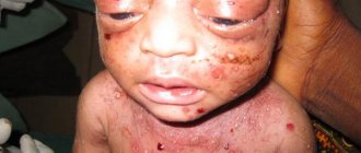
Chickenpox is transmitted not only through the air, but also through household contact. A child receives developmental defects due to intrauterine infection (if the infection occurred in the first trimester). If the infection occurs before childbirth, then there will be a congenital form of this disease, and this is a very dangerous condition.
Listeriosis (a bacterial infection) can develop in a pregnant woman if she eats contaminated cheeses, meats or vegetables. The child eventually develops pneumonia and/or sepsis. Syphilis is transmitted to pregnant women through sexual contact if the partner has been infected. After birth, a child develops this disease with all the ensuing consequences. Tuberculosis, which is also a bacterial infection, is transmitted through the air. The embryo can die in the womb, be born prematurely, with a congenital form of tuberculosis.
Herpes
Herpes is a group of viral infectious diseases that are caused by the herpes simplex virus (HSV). HSV is a DNA virus that is morphologically similar to other viruses of the Herpetoviridae family. There are two types of HSV, with different biological and epidemiological features. Type I affects the mucous membranes of the eyes, mouth, and nose and is one of the causes of severe sporadic encephalitis in adults. Type II in most cases affects the genitals (so-called urogenital herpes). HSV is transmitted by airborne droplets and sexually, as well as transplacentally from the pregnant mother to the fetus. In the case of an advanced chronic course of the disease, herpes of both types can manifest itself as lesions not only of the skin and mucous membranes, but also of the central nervous system, eyes, and internal organs. As with all TORCH infections, when infected with herpes, a person produces antibodies that significantly reduce further progression of the disease, and herpes most often appears only when immunity is reduced (such as type I herpes during a cold).
With primary infection with herpes during pregnancy, especially at its initial stage, when all the organs and systems of the unborn child are formed, the herpes infection can be fatal to the fetus. In this case, the risk of undeveloped pregnancy and miscarriages triples, and the development of deformities in the fetus is possible. If herpes infection occurs in the second half of pregnancy, then the likelihood of congenital fetal anomalies, such as microcephaly, retinal pathology, heart defects, and congenital viral pneumonia, increases. Premature birth may occur. A child can become infected with herpes not only in utero, but also during childbirth, passing through the birth canal of an infected mother. This happens if during pregnancy a woman’s genital herpes worsens, and the rashes are localized on the cervix or in the genital tract.
Primary infection occurs in seronegative patients who have never been infected. Secondary infection is the activation of latent infection or reinfection in seropositive patients.
Most people infected with HSV are asymptomatic, so serological diagnosis is necessary.
IgM antibodies to herpes simplex virus are detected one week after infection, usually indicative of recent or current infection.
IgG antibodies appear 2-3 weeks after infection, and their titer decreases after several months. In patients with relapse of the disease, the IgG antibody titer often does not increase.
Consequences of intrauterine infection
Congenital infection can be acute or chronic. Consequences of acute infection:
- shock
- pneumonia
- severe sepsis
Symptoms of acute intrauterine infection in newborns:
- activity decreasing every day
- long sleep
- poor nutrition
But in most cases there are no symptoms. Long-term consequences of IUI:
- delayed motor development
- mental problems
- visual impairment
- partial or complete deafness
If the infection has penetrated the uterus, the consequences may be:
- stillbirth
- antenatal embryo death
- frozen pregnancy
- miscarriage
If the fetus survives, it will have the following manifestations of IUI:
- Fever
- Skin rashes
- Hydrops fetalis
- Anemia
- Jaundice and enlarged liver
- Pneumonia
- Myocarditis
- Chorioretinitis, cataract
- Micro- and hydrocephalus
Intrauterine infection is dangerous regardless of gestational age. But some pathogens are most dangerous in the first 3 months. A striking example is the rubella virus. Very serious consequences will occur if the mother is infected with chickenpox in the very last week of pregnancy. The consequences can be very different. You can find out about them from your doctor based on the results of ultrasound, tests, gestational age and the detected causative agent of the disease. This means that each case is individual in terms of the course of the disease and in terms of consequences.
Risk groups for intrauterine infection
Women who:
- live in a family with children who go to kindergarten, school, or other institutions
- work in the medical field, having contact with carriers of diseases or sick people
- work in preschool and school children's institutions
- have 2 or more medical abortions
- patients with inflammatory pathologies that occur in a chronic form
- have untimely rupture of amniotic fluid
- have a history of embryonic malformations and antenatal fetal death
- have once given birth to an infected child (one or more)
Symptoms and consequences
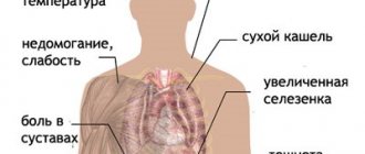
The following are signs that indicate an infection in a pregnant woman:
- cough, shortness of breath
- rash
- lymph nodes are painful and enlarged
- heat
- joint pain and swelling
- runny nose, lacrimation, conjunctivitis
- chest pain
But these symptoms in some cases indicate an allergic reaction. If this is so, then there is no risk of intrauterine infection of the baby. In any case, if one or more of the above symptoms appear, you need to go for a face-to-face consultation with a doctor.
CMV (cytomegalovirus)
CMV (cytomegalovirus) belongs to the group of herpes viruses. It is contracted through sexual contact, through close household contacts, and also through blood (if an operation was performed with dirty instruments or a transfusion from a sick donor). If there is a primary infection of a pregnant woman, the virus enters the placenta, and from there to the baby. The child may have no consequences, which is the case in most cases. But 10 out of 100 children who had sick mothers are born with symptoms of IUI.
The consequences of intrauterine infection with CVM can be miscarriages and stillbirths, as well as:
- sensorineural hearing loss
- underweight at birth
- hydrocephalus
- microcephaly
- pneumonia
- hepatosplenomegaly
- psychomotor development delay
- blindness of various degrees
If there is a severe combined lesion, 1/3 of newborns die in the first 2-3 months after birth. Long-term consequences such as mental retardation, blindness and deafness are also very likely. If the infection is mild, the consequences are less serious. Today there is no medicine that would help with the manifestations of cytomegalovirus in recently born children. If a pregnant woman is infected with CVM, the pregnancy is not terminated because the child may not have symptoms. Doctors prescribe treatment for a pregnant woman to minimize the development of complications.
HSV (herpes simplex virus)
Congenital herpes infection develops if the pregnant woman had HSV, especially type 2 (which is sexually transmitted). Symptoms appear in the first month after birth. Mostly, IUI occurs during primary infection of a pregnant woman. The child picks up the infection in most cases during childbirth, when he passes through the birth canal. But the possibility of transmission of the pathogen through the placenta also needs to be taken into account.
Consequences of herpes for a child:
- lethargy, poor appetite
- miscarriage, stillbirth
- characteristic skin rashes
- fever
- bleeding disorder
- jaundice
- brain damage
- eye damage
- pneumonia
Severe congenital herpes is fraught with disability for the child:
- vegetative state
- mental retardation
- cerebral palsy
Rubella
Rubella is very dangerous for the embryo! It causes various deformities. The highest risk occurs during pregnancy before the 16th week. Consequences of IUI caused by this disease:
- microcephaly
- low birth weight
- miscarriage, stillbirth
- heart defects
- deafness (in half of cases IUI)
- cataract
- skin lesion
- pneumonia
- hepatosplenomegaly
- meningitis and encephalitis
Parvovirus B19
This is the causative agent of infectious epithema. In adults it is mostly unnoticed because it occurs latently. If a pregnant woman is infected with it, she may give birth to a stillborn child, and there is a risk of miscarriage or intrauterine infection of the embryo. Children die in 2.5-10 cases out of 100. It is especially dangerous to become infected with it in the 13-28th week of gestation.
Consequences for a child with IUI:
- swelling
- anemia
- brain damage
- peritonitis
- hepatitis
- myocarditis
Chicken pox
If a patient contracts chickenpox, it is very dangerous for the fetus. If a pregnant woman becomes infected and is about to give birth, there is a risk that her child will die. The fetus becomes infected in 25 out of 100 cases, but symptoms do not always appear.
Congenital chickenpox in children has the following symptoms:
- brain damage
- optic nerve atrophy, eye underdevelopment
- underdevelopment of the limbs
- rash, zigzag scars
- pneumonia
Newborns with IUI are not treated with chickenpox because the symptoms do not progress. If the mother became infected 5 days before the day of birth or later, doctors may advise administering immunoglobulin to the newborn, because antibodies were not transferred to him from the mother.
Hepatitis B
The hepatitis B virus is transmitted mainly through unprotected sexual contact with a sick person. It reaches the fetus through the placenta (“baby place”). It is very dangerous for a pregnant woman to become infected from the 4th to the 9th month of gestation. Consequences of IUI:
- hepatitis B with subsequent recovery
- liver cancer
- carriage and chronic form of hepatitis type B
- acute form of hepatitis (liver failure develops, the child dies)
- psychomotor development delay
- lack of oxygen
- light weight
- miscarriage, stillbirth
HIV infection
The human immunodeficiency virus (HIV) attacks specific immune lymphocytes. Infection occurs mainly through sexual contact. The child becomes infected while arriving in the womb, or passing through the infected genital tract of the mother at birth. If you do not treat a child with a congenital form of HIV, he will not survive even two years, because the virus progresses quickly in a weak body. Death occurs from infections that cannot be fatal in healthy children.
To detect HIV in a newborn, PCR is mainly used. In the first 3-6 months after birth, there is no point in doing an antibody test. It is important to detect HIV in those who are expecting a child. They are given antiretroviral drugs for the entire period, that is, mainly zidovudine is given from the 4th week of gestation. They should not breastfeed their newborn. This greatly increases the baby’s chances of health.
Listeriosis
This disease is caused by the bacterium listeria. It can penetrate the embryo through the placenta, which many bacteria cannot. The routes of infection are described above. A pregnant woman may not have any symptoms of the disease. In some cases there are:
- heat
- diarrhea
- vomit
- flu-like symptoms
Consequences of fetal infection:
- multiple purulent foci, rash
- sepsis
- meningitis
- fever
- refusal to eat
- stillbirth, spontaneous abortion
If symptoms appear in the first 7 days after birth, then children die in 60 cases out of 100. Therefore, if a pregnant woman has an accurate diagnosis of listeriosis, she is prescribed ampicillin for 2 weeks. IUI is also treated if the child actually became infected from the mother.
Syphilis
Primary syphilis in pregnant women, which has not been treated, is transmitted in almost 100 cases out of 100. Out of 10 children, 6 die, while others develop a congenital form of the disease. In a sick pregnant woman, a primary ulcer first forms, and then the disease goes into a latent form, exacerbating from time to time.
Infection of the embryo occurs, even if the mother has a latent disease, from the 4th week of gestation. The consequences of IUI are:
- deafness mental retardation
- damage to the eyes, ears, hands, feet, teeth
- cracks in the skin
- rashes on the skin
- anemia, jaundice in a baby
- premature birth or stillbirth
Toxoplasmosis
It is transmitted to humans mainly from cats, but can also be transmitted from other animals. A pregnant woman can become infected when she cleans up after her pet or eats undercooked meat or dirty vegetables. By the time of pregnancy, most women have already had this disease, so it will not be passed on to the child.
During primary infection during pregnancy, in 50 out of 100 cases, the pathogen overcomes the placental barrier, infecting the embryo. Consequences of IUI for a baby:
- hydro-, microcephaly
- eye damage
- jaundice, enlarged liver and spleen
- encephalitis, meningitis, seizures
- stillbirth, miscarriage
- psychomotor development delay
The progressive increase in the number of cases of intrauterine infection of the fetus is one of the most pressing problems of modern obstetrics and perinatology. This is facilitated by the polyetiology of this pathology, the lack of a clear relationship between the severity of clinical manifestations of infection in the mother and the degree of damage to the fetus, and the multifactorial effect of the infectious agent on the fetus.
Despite increased attention to the problem of intrauterine infection (IUI), many questions remain unresolved. The issues of diagnosis, treatment and prevention of IUI need further research and development. There are still no clear criteria for treatment tactics, and data on the effectiveness of complex therapy have not been summarized.
Epidemiology, etiology and pathogenesis of intrauterine infection
In the development of an infectious process in the fetus, the type of pathogen, its virulence, the route of infection from mother to fetus, the protective reserves of the mother’s body and the ability of the fetus to respond are important [11].
According to modern data [10, 20], the number of cases of IUI varies widely from 6 to 70%. Recently, the structure of infectious morbidity among pregnant women, women in labor and postpartum, as well as the fetus and newborn, has changed [5, 8, 14]. It has been proven that the causative agents of IUI are more than 27 types of bacteria, many viruses, parasites, 6 types of fungi, 4 types of protozoa and rickettsia. Thus, according to a number of researchers [8, 14, 30], the predominant causative agents of antenatal infections are chlamydia (17-50%) and viruses (herpes simplex virus, HSV - 7-47%, cytomegalovirus, CMV - 28-91.6% , enteroviruses - 8-17%). The causative agents of intrapartum infections are group B streptococcus (3-12%), staphylococci (1-9%), fungi of the genus Candida
(3-7%). Associations of pathogens occupy a leading place (75-95%).
It is known that most bacteria exist in nature in the form of specifically organized biofilms [2, 25, 27]. This form of existence creates many advantages for bacteria. Bacteria in biofilms have increased survival in the presence of aggressive substances, immune defense factors, and antibiotics [27]. In this regard, one of the main problems of practical medicine is the problem of treating diseases of microbial origin.
In our study [15, 17], based on the results of bacteriological analysis of the species composition of the vaginal biotope, the strongest influence of Streptococcus faecalis
(p=0.00171),
E. coli
(p=0.01424) and
Staphylococcus epidermidis
(p=0.02714) on the occurrence of intrauterine infection of the fetus. When assessing pathogens identified in the cervical canal of pregnant women by polymerase chain reaction (PCR) and enzyme-linked immunosorbent assay (ELISA), the following was established: in the group without IUI, mycoplasma, chlamydia and ureaplasma accounted for 8%, CMV - 20%, HSV - 36%, candida - 3%, associations - 60%. When analyzing a group of newborns with IUI, the most common pathogens were identified. Thus, mycoplasma, chlamydia and HSV were found in 50%, CMV infection was detected in 45% of cases, ureaplasma (20%) and candida (15%) were less common, associations were observed in 95%. The frequency of detection of pathogenic pathogens in newborns with signs of IUI was higher than in patients without infection.
In the pathogenesis of IUI, the “maternal”, “successional”, and “fetal” stages of development are distinguished [23]:
The “maternal” stage reflects the onset of the infectious process within the lower parts of the urogenital tract.
The “secondary” stage occurs with hematogenous spread of the inflammatory process and occurs with bacteremia and viremia.
During the “fetal” stage, the infectious process spreads to the organs and tissues of the fetus. This occurs when the uteroplacental and placental-fetal antimicrobial barrier fails, the boundary of which is the layer of chorionic epithelium.
The main source of infection in IUI is the child's mother, from whose body the pathogen enters the fetus (vertical transmission mechanism). In this case, infection occurs both through the ascending, transplacental and transovarial routes, as well as through contact and aspiration (directly during childbirth) routes. Moreover, for antenatal infections, the hematogenous route is most typical, and for intranatal infections, the ascending route of infection is most typical [4, 10, 20, 21].
The influence of IUI on the embryo and fetus consists of the influence of a complex of the following factors [22]:
1. Pathological effect of microorganisms and their toxins (infectious disease, fetal hypoxia, delayed fetal development).
2. Violation of the process of implantation and placentation (low placentation, placenta previa).
3. Reduced metabolic processes and immune defense of the fetus.
In the pathogenesis of the occurrence and development of IUI, the duration of pregnancy is of particular importance [13, 22]. A fetus up to 14 weeks of pregnancy does not react to infectious antigens, since it lacks immunocompetent cells, immunoglobulins and does not exhibit immune reactions. With the beginning of the second trimester of pregnancy, the mechanism of action of ascending infection changes due to the fusion of decidua vera
and
deciduas capsularis
into a single complex
deciduas parietalis.
At this time, an ascending infection can penetrate to the fetus from the vagina or cervical canal. From this stage of pregnancy, the internal os of the cervical canal comes into contact with the aqueous membranes of the fetus and, in the presence of infection, microorganisms penetrate into the amniotic fluid. Amniotic fluid acquires antimicrobial properties only after the 20th week of pregnancy, when, in response to exposure to an infectious agent, an inflammatory proliferative reaction develops, limiting further penetration of infection, due to the appearance of lysozyme, complement, interferons, and immunoglobulins [1, 22]. In the third trimester of pregnancy, the antibacterial protection of amniotic fluid increases. During this period, the role of the exudative component predominates in the inflammatory reaction of fetal tissues, when inflammatory leukocyte reactions in the fetus (encephalitis, hepatitis, pneumonia, interstitial nephritis) develop in response to infection [22].
Particularly dangerous with IUI in the second and third trimesters of pregnancy is damage to the fetal brain, which can lead to mental retardation and delayed psychomotor development in children [3, 7]. Intrauterine damage by infectious agents to the structures of the central nervous system in the fetus is accompanied by various severe disorders in the formation of the brain (hydrocephalus, subependymal cysts, cystic degeneration of the brain substance, abnormalities of the cortex, microcephaly). It is also possible to develop ventriculitis (deformation of the choroid plexus, heterogeneity or doubling of reflection from the ventricular ependyma) [11].
Thus, infection of the fetus in later stages of pregnancy does not, as a rule, lead to the formation of gross malformations, but can disrupt the functional mechanisms of cell and tissue differentiation [10, 11, 13].
Changes in the condition of the fetus and the functioning of the fetoplacental system caused by intrauterine infection of the fetus are reflected in the composition and properties of the amniotic fluid [1, 9, 26, 30]. When an infectious agent enters the amniotic fluid, it multiplies unhindered with the subsequent development of chorioamnionitis [1, 9]. The fetus finds itself in an infected environment, which creates favorable conditions for infection of the fetus by contact, i.e. through the skin, mucous membranes, respiratory and gastrointestinal tracts.
The syndrome of “amniotic fluid infection” develops, the mechanism of occurrence of which is presented as follows [20]:
1. When swallowing and aspiration of infected water, a newborn develops signs of intrauterine infection (pneumonia, enterocolitis, vesiculosis, omphalitis, conjunctivitis, etc.).
2. At the same time, microorganisms, spreading through the membranes or between them, reach the basal plate of the placenta (deciduitis). Further spread of the inflammatory reaction leads to the development of chorionitis (placentitis), manifested by leukocyte infiltration of the intravillous space and endovasculitis in the chorionic plate. Vasculitis in the decidua, stem and terminal villi leads to obliteration of blood vessels, the appearance of infarctions, calcifications, massive deposits of fibrinoid, which can manifest as “premature maturation of the placenta.”
3. Polyhydramnios with intrauterine infection is usually secondary in nature and serves as a manifestation of damage to the kidneys or urinary tract of the fetus. The reason for its development is also a change in the ratio of the processes of production and resorption of amniotic fluid by the cells of the amniotic epithelium against the background of amnionitis.
4. In the genesis of the symptom complex of placental insufficiency in IUI, the main role belongs to vascular disorders.
5. Typical manifestations of intrauterine infection are miscarriage and premature birth [24]. Premature development of labor and untimely rupture of membranes are caused by the action of bacterial phospholipases that trigger the prostaglandin cascade and the damaging effect of inflammatory toxins on the membranes.
6. Due to the fact that phospholipases from gram-negative bacteria contribute to the destruction of surfactant in the fetal lungs, the newborn develops respiratory disorders.
In modern literature [4, 9, 29], there are many works devoted to the study of the relationship between immunological parameters and the severity of the infectious-inflammatory process during pregnancy. Increasingly, data appear in foreign and domestic literature on the relationship between bacterial invasion and the synthesis of cytokines by the cells of the amnion, chorion, decidual and fetal tissues [5, 29]. The proliferation of microorganisms in the amniotic fluid leads to an increase in the level of lipopolysaccharides, which activate the synthesis of cytokines by fetal trophoblast cells. In case of IUI, it seems promising to study changes in the cytokine system, which ensures the processes of intercellular cooperation, growth and differentiation of lymphoid cells, hematopoiesis and neuroimmunoendocrine interactions. Cytokines in the preimplantation period and during the development of pregnancy are actively produced by many maternal and embryonic cells, in particular, decidual cells of the uterus and trophoblast cells. It has been established that cell cultures have different abilities to synthesize cytokines under the influence of lipopolysaccharides. Thus, tumor necrosis factor (TNF) is produced by amnion cells, interleukins (IL)-6 and IL-8 are produced by amnion and chorion, and IL-1 is produced only by chorion [9, 11, 28]. According to N.V. Ordzhonikidze [14], from a variety of pro-inflammatory (IL-1, IL-2, IL-6, IL-8, IL-15, TNF, etc.) and anti-inflammatory (IL-4, IL-10, IL-13, transforming growth factor, etc.) cytokines are considered the main markers of the inflammatory process in human tissues and organs: IL-1, IL-6, IL-10, TNF [9, 16, 28].
Clinical characteristics of intrauterine infection
During the preimplantation period, under the influence of an infectious agent, the embryo dies (alterative inflammation) or continues to develop.
Infectious damage to the embryo at 3-12 weeks is usually associated with a viral infection that freely penetrates the chorion. The fetus does not yet have anti-infective defense systems, and during the period of organogenesis and placentation, IUI leads to the formation of malformations (teratogenic) or death of the embryo (embryotoxic effect) [5, 23].
Infectious fetopathy occurs from the 16-27th week of gestation, when the infection in the fetus generalizes and pseudodefects form (myocardial fibroelastosis, polycystic pulmonary disease, hydrocephalus, hydronephrosis). When infected after 28 weeks, the fetus acquires the ability to have a specific local reaction to the introduction of the pathogen, which can result in IUI (encephalitis, pneumonia, hepatitis, interstitial nephritis), preterm pregnancy, intrauterine growth retardation, and fetal death [2, 3, 10].
Currently, the following types of intrauterine lesions in IUI are distinguished [20, 21]:
— blastopathy — with a gestation period of 0-14 days; possible death of the embryo, spontaneous miscarriage, or the formation of a systemic pathology similar to genetic diseases;
— embryopathy — for a period of 15-75 days; characterized by malformations at the organ or cellular levels (true defects), spontaneous miscarriage;
- early fetopathy - at a period of 76-180 days; characterized by the development of a generalized inflammatory reaction with a predominance of alterative and exudative components and the outcome in fibrous-sclerotic deformations of organs (false defects), termination of pregnancy;
- late fetopathy - at 181 days before birth; the development of a manifest inflammatory reaction with damage to various organs and systems (hepatitis, encephalitis, thrombocytopenia, pneumonia) is possible.
IUI often does not have clear clinical manifestations [4, 10, 15, 17]. Rarely do the first signs in a newborn appear immediately after birth; more often they appear during the first 3 days of life. When infected in the postnatal period, symptoms of the infectious process are revealed at a later date. Clinical appearances of congenital bacterial or mycotic skin lesions in a newborn may have the character of vesiculopustulosis [4]. Conjunctivitis, rhinitis and otitis media that appear on the 1st-3rd day of life can also be manifestations of IUI. Congenital aspiration pneumonia can appear on the 2-3rd day of life. From the moment of birth, children exhibit signs of respiratory failure: shortness of breath, cyanosis, often dullness of percussion sound and fine moist rales. The course of intrauterine pneumonia is severe, since as a result of aspiration, large areas of the lung (lower and middle lobes) are excluded from breathing due to obstruction of the bronchi by infected amniotic fluid containing an admixture of meconium and fetal skin flakes. Enterocolitis in newborns occurs as a result of penetration of the pathogen along with amniotic fluid into the gastrointestinal tract. Dyspeptic symptoms usually develop on the 2-3rd day of life. Characterized by sluggish sucking, regurgitation, bloating, hepatosplenomegaly, expansion of the venous network of the anterior abdominal wall, and frequent loose stools. Microbiological examination of intestinal contents revealed a predominance of Klebsiella, Proteus and Pseudomonas aeruginosa. Damage to the central nervous system during IUI in newborns can be either primary (meningitis, encephalitis) or secondary, caused by intoxication. When the choroid plexuses of the lateral ventricles of the brain are damaged, congenital hydrocephalus develops. It is necessary to pay attention to symptoms such as lethargy, poor sucking, regurgitation, delayed recovery or secondary weight loss, delayed healing of the umbilical wound, and the development of omphalitis. Typical symptoms of infectious intoxication in a newborn are respiratory and tissue metabolism disorders. There is a pale cyanotic coloration of the skin with a pronounced vascular pattern. Intoxication is accompanied by impaired excretory function of the liver and kidneys, enlargement of the spleen and peripheral lymph nodes.
Modern methods for diagnosing intrauterine infections
The prevalence of IUI among the causes of unfavorable outcomes and the high level of infection in pregnant and postpartum women necessitate the search for reliable methods for its diagnosis. The nonspecificity of the clinical manifestations of IUI creates diagnostic difficulties, which dictates the need for the combined use of clinical and laboratory research methods. In the last decade, the main methods for diagnosing IUI are bacteriological and immunological [10, 11, 21].
There are 3 stages in the diagnosis of intrauterine infection: 1) diagnosis during pregnancy; 2) early diagnosis at the time of birth; 3) diagnosis during the development of clinical signs of infection in the early neonatal period [12].
Of the non-invasive methods of prenatal diagnosis of IUI, the most informative are ultrasound and Dopplerography [6, 7, 15, 26]. Direct laboratory diagnostic methods (cordocentesis, dark-field microscopy, PCR, ELISA, culture) can detect the pathogen in biological fluids or tissue biopsies of an infected child. Indirect methods for diagnosing IUI include clinical symptoms of the mother, ultrasound, and help to make only a presumptive diagnosis of IUI [10, 17, 21]. Screening tests for IUI in newborns include studies of smears of amniotic fluid, placenta, cultures of umbilical cord blood and stomach contents of the newborn, and sometimes blood cultures [6, 7, 10]. The “gold standard” for post-diagnosis of IUI is histological examination of the placenta, umbilical cord and fetal membranes [4, 14, 23].
Any changes in homeostasis in the mother’s body are reflected in the cellular and chemical indicators of the amniotic fluid, which very subtly characterize the course of the pathological process, and therefore amniotic fluid can serve as an important diagnostic material [1, 6, 16]. According to I.V. Bakhareva [1], the most significant in the diagnosis of IUI is the determination of the antimicrobial activity of amniotic fluid, based on the migration of leukocytes in it when the accumulation of bacteria in the amniotic membrane exceeds 103 CFU/ml. The appearance of a large number of leukocytes in the amniotic fluid, an increase in cytosis due to epithelial cells without detection of microflora may indicate IUI.
Currently, great importance is attached to ultrasound research methods, with the help of which it is possible to determine indirect signs of fetal IUI (polyhydramnios, ventriculomegaly, microcephaly, hepatomegaly, increased thickness of the placenta, fine suspension in the amniotic fluid) and structural changes in various organs [4, 11, 18 , 26].
We have developed the necessary list of diagnostic measures for the purpose of early detection of IUI [19].
The complex of examination of pregnant women included:
1. General clinical and biochemical tests of blood and urine with determination of standard indicators.
2. Determination of pathogens of the TORCH complex using PCR in vaginal smears and amniotic fluid.
3. Determination of antibodies in the blood to chlamydia, mycoplasma and ureaplasma, CMV and HSV using ELISA.
4. Carrying out an amine test, pH measurement of vaginal contents.
5. Bacterioscopic examination of the contents of the vagina, cervical canal and urethra.
6. Bacteriological examination of the maternal surface of the placenta, amniotic fluid, and intestinal contents.
7. Determination of the level of pro-inflammatory (IL-1β, TNF) and anti-inflammatory (IL-10) cytokines in amniotic fluid, maternal venous blood serum and fetal cord blood.
8. Ultrasound scanning of the fetus, amniotic fluid and placenta.
9. Histomorphological examination of the placenta.
The complex of examination of newborns included:
1. Apgar score, measurement of body weight and length at birth, dynamics of body weight gain until discharge from the maternity hospital.
2. Determination of antibodies in the blood to chlamydia, mycoplasma and ureaplasma, CMV and HSV using ELISA.
3. Bacteriological examination of scrapings from the conjunctiva, posterior wall of the pharynx and vulva.
4. Identification of clinical signs of IUI together with a neonatologist.
A comprehensive study of the species composition of microorganisms in the birth canal, amniotic fluid, placenta, newborn, determination of antigens and antibodies to the putative pathogen in umbilical cord blood and amniotic fluid, histological examination of the placenta allows us to determine the route of infection of the child, the nature of the pathogen and clarify the scope of additional diagnostic tests for IUI, and also determine treatment and preventive measures in the early neonatal period.
TORCH infections
The following are called torch infections:
- cytomegalovirus
- rubella
- toxoplasmosis
- herpes
- tuberculosis
- syphilis, etc.
They are dangerous if they infect a pregnant woman. Some occur in a hidden or erased form, so they need to be identified by analysis. Therefore, at the stage of planning conception, the couple is tested for TORCH infections. If no antibodies to the rubella pathogen are detected in the expectant mother's body, she is vaccinated. Also, during pregnancy it is better not to come into contact with cats, so as not to become infected with toxcoplasmosis.
Toxoplasmosis
Toxoplasmosis is a widespread disease caused by the intracellular protozoan parasite Toxoplasma gondii. The primary host of Toxoplasma, in whose body this parasite multiplies, is the domestic cat, which most often becomes the source of human infection. In addition, human infection can also occur through food contaminated or contaminated with parasite oocysts. The disease can be transmitted transplacentally to the fetus from an infected mother. Cases of infection transmission through blood transfusion and organ transplantation have been described.
Toxoplasmosis affects almost 30% of people in the world. In adults, toxoplasmosis is often asymptomatic; sometimes there may be headache, sore throat, asthenia, and in rare cases lymphadenitis. In exceptional cases, toxoplasma can cause myocarditis, hepatitis, pneumonia, meningoencephalitis, and eye damage. After the illness, stable immunity to toxoplasma is developed.
Toxoplasmosis is a serious danger when a woman is initially infected during pregnancy. If a woman suffered from the disease before pregnancy (at least six months), toxoplasmosis does not threaten her unborn child, but if a woman is infected during pregnancy, then much depends on at what stage of pregnancy toxoplasma entered the pregnant woman’s body. The most dangerous infection is considered to be toxoplasmosis in the first trimester. In these cases, congenital toxoplasmosis often leads to the death of the fetus or the development of severe damage to the eyes, liver, spleen, and nervous system of the child.
The frequency of fetal infections varies depending on at what stage of pregnancy the mother was infected:
- less than 5% when the mother is infected in the first trimester;
- more than 60% when the mother is infected in the third trimester.
If the mother becomes infected later in pregnancy, the risk of transmission of the infection to the fetus is very high, but the risk of severe damage to the fetus is reduced. If a woman has not had toxoplasmosis, then infection during pregnancy can be prevented by observing basic hygiene rules:
- During pregnancy, there should be no contact with cats, especially young ones, because cats infected with toxoplasmosis also develop immunity to it as they age.
- Avoid working with soil in the garden; if you cannot avoid it completely, then you should only work with gloves.
- All vegetables, fruits, and herbs must be thoroughly washed before use.
- Avoid contact with raw meat; all meat dishes must be thoroughly boiled or fried.
The diagnosis of toxoplasmosis is made on the basis of clinical data and laboratory examination data (determination of antibodies to Toxoplasma gondii in the blood).
IgM antibodies to Toxoplasma gondii appear in the blood 2-4 weeks after infection and disappear after 3-9 months. Next, antibodies of the IgG class appear, and their titer gradually increases, and 2-5 months after infection reaches a peak.
Diagnosis of intrauterine infection
Pregnant women undergo a blood test that can detect hepatitis B and C, as well as syphilis. Vaginal smears are regularly taken to check microflora. The PCR method is used, which makes it possible to detect viruses circulating in the blood, if any.
Ultrasound is safe for pregnant women. It does not indicate the presence of infection, but makes it possible to detect defects caused by infection and intrauterine growth retardation. Ultrasound is also a control method when performing cordocentesis. IUI is suspected if an ultrasound reveals the following signs:
- formed developmental defects
- high or low water
- swelling of the placenta, amniotic bands
- intrauterine growth restriction syndrome
- enlargement of the abdomen and expansion of the renal collecting system
- enlargement of the heart, liver and spleen
- multiple calcium deposits in the brain, liver, intestines
- enlargement of the ventricles of the brain
The seroimmunological diagnostic method is based on the determination of immunoglobulins. It is recommended for women from the risk groups listed above. Sometimes methods such as cordocentesis and amniocentesis are necessary. In the first method, blood is taken from the umbilical cord to detect any infection there. Amniocentesis means the examination of amniotic fluid.
If the doctor suspects IUI, a newborn is tested for biological fluids, such as saliva or blood.
What will a fetal ultrasound show?
Ultrasound examination (ultrasound) of the fetus during gestation helps the specialist to timely detect the presence of various pathologies in the development of the unborn child. These are, for example, oligohydramnios or polyhydramnios, a large belly, enlarged internal organs, for example, the liver, spleen. Ultrasound diagnostics can also detect calcification deposits in the intestines, liver, and brain. In our medical diagnostic and treatment center in Makhachkala, experienced ultrasound diagnostic specialists will conduct a high-quality study that will confirm or exclude pathology with high reliability.
The ultrasound markers of intrauterine infection discovered during the study are a real help in assessing the condition of the fetus and predicting further pregnancy management tactics. If possible, doctors will take measures at an early stage to eliminate any detected abnormalities. If this is a condition that is incompatible with life, then a decision is made that it is better to terminate the pregnancy.