Toxocariasis
is a disease caused by helminths of the nematode group (roundworms). The symptoms are very extensive, as they depend on which organs the larvae have entered. In the acute period, the signs of the disease are similar to those of other helminthiases.
Toxocariasis is quite common in all countries. Most often, it affects children who become infected while playing in the sand and with animals, and adults in the course of their professional activities. The risk group includes veterinarians, dog handlers, agricultural workers, and utility workers.
tests
Definition of disease
Toxocariasis is the migration of animal helminth larvae into the human body. The source of the disease, toxocara, was discovered by the German scientist Werner in 1782. Only in 1950 was infection by these helminths identified as a separate disease. Toxocara eggs can be found in soil and contaminated water. The main carriers are dogs, cats, other domestic and wild animals.
Since almost all invasions are characterized by a long course in most cases with mild manifestations, it can be argued that almost 90% of the population are infected or have been infected at different times. According to WHO research, helminthiasis ranks 4th in the world ranking in terms of the degree of damage to human health.
Toxocara: treatment
In modern medicine there is still no established method of treating parasites. Doctors do not know how to treat toxocariasis, therefore the toxocariasis caused by them is not subject to official registration. Drugs that kill migratory larvae are not effective against protected granulomas. Due to the fact that medicine is powerless against this parasite, it has to argue that toxocara is not so dangerous and harmful to the body. In fact, their activity (especially in hosts with weak immunity) can be fatal.
Do not ignore Toxocar under any circumstances. Preparations based on natural ingredients will help remove them from the body and cleanse them of toxic waste products. It is precisely these antiparasitic agents that are being developed. They free the body from Toxocora larvae and cysts, promote the restoration of damaged organs and tissues, and replenish the lack of microelements.
Causes of toxocariasis
Infection with toxocariasis occurs when Toxocara eggs enter the human body with food and water, through household, contact, and fecal-oral routes. Infection is possible through contact with animals, especially homeless ones, and soil contaminated with eggs. The most common sources of infection are puppies, pregnant and lactating bitches. Cockroaches contribute to the spread of toxocariasis in everyday life; they absorb helminth eggs and subsequently release some of the living individuals.
There are currently 2 types of parasites known:
- T. canis - parasitic in animals of the canine family (dogs, wolves, arctic foxes, foxes);
- T. mystax are parasitic on animals of the cat family (cats, lynxes, panthers, lions).
Humans are not a natural host for the parasite. Therefore, most of the Toxocara die in the human body due to unsuitable conditions for survival. They do not reach sexual maturity. Humans are a dead-end branch; it is impossible to become infected from other people.
Diagnosis of toxocariasis
Disparate symptoms complicate making an accurate diagnosis. When pulmonary syndrome develops, doctors treat pneumonia; when skin rashes appear, they suspect a food or drug allergy, but the true cause of the patient’s serious condition - the presence of parasites in the body - is extremely rarely established. Often patients are mistakenly diagnosed with granulomatosis or a tumor, when in fact it is not a neoplasm, but a Toxocara larva surrounded by infiltrates. In more than 83% of cases, doctors mistakenly diagnose an “eye tumor,” while the cause of vision impairment is nematodes.
To determine the parasitic load, a blood test is taken from the patient for toxocariasis, but it is not 100% reliable. The Optisalt company has developed the Optisalt IridoScreen device, which can be used to determine the parasitic load on the body based on the iris of the eyes. Scientists have proven that each organ has its own projection on a certain segment of the iris, to which information about its condition is transmitted using nerve impulses.
Pathogenesis
The causative agent of toxocariasis is roundworms of the Nematoda class. Two species of Toxocara have been discovered; T. canis, parasites of the canine family, can be found in humans. The likelihood of infection with T. mystax has not been established. The pathogen enters the human body in the egg phase with a living larva (cyst), with an average size of 60 microns.
In the digestive tract, the shell of the eggs disintegrates. If a person’s immunity is weakened, the larvae (larvae) become active and begin their migration. Through the bloodstream they penetrate into various tissues, systems and organs. The next stage is the formation of lesions: inflammation, hemorrhage, necrosis. The antigenic effect of larvae leads to allergic reactions.
Granulomas form in the areas where the larvae invade. They are surrounded by fibrous tissue, as a result they cannot receive more nutrients and die. However, in such a short life, the larvae manage to have a significant negative impact on the liver, lymph nodes, lungs, myocardium, and brain. This can lead to the development of pneumonia, pancreatitis, respiratory failure, cause hepatitis or meningitis. Infestations can lead to partial or complete loss of vision and other diseases.
Consequences of toxocariasis
If you seek medical help in a timely manner, as well as with effective and adequate treatment, the prognosis for toxocariasis is favorable.
The development of complications is possible with severe disease, as well as with prolonged ignoring of symptoms and untimely initiation of therapy. The main adverse effects that can occur with toxocariasis:
- one-sided blindness (can occur in the ocular form of the disease);
- complications from the respiratory system, namely the development of severe pneumonia (pneumonia);
- hemorrhages (hemorrhages) and tissue necrosis may occur due to the traumatic effect of the larvae on body tissue.
Classification
In the international classification, toxocariasis is assigned the code B83.0.
The disease is classified according to the following characteristics:
Type of disease development and severity of symptoms:
- Typical form.
- Atypical form (asymptomatic, with erased symptoms).
Form of defeat:
- visceral,
- ophthalmic,
- cutaneous,
- neurological,
- muscles affected
- the thyroid gland is affected,
- lymph nodes are affected.
Severity of illness:
- light;
- medium-heavy;
- severe form.
Nature of the disease:
- Smooth,
- Unsmooth (with complications, exacerbation of existing diseases).
Duration:
- Acute (up to 3 months from the moment of infection).
- Chronic, lasting more than 3 months.
Visceral form
The most common form of toxocariasis is visceral, diagnosed in 90% of cases. Characterized by low-grade fever, intoxication syndrome, and decreased appetite. More than half of patients have a nighttime, unproductive cough. The invasion is accompanied by disturbances in the gastrointestinal tract, including flatulence, diarrhea, and vomiting.
Ophthalmic toxocariasis
The ocular form is the second most frequently diagnosed, but still quite rare. Granulomas are found in the posterior and peripheral parts of the eyeball. This leads to inflammation of the choroid, vitreous abscesses, inflammation of the optic nerve and other abnormalities caused by the migration of larvae. As a rule, only one eye is affected; double damage is extremely rare. Ophthalmic toxocariasis leads to decreased vision, strabismus, and the formation of a dull (opaque) white lump (leukocoria).
Neurological form
Characterized by convulsions, paralysis, behavior disorder, muscle movement disorders, muscle pain. The neurological form is extremely rare and leads to meningitis or meningoencephalitis.
Damage to the thyroid gland
Symptoms of larvae penetration into the thyroid tissue are expressed in its enlargement. The lymph nodes in the submandibular and neck area also increase in volume. The liver and spleen are affected. Lymph nodes are painful when palpated, but no inflammatory changes occur.
Forms of toxocariasis
Eye shape
The larvae are found in the tissues of the eye. A rare form of the disease, manifested by chronic inflammatory processes - keratitis, neuritis, uveitis. Active toxocariasis causes blurred vision and strabismus. Most often one eye is affected.
Neurological form
Occurs when the brain is damaged by larvae. An even rarer form develops mainly in people with immunodeficiency. It manifests itself as movement disorders, convulsions, epileptic seizures, and mental disorders.
Cutaneous form
It can only be active - this is the migration of larvae inside the skin. It is rare and is manifested by the visible movement of larvae under the skin.
Visceral toxocariasis
The larvae are found in internal organs, most often the liver, intestines, and lungs. This is the most common form of the disease.
Symptoms
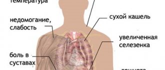
The clinical picture of toxocariasis is varied, since the symptoms are directly related to the affected organs. The following symptoms are common to all forms of the disease:
- Temperature with chills from subfebrile in mild cases to +39 C, can last up to 3 weeks.
- Headaches of moderate intensity.
- Intoxication syndrome: loss of appetite, nausea, non-localized abdominal pain, diarrhea.
- Significant increase in the size of lymph nodes, liver, spleen.
- Respiratory syndrome: from mild night cough to bronchial obstruction, pneumonia, signs of suffocation, cyanosis.
- Skin syndrome, expressed in allergic reactions, urticaria, papular rashes. More often they appear on the skin of the lumbar region and extremities.
In the acute form of central nervous system damage, headaches, insomnia, and convulsions are observed. The pathological condition can lead to meningoencephalitis, myelitis, arachnoiditis, paresis and paralysis. In some cases, mental disorders are observed. Patients with ocular toxocariasis complain of deterioration in the quality of vision, the appearance of passing “dots” in front of the eyes and a blind spot that does not pass.
Symptoms are recurrent, on average they appear for 1 - 3 weeks, then go away, and after some time they make themselves felt again.
Treatment of the disease
If infection is suspected, it is necessary to undergo a set of diagnostic procedures described above, after which treatment will be prescribed.
Self-administration of medications is strictly prohibited, as it can lead not only to distortion of symptoms, but also to a worsening of the condition.
Treatment of toxocariasis is carried out in three stages:
- Preparatory.
Includes relieving intoxication, maintaining affected organs, and stabilizing the patient’s condition. It is performed using physiotherapeutic procedures using MAG-Expert, IONOSON-expert devices.
- Basic.
Taking antiparasitic drugs. Drugs and dosage are determined individually by the attending physician.
- Recovery.
Removal of parasites through intraduodenal lavage, a course of physiotherapy using MAG-Expert, IONOSON-expert devices. Taking medications to reduce the risk of an allergic reaction to the parasite's toxins. The drugs are prescribed by a doctor.
A complete recovery can only be achieved by strictly following all the recommendations of the attending physician and going through all the above stages of treatment.
Diagnostics
Diagnosis of toxocariasis is carried out by an infectious disease specialist. It is necessary to interview the patient to identify the route of infection. Are there dogs in the house, have you had contact with stray animals? Is it possible to accept the possibility of consuming foods and water containing Toxocara eggs?
Laboratory diagnostics include the following methods:
- Biochemical - confirmation of nosology by detecting increased IgE antibodies, decreased albumin levels, hypergammaglobulinemia. A study can also be conducted to determine changes in the data of the hepatic and pancreatic complex.
- The serological ELISA method also allows you to confirm the diagnosis.
- Hematological - aimed at identifying the growth of eosinophils, an increase in leukocytes, a decrease in hemoglobin levels, and an increase in ESR. The method allows you to determine the severity of the disease.
- Sputum microscopy is used to identify the form of the disease.
To clarify the clinical form of toxocariasis and determine the severity of the disease, instrumental studies are prescribed:
- X-ray;
- Ultrasound of the peritoneum;
- external respiration testing.
Electrocardiography, computer and magnetic resonance imaging are performed. Also, if necessary, the reactivity of the bronchi can be studied, and ophthalmological, neurological and histomorphological studies of the affected organs and tissues can be performed.
Toxocariasis of the eye
If ophthalmic toxocariasis is suspected, an ophthalmological examination is performed. Its goal is to identify Toxocara larvae in the macula, optic nerve area, and vitreous body. Sometimes during examination you can record the movements of the parasite. The examination reveals not only the larvae themselves, but the degree of changes that have occurred: hemorrhages, fibrosis, retinal detachment.
Criteria for confirming the diagnosis
- Sort by diagnostic power score from highest to lowest:
- Eosinophilia in peripheral blood.
- Increases in total protein and IgE with a decrease in albumin volume.
- Detection of IgG antibodies in a titer of 1:800.
- An increase in ESR with a decrease in the level of hemoglobin and red blood cells.
- Detection of increased levels of liver transferases, alkaline phosphatase, and direct bilirubin amylase in the blood serum.
- Detection of increased amylase levels in urine.
- Detection of Toxocara larvae in sputum.
These data also help determine the severity of the disease. For example, high leukocytosis indicates the presence of complications along with severe eosinophilia and increased amylase in the urine.
The tasks of an infectious disease doctor also include differential diagnosis. First of all, it is necessary to exclude ascariasis, opisthorchiasis, and strongyloidiasis. If ophthalmic toxocariasis is suspected, it is necessary to exclude retinoblastoma, and allergic skin rashes from a reaction to allergens and insect bites.
TOXOCAROSIS: a modern approach
Toxocariasis is a parasitic disease caused by the migration of roundworm larvae of canines (T. canis). It is characterized by a long-term relapsing course and multiple organ lesions of an immunological nature. The causative agents of toxocariasis can also be the larvae of other roundworms - cats (T. mystax), cows, buffalo (T. vitulorum). However, the role of these pathogens in human pathology has been practically unstudied.
- Biology of the pathogen
Sexually mature forms of T. canis are large dioecious worms 4–18 cm long, localized in the stomach and small intestine of animals (dogs). The intensity of infestation in dogs can be very high, especially in young animals. The average lifespan of sexually mature individuals is 4 months, the maximum is 6 months. The female parasite lays more than 200 thousand eggs per day. 1 g of feces can contain 10,000-15,000 eggs, so millions of eggs end up in the soil, thereby creating a high risk of toxocariasis infection.
Toxocara eggs are round in shape, larger than roundworm eggs (65-75 microns). The outer shell of the egg is thick, dense, and finely lumpy. Inside the egg is a dark blastomere.
The development cycle of the pathogen is as follows. The released Toxocara eggs fall into the soil, where, depending on the humidity and temperature of the soil, they mature in 5-36 days, becoming invasive. The eggs remain infective in the soil for a long time, and in compost for several years.
The life cycle of Toxocara is complex. There is a main cycle and two auxiliary options. The main cycle occurs according to the following scheme: final host (canids) - soil - final host (canids). Transmission of infection occurs through the geo-oral route. The auxiliary cycle (option 1) occurs transplacentally, in this case the parasite in the larval stage passes from the pregnant female to the fetus, in whose body it completes migration, reaching the sexually mature stage in the intestine of the puppy. The infested puppy becomes a functionally full-fledged definitive host, a source of invasion.
The auxiliary cycle (option 2) is carried out along the chain: the final host (canids) - soil - paratenic host. The paratenic (reservoir) host can be rodents, pigs, sheep, birds, and earthworms. Humans also act as a paratenic host, but are not included in the infection transmission cycle, being a biological dead end for the parasite. Further development of the pathogen occurs under the condition that the paratenic host is eaten by a dog or other definitive host. The mechanism of transmission of invasion in this variant is geooral - xenotrophic.
Depending on the age of the host, different migration routes for Toxocara larvae are implemented. In young animals (puppies up to 5 weeks), almost all larvae complete migration, reaching sexually mature forms in the intestines and releasing eggs into the external environment. In the body of adult animals, most of the larvae migrate to somatic tissues, where they remain viable for several years. During pregnancy and lactation in pregnant bitches, migration of larvae resumes. Migrating larvae enter the fetus through the placenta. The larvae remain in the liver of prenatally infested puppies until birth, and after birth, the larvae from the liver migrate to the lungs, trachea, pharynx, esophagus and enter the gastrointestinal tract, where after 3-4 weeks they reach the mature stage and begin to release eggs into the external environment. Lactating bitches can also transmit the infection to puppies through milk.
In humans, the development cycle of the pathogen, its migration is carried out as follows. From Toxocara eggs that fall into the mouth, then into the stomach and small intestine, larvae emerge, which penetrate the blood vessels through the mucous membrane and migrate through the portal vein system to the liver, where some of them settle, become encysted or surrounded by inflammatory infiltrates, forming granulomas. Some of the larvae pass through the liver filter through the hepatic vein system, enter the right heart and, through the pulmonary artery, into the capillary network of the lungs. In the lungs, some of the larvae are also retained, and some, having passed through the filter of the lungs, are carried through the systemic circulation to various organs, settling in them. Toxocara larvae can be localized in various organs and tissues - kidneys, muscles, thyroid gland, brain, etc. In tissues, the larvae remain viable for many years and periodically, under the influence of various factors, resume migration, causing relapses of the disease.
- Geographical distribution and epidemiology
Toxocariasis is a widespread invasion; it is registered in many countries. Carnivore infestation rates are high in all countries of the world. The average incidence of intestinal toxocariasis in dogs examined on various continents is over 15%, but in some regions it reaches 93% in some animals. According to seroepidemiological studies, from 2 to 14% of examined practically healthy individuals in various foci of toxocariasis have positive immunological reactions to toxocariasis. The prevalence of invasion in different regions is not precisely known, since toxocariasis is not subject to mandatory registration. It is quite obvious that toxocariasis has a wide geographical distribution, and the number of patients is much higher than officially registered.
| Toxocariasis is widespread and is reported in many countries. The average incidence of intestinal toxocariasis in dogs examined on various continents is over 15%, but in some regions it reaches 93%. According to seroepidemiological studies, from 2 to 14% of examined practically healthy individuals in various foci of toxocariasis have positive immunological reactions to toxocariasis |
The main source of infection for humans are dogs, especially puppies. Infection occurs through direct contact with an infested animal whose fur is contaminated with infective eggs, or through ingestion of soil containing Toxocara eggs. Children are especially susceptible to infection while playing in the sand or with a dog. The greatest risk of infection is in children suffering from geophagia. Adults become infected through household contact with infected animals or during professional activities (veterinarians, dog breeders, utility workers, drivers, diggers, etc.). Humans can also become infected by eating raw or poorly thermally processed meat from paratenic hosts. Cases of infection with toxocariasis from eating lamb liver have been described. The possibility of transplacental and transmammary transmission of the infection in humans cannot be ruled out.
- Pathogenesis and pathological anatomy
The pathogenesis of toxocariasis is complex and is determined by a complex of mechanisms in the parasite-host system. During the migration period, the larvae injure blood vessels and tissues, causing hemorrhages, necrosis, and inflammatory changes. The leading role belongs to the immunological reactions of the body in response to invasion. Excretory-secretory antigens of larvae have a sensitizing effect with the development of immediate and delayed types of reactions. When the larvae are destroyed, somatic antigens of the larvae enter the human body. Allergic reactions are manifested by edema, skin erythema, increased resistance of the respiratory tract to inhaled air, which is clinically expressed by the development of attacks of suffocation. Mast cells, basophils, and neutrophils take part in allergic reactions, but eosinophils play the main role. The proliferation of eosinophils is regulated by T-lymphocytes with the participation of mediators of inflammatory reactions secreted by sensitized lymphocytes, neutrophils, and basophils. The resulting immune complexes attract eosinophils to the lesion. Sensitized T-lymphocytes accumulate around Toxocara larvae, macrophages and other cells are attracted - a parasitic granuloma is formed.
The pathomorphological substrate of toxocariasis is granulomatous tissue damage expressed to varying degrees. With intensive invasion, severe granulomatous lesions of many organs and systems develop, which can become chronic with repeated infections. With toxocariasis, numerous granulomas are found in the liver, lungs, pancreas, myocardium, lymph nodes, brain and other organs.
- Clinical picture of toxocariasis
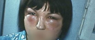
Clinical manifestations are determined by the intensity of invasion, the distribution of larvae in organs and tissues, the frequency of reinvasion and the characteristics of the human immune response. The symptoms of toxocariasis are not very specific and are similar to the clinical symptoms of the acute phase of other helminthiases. The disease usually develops suddenly and acutely or after a short prodrome manifests itself in the form of mild malaise. A temperature appears - low-grade in mild cases and high up to 39°C and above, sometimes with chills - in severe cases of invasion. Skin rashes in the form of urticaria or polymorphic rash, sometimes Quincke's edema, may be observed. In the acute period, pulmonary syndrome of varying severity is observed: from mild catarrhal symptoms to acute bronchitis, pneumonia, and severe attacks of suffocation. Pulmonary syndrome is especially severe in young children. X-ray reveals an increase in the pulmonary pattern, “volatile” infiltrates, and a picture of pneumonia. Along with this, some patients experience an enlargement of the liver, and sometimes the spleen. Lymphadenopathy is more pronounced in children. Sometimes abdominal syndrome occurs in the form of attacks of abdominal pain and symptoms of dyspepsia. Possible development of myocarditis, pancreatitis. There are known cases of damage to the thyroid gland, manifested by symptoms of a tumor. Possible damage to muscle tissue with the development of painful infiltrates along the muscles. When the larvae migrate to the brain, symptoms of central nervous system damage develop: persistent headaches, epileptiform seizures, paresis, and paralysis. In children, the disease is accompanied by weakness, mild excitability, and sleep disturbances.
The most characteristic laboratory indicator is an increased level of eosinophils in the peripheral blood. The relative level of eosinophilia can vary widely, reaching in some cases 70 - 80% or more. The content of leukocytes increases (from 20x109 to 30x109 per 1 l). When examining bone marrow punctate, hyperplasia of mature eosinophils is revealed. Children often experience moderate anemia. Some researchers note a direct correlation between the severity of clinical manifestations of invasion and the level of eosinophilia and hyperleukocytosis in peripheral blood. A characteristic laboratory sign is also acceleration of ESR, hypergammaglobulinemia. In cases of liver damage, increased bilirubin and hyperfermentemia are observed.
In the chronic stage of the disease, acute clinical and laboratory signs fade. The most stable laboratory indicator remains hypereosinophilia of peripheral blood.
There are subclinical, mild, moderate and severe forms of toxocariasis. So-called asymptomatic blood eosinophilia is possible, when there are no obvious clinical manifestations of invasion, but along with hypereosinophilia, antibodies to T.canis antigens are detected.
One of the most serious problems associated with toxocariasis is its relationship with bronchial asthma. Seroepidemiological studies have established that patients with bronchial asthma often have antibodies to T.canis antigens of the Ig G and Ig E classes. Depending on the severity of the parasitic process, its duration and the duration of clinical manifestations of bronchial asthma, one or another class of immunoglobulins predominates. There are clinical observations indicating an improvement in the course of bronchial asthma or recovery after the elimination of toxocariasis invasion.
- Diagnostics
A parasitological diagnosis is rarely established and only by the presence of characteristic granulomas and larvae in the tissues and their identification during the study of biopsy and sectional material. This is possible with a puncture biopsy of the liver, lungs, or surgery. Typically, the diagnosis of toxocariasis is established on the basis of epidemiological history, clinical symptoms and hematological manifestations. Immunological reactions are also used to detect antibodies to Toxocara antigens. Typically, ELISA is used with the secretory-excretory antigen of second-instar Toxocara larvae. Currently, a commercial diagnosticum is being produced in Russia. An antibody titer of 1:400 or higher (in ELISA) is considered a diagnostic titer. An antibody titer of 1:400 indicates invasion, but not disease. An antibody titer of 1:800 or higher indicates toxocariasis. Practice shows that a direct correlation between the level of antibodies and the severity of clinical manifestations of toxocariasis does not always exist. There is not always a correlation between the level of antibodies and hypereosinophilia in the blood.
When making a diagnosis and determining indications for specific therapy, it should be taken into account that toxocariasis occurs cyclically with relapses and remissions, and therefore significant fluctuations in clinical, hematological and immunological parameters in the same patient are possible.
M. I. Alekseeva et al. (1984) developed an algorithm for diagnosing toxocariasis, based on scoring the significance of clinical symptoms and comparing clinical, epidemiological and laboratory parameters. This method may be promising when conducting mass population surveys.
Differential diagnosis is carried out with the migratory stage of other helminth infections (ascariasis, opisthorchiasis), strongyloidiasis, eosinophilic granuloma, lymphogranulomatosis, eosinophilic vasculitis, metastatic pancreatic adenoma, hypernephroma and other diseases accompanied by an increased content of eosinophils in the peripheral blood. It should be borne in mind that in patients with systemic lymphoproliferative diseases and serious disorders in the immune system, immunological reactions may be false positive. In these cases, a careful analysis of the clinical picture of the disease is necessary.
| With intensive invasion, severe granulomatous lesions of many organs and systems develop, which with repeated infections can become chronic |
Ocular toxocariasis. The pathogenesis of this form of toxocariasis is not completely clear. There is a hypothesis about selective damage to the eyes in individuals with low-intensity invasion, in which a sufficiently pronounced immune response of the body does not develop due to the weak antigenic effect of a small number of Toxocara larvae entering the body.
This form of toxocariasis is most often observed in children and adolescents, although cases of the disease have also been described in adults.
Toxocariasis is characterized by unilateral eye damage. The pathological process develops in the retina, affecting the lens and sometimes the periorbital tissue. An inflammatory reaction of a granulomatous nature is formed in the tissues of the eye. The pathological process is often mistaken for retinoblastoma, and the eye is enucleated. Morphological examination reveals eosinophilic granulomas, sometimes Toxocara larvae.
Clinically, eye damage occurs as chronic endophthalmitis, chorioretinitis, iridocyclitis, keratitis, papillitis. Ocular toxocariasis is one of the common causes of vision loss.
Diagnosis of ocular toxocariasis is difficult. The eosinophil count is usually normal or slightly increased. Specific antibodies are not detected or are detected in low titers.
- Treatment of toxocariasis
Not developed enough. Antinematode drugs are used - thiabendazole (Mintezol), mebendazole (Vermox), medamine, diethylcarbamazine. These drugs are effective against migrating larvae and are not effective enough against tissue forms located in granulomas of internal organs.
Mintezol (thiabendazole) is prescribed in doses of 25-50 mg/kg body weight per day in three doses for 5-10 days. Side effects occur frequently and are manifested by nausea, headache, abdominal pain, and a feeling of disgust for the drug (the drug is not currently available in the Russian pharmacy chain).
Vermox (mebendazole) is prescribed 200-300 mg per day for 1-4 weeks. Adverse reactions are usually not observed.
Medamin is used at a dose of 10 mg/kg body weight per day in repeated cycles of 10–14 days.
Diethylcarbamazine is prescribed in doses of 2 - 6 mg/kg body weight per day for 2 - 4 weeks. (Currently, the drug is not produced in Russia, nor is it purchased abroad. - Ed.)
Albendazole is prescribed at a dose of 10 mg/kg body weight per day in two doses (morning - evening) for 7 - 14 days. During treatment, monitoring of blood tests (possibility of developing agranulocytosis) and aminotransferase levels (hepatotoxic effect of the drug) is necessary. A slight increase in aminotransferase levels is not an indication for discontinuation of the drug. In case of increasing hyperenzymemia and the threat of developing toxic hepatitis, discontinuation of the drug is required.
Criteria for the effectiveness of treatment: improvement in general condition, gradual regression of clinical symptoms, decrease in the level of eosinophilia and titers of specific antibodies. It should be noted that the clinical effect of treatment outpaces the positive dynamics of hematological and immunological changes. In case of relapse of clinical symptoms, persistent eosinophilia and positive immunological reactions, repeated courses of treatment are carried out.
The prognosis for life is favorable, however, with massive invasion and severe multiple organ lesions, especially in persons with impaired immunity, death is possible.
- Prevention
Includes personal hygiene and teaching children sanitation skills.
An important preventive measure is timely examination and deworming of dogs. The most effective pre-imaginal treatment is for puppies aged 4-5 weeks, as well as for pregnant bitches. Antinematode drugs are used to treat dogs. It is necessary to limit the number of stray dogs and equip special areas for walking dogs.
It is necessary to improve sanitary education among the population, provide information about possible sources of invasion and routes of its transmission. Persons whose occupations have contact with sources of invasion (veterinarians, dog breeders, diggers, and others) require special attention.
Note!
- Dogs are the main source of infection for humans.
- Children are especially susceptible to infection when playing in the sand or with a dog.
Treatment
Therapy for toxocariasis can be carried out in a hospital or on an outpatient basis, depending on the nature and severity of the disease. Patients with manifest and atypical forms are subject to mandatory hospitalization, in the presence of complications. The activities are aimed at achieving the following tasks:
- stopping the development of pathology;
- prevention of complications;
- preventing the formation of residual and recurrent phenomena.
The treatment method depends on the clinical picture. First of all, anthelmintic drugs are prescribed to stop the migration of larvae.
For the purpose of detoxification, solutions consisting of potassium, magnesium, calcium, sodium, sodium acetate chlorides and containing dextrose are administered. Potassium and sodium chloride also helps restore electrolyte balance.
Healing is the complete disappearance of symptoms, a decrease in the level of antibodies and eosinophils in the blood, and the absence of signs of damage to other organs. Dispensary observation is recommended once every 2 months for a year. Children are seen by a pediatrician, adults by a general practitioner, family doctor, or infectious disease specialist. The list of laboratory tests used during the observation period includes a clinical blood test, coprological studies, and others, depending on the affected organs.
Preventive actions
In recent years, every case of toxocariasis has been registered with the SES. Prevention of toxocariasis comes down to the following. One of the main activities is the examination of dogs and their timely deworming. Equally important is the limitation of special areas for their walking and maintenance of these areas in proper hygienic condition. It is still important to observe the rules of personal hygiene: washing hands after contact with soil or contact with animals, careful handling of greens, vegetables, and food products that may contain soil particles. And in the USA they even issued a postage stamp containing information about the need to immediately clean up dog feces in order to reduce the incidence of toxocariasis.
Infectious disease doctor-gastroenterologist Pugaeva S.V.
Complications
Among the most common complications after suffering toxocariasis are:
- asthma of bronchitis type;
- Chronical bronchitis;
- epilepsy;
- retinal detachment;
- one-sided blindness.
In the acute phase of the disease, larvae located subcutaneously can cause the formation of infiltrates, abscesses, and phlegmons. Due to weakened immunity, a bacterial infection may occur. Damage to the lungs is dangerous, which causes the development of pneumonia, respiratory failure, which, if unfavorable, can cause death.
The degree of development of complications depends not only on which organs are affected, but also on the number of larvae. Extensive parasite infestations cause multiple organ failure. The patient requires urgent resuscitation care and the risk of death is high.
If infection occurs in a pregnant woman, there is a high probability of damage to the fetus. Toxocariasis leads to miscarriages, premature births and various intrauterine growth retardations.
Treatment of toxocariasis
Treatment of this disease is aimed at eliminating the cause of the disease - Toxocara larvae. For this purpose, specific anthelmintic therapy is used. In addition, drug treatment includes antihistamines, antipyretics, and in severe cases, corticosteroids are used. If a child has pulmonary syndrome with obstruction, bronchodilators are prescribed to relieve these manifestations. Antibacterial drugs are used only when a secondary bacterial infection occurs.
All drugs for the treatment of toxocariasis are prescribed to a child exclusively by a doctor; self-medication is unacceptable and dangerous to the life and health of the child.
Prognosis and prevention
For mild and moderate forms of toxocariasis, the prognosis is favorable. Complete recovery without complications is observed in 90% of patients. In 5%, the development of iatrogenic complications is observed, in 5% new diseases caused by toxocariasis develop, and relapses are noted.
Prevention consists of carrying out general sanitary measures to protect the environment. First of all, it is necessary to create special walking areas for dogs. Feces must be disposed of and not left on the ground. Also, dog owners must undergo regular deworming, and street stray animals must be caught.
Particular attention should be paid to hygiene. It is necessary to wash your hands thoroughly after returning home and avoid contact with other people's animals. You cannot eat food on the street if you cannot wash or sanitize your hands. Vegetables and fruits should be well washed. Children need to be taught hygiene skills from childhood. While playing in the sandbox, it is important to prevent sand and soil from being eaten; afterward, it is equally important to wash the baby.
If there is a sick person in the house, all family members must be examined. The premises should be disinfected and individual, disposable care products should be provided to the patient.
Diagnosis and treatment
An epidemiological history plays a significant role in making the diagnosis of toxocariasis. Having a dog in the family or close contact with animals on the street, or a child’s habit of biting his nails increases the likelihood of contracting toxocariasis. Thus, the following risk groups can be distinguished:
- Age - children 3-5 years old, often playing in the sandbox
- Professionals - veterinarians and dog handlers, utility workers, grocery store sellers
- Behavioral - people who do not follow personal hygiene rules
- Others - owners of personal plots, hunters
The problem of specific treatment of toxocariasis cannot yet be considered solved.
Good results are obtained with the use of drugs such as Mintezol, Vermox (Mebendazole), Nemozol, Ditrazine, prescribed according to a specific regimen and age-specific dosage with mandatory repeated courses. Along with anthelmintic drugs, antihistamines (Fenkarol, Fenistil, Claritin, etc.) are also used, since allergic reactions may occur as a result of treatment with mass death of Toxocara larvae. When the temperature rises, which often happens with this disease, antipyretic drugs are prescribed that have minimal contraindications in childhood: Nurofen for children, Paracetamol. Subsequently, all children who have suffered toxocariasis are monitored by a pediatrician, and the blood of such patients is examined for specific markers.
Advantages of JSC "SZTsDM"
You can take tests to identify Toxocara larvae in one of the laboratories of the SZTsDM. You can undergo a full examination in the comfortable conditions of the center:
with the latest diagnostic equipment;
with qualified, friendly employees;
without queues, with the possibility of a specialist visiting your home.
The laboratories are located in places with convenient transport accessibility in Pskov, Veliky Novgorod, Kaliningrad, St. Petersburg and other cities of the Leningrad region. Regardless of where the tests are taken, you are guaranteed quick availability of results, which can be obtained in several ways.
For more detailed information, please contact our managers by phone.
Development of the disease
Once swallowed, eggs enter the intestines. Here the larvae emerge from them and are carried through the bloodstream to the organs. The body's immune response suppresses the activity of the larvae. Capsules are formed around them, and Toxocara larvae are dormant in them. Such asymptomatic carriage can last for decades, while the parasite does not have any negative effect on the human body.
When the immune defense weakens, the larvae become activated and begin to migrate to different organs. At the same time, toxocara causes allergic reactions, as well as mechanical damage to tissues and an inflammatory process.
Clinical example
I have not yet encountered ocular toxocariasis; after all, the form is quite rare. But the visceral one had to be treated. A 56-year-old woman was sent to me for consultation. She was treated by a dermatologist for about six months for chronic urticaria. Periodically there was discomfort in the abdomen and occasional diarrhea. In a general blood test, the level of eosinophils was from 15 to 25 over six months. The dermatologist prescribed a test for helminthiases for the patient and an antibody titer to Toxocara was detected at 1:1600.
During the interview, it turned out that the patient has a dacha where she spends all the summer months and part of the autumn. She doesn't have dogs, but her neighbors in the country do. Considering all this data, it was possible to make a diagnosis of toxocariasis without a doubt. The patient completed the course of treatment, and two weeks after its completion, she donated blood for antibodies. At the follow-up appointment, she noted the disappearance of rashes and itching. The antibody titer decreased to 1:400. For control, I scheduled her to donate blood again in three months.
As a conclusion, let me remind you once again that we do not live in India, Africa or Thailand. The population of Russia is not completely infected with helminths. We do not need to take prophylactic anthelmintic drugs.
Prevention of toxocariasis is the treatment of pets and careful adherence to personal and food hygiene. Only an infectious disease specialist can diagnose and treat toxocariasis.
Be healthy.
