Characteristics of pinworms
Today, all helminths can be divided into 3 classes. There are flatworms, tapeworms and roundworms. Pinworms belong to the class of nematodes (roundworms).
The disease in humans is caused by pinworms of the genus Enterobius vermicularis. Pinworms have a small, elongated body. The length of females reaches 10-12 mm, and males - 2-5 mm.
These organisms live in humans for 3-4 weeks, after which they die.
Pinworms live in the lower part of the small intestine, cecum and ileum. A special feature of these roundworms is the ability to actively move and exit the anus onto the human skin. In this case, the female lays several thousand eggs in the perianal area.
This happens at night when the anal sphincter relaxes. After laying, the pinworm dies. The second distinctive feature of these helminths is the high contagiousness of enterobiasis. Eggs can be transmitted from one person to another through close contact (a simple handshake).
This explains the outbreak of enterobiasis in children's institutions and adult groups.
The development cycle of pinworms is quite simple. Human infection occurs through contact, food, household or through self-infection. Eggs from dirty hands or food enter the human intestines.
Soon the eggs hatch into larvae that attach to the intestinal wall. After a month they can lay eggs.
After laying eggs, a person experiences itching, scratches the skin, and the eggs are brought back into the oral cavity with their hands.
Enterobiasis ranks 1st among all helminthiases in terms of prevalence. The main source of infection is a sick person. He is also the definitive host of pinworms.
Factors in the transmission of eggs can be food, various toys and household items, and contaminated hands. Eggs can be inhaled along with dust, but this mechanism of transmission is of minor importance.
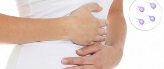
With the development of enterobiasis, the following symptoms may be observed:
- itching in the anal area;
- nausea;
- vomit;
- loss of appetite;
- bowel dysfunction;
- pain in the abdominal area;
- rash;
- difficulty urinating (infantile enuresis);
- development of inflammatory diseases of the genital organs (in girls);
- fatigue;
- irritability.
The incubation period from the moment of infection is 3-6 weeks. Complications of this disease include purulent inflammation of the skin, paraproctitis, vulvovaginitis, enterocolitis, acute appendicitis, peritonitis, salpingitis.
Symptoms of pinworms
Among the symptoms of pinworms that may indicate an infection, the most common is persistent itching around the anus.
Patients are often irritable and have trouble concentrating. They often complain of abdominal pain and headaches. Teeth grinding is also a characteristic symptom that many parents pay attention to.
Pinworms may also be accompanied by:
- loss of appetite;
- weight loss;
- pale skin;
- dark circles under the eyes.
Dark circles under the eyes
Weight loss
It is worth noting that women may experience itching around the vagina. Pinworms can penetrate the female genitals and cause inflammation. There are also known cases of appendicitis caused by pinworms. In adults, the symptoms of pinworms are not very different from those in children. However, they are often much less serious.
Human roundworm
Another prominent representative of roundworms is the roundworm. These helminths are large in size. Females can reach a length of 40 cm, males - up to 25 cm. Most often, parasites live in the small intestine. Roundworms are classified as geohelminths.
This means that their development cycle does not require intermediate hosts. A sick person excretes ascaris eggs along with feces, which then fall into the soil. This is where the larvae develop. The soil has optimal temperature and humidity for this.
It takes about 2 weeks for the eggs to become infective.
Human infection occurs through the fecal-oral mechanism (through food, water and dirty hands). In the stomach, the shells of the eggs are destroyed and the larvae emerge.
They live in the intestines, often leading to injury and obstruction. Sometimes the larvae are carried through the bloodstream to various organs (heart, lungs, brain, sinuses).
It is important that the development of larvae does not necessarily have to take place in the soil. Autoinvasion (self-infection) often occurs.
Examination of stool for parasites (giardia, opisthorchid, roundworm, pinworm, whipworm)
Helminths
Helminths (colloquially worms) are the general name for parasitic worms that live in the human body. Helminthiasis is a series of diseases caused by parasitic worms. The causative agents of helminthic infection are:
- representatives of roundworms of the class Nematoda (nematodes);
- flat tapeworms (cetodes);
- fluke worms (Trematoda, trematodes).
To preserve their species, representatives of all classes of helminths leave the host’s body to reproduce. At the same time, to carry out intermediate stages of development, they move into the body of a new host or remain in the external environment. The movement of parasites from one host to another is carried out by vectors:
- mechanical (for example, arthropods (in particular houseflies), carrying parasite eggs over considerable distances);
- specific – intermediate hosts, in whose body one of the development cycles of the parasite occurs.
The transfer can be:
- contact (active), in which the penetration of parasites occurs through damaged/undamaged skin and mucous membranes;
- food (passive), when ingesting larvae or eggs of parasites with food or water.
The roundworm (Ascaris lumbricoides or stringworm) is a nematode (roundworm) of the order Spirurida that lives in the small intestine. Infection with roundworm differs from infection with other parasites in that fresh helminth eggs do not pose a danger to the owner. In order for them to become infectious, they need to ripen in the soil for 10-15 days, and sometimes for a month. Moreover, they can remain there for three years without dying due to significant drops in temperature and without reacting to exposure to ultraviolet rays.
The presence of adult parasites of both sexes in the body makes a person a source of infection for others. Fertilized females lay about 200 thousand eggs within 24 hours, which are excreted along with feces. If favorable conditions exist, their development begins.
When mature eggs enter the upper part of the small intestine of a new host, larvae begin to emerge from them. Migration follows: through the intestinal wall through a vein, through the vascular system of the liver and lungs, the parasite larvae enter the bronchi, and then into the lungs. Using the epithelium of the respiratory tract as a “ladder”, they rise to the pharynx, from where they are swallowed back into the stomach and returned to the intestines. There they develop (usually this period lasts up to 75 days), after which the larvae turn into adults capable of reproduction.
An adult worm lives on average for about a year, then dies and leaves the body in feces. Consequently, perennial ascariasis is caused by repeated reinfection with the parasite. Infection can occur through food, water or household items. An additional possibility of spreading infection is created by the consumption of greens, vegetables and fruits from land plots fertilized with feces that have not undergone disinfection treatment.
Ascariasis is a two-phase infection. The early phase is migratory and occurs during the advancement of larvae. The late phase is characterized by the presence of mature worms in the intestines and the development of complications. The symptoms of ascariasis are identical to those of pulmonary diseases: the infection may be accompanied by a dry cough, difficulty breathing, and chest pain. In addition, an increase in body temperature (up to 38⁰), the appearance of bright pink, very itchy blisters, headache, and weakness are possible. At the same time, the level of leukocytes and eosinophils in the blood increases significantly. Chronic ascariasis is accompanied by nausea, abdominal pain, intestinal dysfunction, and sleep disturbances. With a complicated course of the disease, pancreatitis develops, intestinal patency is impaired, and appendicitis becomes inflamed.
Pinworms (Enterobius vermicularis or Oxyuris vermicularis) are small roundworms, causative agents of enterobiasis, which got their name due to the presence of a pointed end in the female. The location of pinworms is the lower part of the small intestine, the cecum (the appendage at the junction of the small intestine with the large intestine, the first section of the large intestine) and the appendix (a blindly ending tubal formation - the appendage of the cecum). The worms feed on the contents of their intestines.
At night, pinworms crawl from the rectum to the exit of the anus to lay eggs, which require oxygen to develop. One individual is capable of laying from 5 to 15 thousand of them. After this, the females die. Eggs mature within 4-6 hours, which is accompanied by discomfort and severe itching in the anus.
When scratching this area, eggs get under the nails and, in the absence of proper hygiene, into the mouth, and then back into the intestines. Adults are formed inside the body within 10-12 days. Then the cycle repeats. The source of enterobiasis is a person infected with pinworms.
Under the influence of waste products and, especially, the remains of parasites, the body is poisoned, allergic reactions occur, which causes an increase in the level of eosinophils (a subtype of white blood cells secreted by the immune system during the development of allergies or infection with parasites). In addition, the disease may be accompanied by metabolic disorders and vitamin deficiency. Pinworm larvae that attach to the skin cause the development of inflammatory processes. Also characteristic features of enterobiasis are intestinal dysfunctions:
- motor – aimed at mixing and promoting its contents;
- secretory - a violation of the release into the lumen of the gastrointestinal tract of secretions involved in digestion.
Severe perianal itching can cause anxiety, worsening or complete loss of appetite, weight loss, irritability, and insomnia. In children with severe and prolonged enterobiasis, deviations in mental development may occur.
Opisthorchiasis is another special case of helminthiasis. Its causative agent is opisthorchid , a small flatworm of the trematode class of the family Opistorhidae. The source of human infection is cyprinids infected with worm larvae that have not undergone sufficient heat treatment. Infection is also possible when cutting infected fish. The location of parasites in the human body is the bile ducts of the liver and the pancreatic ducts. The incubation period for infection is approximately 2-3 weeks. The disease may occur without symptoms, or may be accompanied by weakness, jaundice, fever, and chills. One of the reasons for the development of the disease is the ingestion of waste products of worms into the body (they cause an allergic reaction). In addition, health problems arise due to:
- mechanical effects of parasites on the walls of the pancreatic ducts, as well as on the ducts and the wall of the gallbladder;
- irritation of elements of nerve tissue located in these ducts;
- creating favorable conditions for the development of secondary biliary tract infections;
- proliferation of the glandular epithelium of the bile and pancreatic ducts.
The disease is often asymptomatic, which increases the risk of developing its chronic form, which can last for many years. The most characteristic consequences of opisthorchiasis infection are damage to the pancreatic ducts and chronic cholangitis (chronic inflammation of the bile ducts). As a result of a complicated course of the disease, the following diseases can develop:
- purulent cholangitis;
- pancreatitis (inflammation of the pancreas);
- cholelithiasis;
- cholangiocarcinoma (a cancerous tumor formed from mutant cells of the bile duct epithelium).
The most common cause of the development of chronic opisthorchiasis is repeated infections, causing relapses of inflammatory processes. With a single infection, the likelihood of developing severe complications is much lower.
Whipworm is a representative of roundworms (nematodes) 2-5 cm long, which parasitizes only in the human body. It is the causative agent of the disease trichocephalosis. The worm owes its name to its unusual shape: the front part of its body is much thinner than the back, and resembles a thread in appearance. It contains only the esophagus, and the remaining organs are located in the back. Whipworm eggs are transparent, barrel-shaped, with caps at the ends.
The female worm lays from 2 to 10 thousand eggs during the day, which are excreted from the intestinal lumen along with feces. When eggs fall into moist soil, larvae begin to develop. The optimal temperature for this process is +25-30°C. Development lasts 3-4 weeks, after which the eggs become invasive. Human infection is caused by ingestion of eggs with larvae, which can be on vegetables, berries, fruits, and also in water. In the human body, under the influence of digestive enzymes, the egg shell dissolves and the larva is released. The process of her reaching puberty continues for several weeks. The parasite lives for about 6 years.
The worm’s habitat is the initial section of the large intestine, where it “drills” deeply into the wall with its thin end. The parasite feeds on the host's blood and tissue fluid. Violation of the integrity of the intestinal wall can cause the development of an inflammatory process and, with massive invasion, the addition of a secondary infection at the site of damage (in particular, cause the development of pathological processes in the appendix). Also, a significant increase in the number of parasites can lead to anemia. The waste products of the worm cause poisoning of the body. The most common complaints of patients with trichuriasis are abdominal pain, headaches, and dizziness.
Diseases caused by parasite infection have a number of common symptoms. In particular, any of them can occur in acute or chronic form.
Acute helminthic infestation is usually characterized by:
- general allergic reaction;
- fever;
- leukocytosis (increased levels of white blood cells in the blood);
- eosinophilia (increased concentration of eosinophils (a subtype of leukocytes) in the peripheral blood more than 500/μl).
Symptoms of the chronic form of infection depend on the number of helminths and their location. In the vast majority of cases, infection with worms is accompanied by metabolic disorders, abnormalities in neuropsychic activity, and weakened immunity.
Protozoa
The Protozoa group includes microscopic unicellular organisms (which, however, can unite into multicellular colonies). Although their body is represented by only one cell, the protozoa are absolutely full-fledged organisms with all the characteristic features - their life activity is coordinated, orderly, which is confirmed by:
- the process of nutrition and the release of decay products;
- carrying out the reproductive function.
Protozoa differ from prokaryotic bacteria (single-celled living organisms that do not have a formed cell nucleus and other internal membrane organelles) by the presence of a cell nucleus surrounded by a membrane and containing DNA molecules.
For most protozoa, as well as for animals, food comes from ready-made organic substances present in their environment. However, some protozoa, like plants, contain chlorophyll and, using solar energy, are able to convert inorganic substances into organic ones (carbohydrates). In connection with this division, protozoa were classified either as plants or animals, or as a separate group. Ultimately, they, together with a number of organisms, form a group of protists (Protista), within which they are separated into an independent subgroup.
To date, about 30,000 species of protozoa have been described. There are many of them that are dangerous to humans, but three are recognized as the most significant:
- lamblia (Lamblia intestinalis);
- dysentery amoeba (Entamoeba histolytica);
- balantidia (Balantidium coli).
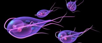
Giardia (Giardia intestinales, also known as Giardia lamblia or Giardia duodenalis) is the most common single-celled parasite that is found almost anywhere on the planet, both in humans and animals. Giardia is protected on the outside by a shell that allows them to survive in the external environment for a long time. Infection with the parasite occurs through the oral-fecal route, through dirty hands, through water (the presence of Giardia in it is the most common cause of infection) and contaminated products.
Giardia is the causative agent of the pathology - giardiasis and lives mainly in the duodenum and bile ducts of the host. There they can move freely using flagella or attach to the epithelium through an adhesive disk, which is located in their lower part. Up to a million lamblia can stick to 1 cm^2. In this case, a violation of the villi occurs, which disrupts the absorption of nutrients and provokes the development of inflammation of the mucous membrane.
The most characteristic manifestations of the disease are diarrhea with watery stools and stomach cramps. The onset of symptoms is observed 7-14 days after infection. The active phase usually lasts from 2 to 6 weeks. If the infection affects the bile ducts, the disease is accompanied by jaundice.
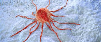
Dysenteric amoeba is the causative agent of amoebiosis. It is distributed everywhere, but especially widely in the tropical zone. Infection with the parasite most often occurs through ingestion of cysts - a temporary form of existence of the infectious agent, in which it can exist for several years.
Amebiasis is a deadly disease, from which, according to available data, about 100 thousand people die every year. Once in the human blood, the amoeba is able to penetrate the tissues of the liver, lungs, kidneys, brain, heart, spleen, and genitals. The food for this protozoan is epithelial cells, food fragments, bacteria, erythrocytes, and leukocytes.
Dysenteric amoeba lives in the intestines and has two forms - tissue and luminal. In countries with temperate climates, it usually remains in the intestinal lumen, so the infection does not manifest itself visibly. In subtropical and tropical climates, the amoeba becomes active and begins to attack its walls. The reasons for the transition of amoebas to a pathogenic tissue form remain unclear, but it is known that it can attack in several ways. First of all, the amoeba secretes substances that dissolve the mucous membrane, which allows it to kill the cells of the host body. The parasite presumably kills cells in one of the following ways:
- by launching the process of apoptosis (programmed death) in them, which occurs in a few minutes;
- through trogocytosis (gnawing off pieces from the cell), which today explains the amoeba’s ability to penetrate the blood, liver and other areas of the body.
Balantidia are the largest protozoa that parasitize the human body - they are much larger than the intestinal cells among which balantidia live. They are the causative agent of an infectious disease - balantidiasis. Infection with the parasite occurs through consumption of food and/or water contaminated with its cysts.
Balantidium is essentially a ciliate (the only one of all protozoan parasites). It lives in areas where pigs live, which are its main carriers. The habitat of balantidia in the human body is the submucosal layer of the large intestine, although the parasite is also able to penetrate the epithelium of the lungs. Balantidia feed on bacteria, pieces of food and fragments of the epithelium of the host organism. They can cause both mild colitis and severe illness with the formation of ulcers in the walls of the colon, which are accompanied by severe diarrhea with blood and mucus. Deaths from balantidiasis are extremely rare, but severe disease leads to chronic exhaustion.
Manifestations of ascariasis
The development cycle of these worms is about 3 months. This period is the incubation period of the disease. Clinical symptoms of ascariasis are varied. They include:
- the appearance of a rash;
- skin itching;
- slight increase in body temperature;
- decreased appetite;
- nausea;
- flatulence;
- abdominal pain;
- hypersalivation;
- drowsiness;
- weakness;
- irritability.
In severe cases of ascariasis, signs of intestinal obstruction are possible: pain, bloating, constipation. In some cases, adult helminths can be excreted in the feces. Less commonly, ascariasis affects other organs. If roundworms are localized in the eye, oculomotor disorders and hemorrhages are formed. With pulmonary localization, suffocation may occur.
Clinical picture of enterobiasis
The most important symptom of a helminthic infection is itching in the anus ; it occurs mainly at night or in the early morning hours. The laid eggs irritate the skin in the anal area. Pinworms in children are accompanied by unbearable itching and the child cannot stop trying to stop scratching the skin. In this regard, scratches and scratches appear. Sometimes there is a violation of the integrity of the skin. This may be accompanied by an additional infection and symptoms of inflammation appear in the areas of scratching: hyperemia, swelling and soreness.
When a large number of parasites remain in the body for a long time, night sleep is disturbed due to a certain time of egg laying and the occurrence of specific symptoms at night.
Treatment and prevention of ascariasis and enterobiasis
Treatment of ascariasis involves taking anthelmintic drugs. These include tablets Albendazole, Vermox, Levamisole. Therapy for ascariasis includes following a diet (table No. 13 is prescribed), taking enzyme preparations, probiotics and prebiotics, vitamins, and antihistamines.
Compliance with the rules of personal hygiene (regular washing of hands and body, washing, washing and ironing clothes, cutting nails, wearing thick underwear, changing bed and underwear) and wet treatment of household items is of great importance. Preventive measures against ascariasis and enterobiasis include thoroughly washing fruits and vegetables, limiting contact with infected people, and boiling water.
Thus, pinworms and roundworms are the most significant helminths from an epidemiological perspective.
What pinworms and their eggs look like
Pinworms are oblong-shaped worms, round in cross-section.
In the center their body is widened and narrows closer to the edges. Their color ranges from white to grayish-yellow. The size of the pinworm depends on gender. Females are usually larger than males. The size of females ranges from 8 to 13 mm, males are 3-5 mm long. Pinworms can be seen in stool after taking anthelmintic medications. Pinworm eggs are asymmetrical in shape and have a double shell, but they are so small that they cannot be seen with the naked eye.
Differences between roundworms and pinworms
Most often, these parasites have the same method of infecting the human body. If you do not wash your hands after using the toilet, after going outside, or before eating food, then you are likely to introduce parasites into your body. But there are some differences in infection with worms, since roundworms are quite tenacious and can be found on different objects and in different places. For example, in raw, untreated water or on objects in an unsanitary environment. They remain on fruits and vegetables if they are not washed properly. It is also worth remembering that worm larvae can be introduced through food if it is poorly processed or has not undergone heat treatment at all.
It is not always possible to become infected with helminths from another person, since the larvae can remain on underwear or bedding. Therefore, we must not forget that simply taking a course against worms is not enough. It is definitely recommended to change your underwear and bed linen. In addition, eggs can be carried by insects that come into contact with food, such as flies or cockroaches. Moreover, eggs can be inhaled into the lungs after inhaling dust.
Treatment of pinworms
When pinworms are detected in the body, the doctor selects the optimal treatment based on the individual characteristics of the child, his age, body weight, as well as the volume and location of the lesion.
Therapy should be aimed at combating parasites of all phases of development (eggs, larvae, adults), eliminating the consequences of pinworm activity, as well as relieving itching and other unpleasant symptoms of the disease. For this purpose, anthelmintic drugs are first selected. Medicines can come in various forms (tablets, suspensions, suppositories), which allows them to be used to treat children of all ages. Specialists at the SM-Doctor clinic for children and adolescents strongly do not recommend choosing an anthelmintic drug without consulting a specialist. This may lead to intoxication and cause other side effects.
Taking an antiparasitic drug alone is not enough. To eliminate the consequences of pinworms in the body, the following groups of drugs are needed:
- Sorbents are prescribed to remove parasites and their metabolic products from the body.
- Probiotics – recommended in the presence of intestinal dysfunction caused by helminths.
- Antihistamines are prescribed if enterobiasis is accompanied by allergic reactions.
It should be remembered that to avoid re-infection, all members of the patient’s family should undergo treatment. A number of hygiene procedures are also required:
- Change of linen followed by washing and hot ironing.
- Heat treatment of toys and other objects with which the child has been in contact.
- Regular wet cleaning of the premises.
To confirm the fact of recovery, the child must be re-examined for enterobiasis 2 weeks after the therapy.
Ways of infection with ascariasis
The main difference in infection mechanisms is that the eggs mature in different environments. For pinworms, these are the folds of the perianal zone of an infected person. From here they enter the mouth and then the intestines of the same person. Or into another person's mouth and intestines.
The method of infection by roundworms is determined by the fact that in order for their eggs to mature, they need to lie in the soil. Leaving the human intestines along with feces, the embryos of the parasite end up in the ground. After lying there at a certain temperature (1336 C) and humidity (not lower than 4%), they become ready for invasion. The optimal temperature for eggs is 24 C, at which maturation occurs in two weeks.
This method of infection is called fecal-oral. Eggs are capable of infecting humans only after they have remained and matured in the ground.
The principles of infection with roundworms in humans can be different.
- When eating thermally unprocessed food.
- When eating poorly washed fruits and vegetables.
- Drinking raw, unboiled water from open reservoirs and water pipes.
- When swimming in open waters.
- From pets that have parasite germs on their fur.
- From flies and other insects. Crawling on uncovered food, they infect it.
- Upon contact with things and interior elements that could somehow get worm eggs.
Contributes to parasite infection:
- working with soil in the garden and vegetable garden, especially if feces are used to fertilize them;
- improperly constructed cesspools in private farmsteads.
What are pinworms and why are they dangerous?
Pinworms are representatives of roundworms.
This type of helminth is dangerous and can lead to many negative consequences in the body:
- disrupt their own beneficial intestinal microflora due to the production of their metabolic products and the formation of harmful toxic substances;
- in girls, young women and women, due to the close anatomical location of the anus and vagina, pinworms can spread to the female genital organs, causing gynecological inflammatory diseases;
- as the number of parasites in the body increases, they populate different parts of the intestine; if they enter the cecum, where the appendix is located, it may become inflamed and appendicitis may begin;
- helminths are able to rob the body by feeding on blood, the level of red blood cells and iron decreases, and as a result anemia occurs. This condition is accompanied by lethargy, drowsiness, fatigue, lethargy, brittle hair and nails, pale complexion and skin;
- When parasites enter a child's growing body, they affect the child's immune system, he gets sick more often, and lacks vitamins and microelements.
When pinworms enter the human body, they parasitize primarily in the intestines.
Similarities
The main similarity between these parasites lies in their structure. Like pinworms, roundworms are also roundworms and nematodes. They have the same light color. There are also other similarities:
- Both types of parasites are localized in the intestines and affect other organs to a much lesser extent;
- Both parasites have the same transmission pattern - exclusively fecal-oral, that is, for the development of invasion, you need to ingest a certain number of parasite eggs;
- The transmission routes are the same. These are unwashed vegetables and fruits, raw meat, contaminated water, contact with pets or contaminated surfaces;
- They have some similarities in symptoms. Pinworms and roundworms cause mainly intestinal symptoms, intoxication and an allergic reaction;
- There are similarities in the parasite's life cycle. In particular, the peculiarity is that the invasion develops with constant repeated self-infection;
- In both species, males are much smaller than females;
- Both species are present in the intestines in entire colonies.
It is important to take into account that mixed invasions are currently widespread, when several types of parasites are present in the intestines at the same time. Roundworms and pinworms may well exist together in the body, causing the characteristic symptoms of both invasions.
Description of the disease
Damage to the human body by pinworms is called enterobiasis.
This is the most common parasitic disease in childhood. Penetrating into the body, the worms are localized in the lower parts of the small intestine, cecum and colon. Adult females who have reached sexual maturity are able to exit through the anus to lay eggs between the folds of skin around the anus. Pinworms have distinctive features of their external structure. Females have a “tail”, that is, a pointed end (hence the name of the worms - pinworms). It can injure the intestinal mucosa, causing severe itching in the perianal area. Males are smaller than females, their “tails” are bent in a spiral. The disease causes significant discomfort to the child. Constant itching (intensified in the evening and at night) makes it impossible to sleep peacefully; the waste products of pinworms poison the body, disrupting the functioning of the gastrointestinal tract. The baby loses his appetite and a sharp decrease in body weight begins.
If a child is suspected of having enterobiasis, parents should promptly consult a gastroenterologist or infectious disease specialist. Competent specialists are able to determine whether a child has pinworms and help to quickly and effectively cope with the disease.
Prevention
- Mandatory drug therapy for persons with a confirmed diagnosis and persons in close contact with the patient.
- Proper sanitary and hygienic education of children from an early age. Children should be taught to wash their hands with soap after using the toilet, before eating and after going outside. Wash vegetables and fruits before eating.
- Change underwear daily. Don’t be shy and don’t hide the symptoms of enterobiasis from parents, talk right away to exclude advanced forms of the disease.
- In crowded places such as a kindergarten or school, where there was a child with enterobiasis, it is necessary to carry out deworming for preventive purposes.
- In the house where a patient with enterobiasis lives, carry out general cleaning, washing the floors with disinfectants.