What provokes / Causes of Trichinosis:
Trichinella are small, almost thread-like helminths (thrix - hair), covered with a transversely striated cuticle. The body of T. spiralis is rounded, somewhat narrowed towards the anterior end. The length of a sexually mature male is 1.2-2 mm with a width of 0.04-0.05 mm. The length of a sexually mature female before fertilization is 1.5-1.8 mm; after fertilization, its length increases to 4.4 mm.
The existence of two types of foci of trichinosis has been established: natural and synanthropic. Natural foci are primary in origin. Trichinella can parasitize the body of 57 species of wild and domestic animals; the circulation of the pathogen is based on nutritional connections. In these foci, parasites circulate among wild animals (wild boars, badgers, raccoon dogs, brown and polar bears, foxes, martens, minks, ferrets, etc.), marine mammals (whales, harbor seals) due to predation or eating carrion. In synanthropic foci, Trichinella circulates among domestic animals (pigs, cats, dogs), rodents (mice, rats), also by eating each other or carrion. In addition, synanthropic foci are replenished by hunting trophies - trichinosis wild animals.
There is a direct and inverse relationship between natural and synanthropic foci. Invasion from natural foci is introduced into synanthropes in two ways: by humans, who hunt infected wild animals and feed their remains to domestic animals, and by wild synanthropes (rats, mice), which migrate to natural foci in the spring and return back in the fall. As a result, mixed natural-synanthropic foci are created.
In the muscles of animals, the larvae remain infective for years, and in cadaveric material they die under the influence of very high or low temperatures (-40, -50°C) and can tolerate conditions in the Arctic zone.
The source of infection for humans is domestic and wild animals affected by trichinosis. Most often these are pigs, wild boar, brown and polar bears, nutria, badgers, foxes, and for some nationalities - dogs.
The mechanism of infection is oral. People's susceptibility to trichinosis is very high. In order to get a serious illness, it is enough to eat 10-15 g of trichinosis meat. Infection usually occurs when eating raw or undercooked meat from animals affected by trichinosis, most often meat, lard, ham, bacon, brisket, brisket, sausage made from infested pork, as well as meat from wild animals affected by trichinosis (bear, wild boar, badger ).
The incidence of trichinosis is usually group in nature. Members of the same family, persons participating in the same festive feast, hunting meal, and those who used the meat of the same trichinosis animal that did not undergo preliminary sanitary and veterinary control become ill.
Trichinella larvae die when the temperature inside a piece of meat reaches at least 80°C. Salting and smoking meat does not affect encapsulated larvae.
The seasonal nature of group outbreaks has been established. In synanthropic foci, they are in most cases associated with the autumn period - the period of slaughtering pigs and procuring meat products.
Outbreaks of trichinosis in natural foci are associated with the hunting season - the autumn-winter period. Due to existing poaching, they can occur at any time of the year.
The formation of foci of trichinosis is facilitated by improper management of pig farming: free keeping of pigs, their wandering, access to pigsties for rodents, cats, and dogs.
Life cycle of Trichinella
Trichinella are viviparous helminths. An important biological feature is also that the same organism becomes first the definitive and then an intermediate host. Trichinella has a variety of hosts other than humans. They parasitize many mammals - pigs, wild boars, bears, wolves, foxes, badgers, dogs, cats, as well as rodents, insectivores and marine mammals.
In the sexually mature stage, helminths parasitize in the wall of the small intestine, and in the larval stage - in the striated muscles, except for the heart muscle.
A person becomes ill with trichinosis by eating pork or wild animal meat contaminated with encapsulated larvae. During the digestion process, the larvae are released from the capsules and within an hour they penetrate the mucous membrane, reaching the submucosal layer of the small intestine.
After a day they turn into males and females. Sexually mature individuals, using the head stylet, are attached to the intestinal mucosa, where they then copulate.
In the body of various animals, the female Trichinella parasitizes from 10 to 56 days, giving birth to from 200 to 2000 live larvae. During the entire period of parasitism in the human intestine (no more than 42-56 days), one female gives birth to an average of 1,500 larvae.
The larvae penetrate through the intestinal mucosa into the lymphatics, then into the blood vessels and are carried throughout the host’s body by the blood stream. On the 5-8th day, the larvae enter the skeletal striated muscles. With the help of the hyaluronidase they secrete, they penetrate into the sarcolemma of the muscle fiber, where their further development occurs, already in the body, which has become an intermediate host for the pathogen. 18-20 days after infection, the larva in the muscles lengthens to 0.8 mm, reaches the invasive stage and begins to curl up.
Consequences and complications
Trichinosis is a deadly disease that leads to various organ damage, including the liver, kidneys, and severe complications usually occur in the 3-5th week of the disease. These include:
- trichinosis myocarditis , which can provoke the development of acute cardiovascular failure ;
- pneumonia - usually characterized by a diffuse increase in the vascular pattern and the presence of pleural lesions;
- meningoencephalitis;
- infectious-toxic shock;
- abdominal syndrome;
- paralysis and paresis are the result of nonspecific vasculitis and diffuse focal granulomatosis in the brain and/or spinal cord;
- thrombosis of large vessels.
Pathogenesis (what happens?) During Trichinosis:
The pathogenesis of trichinosis is complex, representing a complex of pathological reactions, the trigger of which is the pathogen.
As is known, the entire biological cycle of Trichinella takes place in the body of one host, in this case, a person, in which successive stages of helminth growth have different localizations: the infective larva in the lumen, and then in the mucous membrane of the small intestine; a growing and then adult individual in the tissue of the small intestine; migratory larva - in the bloodstream and lymph; muscle larva - in striated muscles. As a result, the products of metabolism and partial decay, especially of larval and growing individuals, enter directly into the tissues. They are parasitic antigens with high sensitizing activity.
The allergic nature of trichinosis underlies its pathogenesis. N. N. Ozeretskovskaya distinguishes three phases of development of the pathological process: enzymatic-toxic (1-2 weeks after infection), allergic (from the end of the 2nd -3-4 weeks after infection) and immunopathological.
The enzymatic-toxic phase is associated with the penetration of invasive Trichinella larvae into the intestinal mucosa and the formation of adult helminths, under the influence of enzymes and metabolites of which an inflammatory reaction develops in the intestine.
The second, the allergic phase of trichinosis, is characterized by the occurrence of general allergic manifestations in the form of fever, myalgia, edema, skin rashes, conjunctivitis, catarrhal pulmonary syndrome, etc. By the end of the first week, mature adult Trichinella begin to hatch young larvae, which migrate through the lymph and blood to striated muscles. The suppressed protective reaction of the host due to the immunosuppressive effect of adult parasites does not prevent the active circulation of larvae.
However, by the end of the second - third week of the disease, the level of specific antibodies in the serum of the infected person increases and a violent allergic reaction develops.
Violent allergic inflammation in the small intestine contributes to the death of adult Trichinella, the formation of granulomas around Trichinella larvae in the muscles, from which fibrous capsules are subsequently formed that prevent the entry of parasite antigens into the host body.
The severity of immunological reactions depends on the dose of the antigen and the immunoreactivity of the host organism, on the degree of adaptation of the parasite to the host. An increase in the dose of infection causes an increase in the intensity of intestinal and muscle invasion, which in turn leads to an increase in the severity of the disease and inhibition of immunological processes. This is accompanied by systemic damage to organs and tissues as a result of sensitization of the body not only by the metabolic products of helminths, but also by the decay products of damaged or destroyed host tissues. This phase is manifested by fever, muscle pain, swelling, conjunctivitis, and respiratory problems.
The immunopathological phase of trichinosis, usually associated with intense infection, is characterized by the appearance of allergic systemic vasculitis and severe organ damage.
Nodular infiltrates occur in the myocardium, brain, lungs, liver and other organs. Trichinosis is complicated by severe allergic diffuse focal myocarditis, meningoencephalitis, focal pneumonia and other equally severe organ lesions, which can be combined with each other, accompanied by high fever, severe muscle pain, skin rashes, and the spread of edema.
By 5-6 weeks after infection, the inflammatory process in parenchymal organs is replaced by dystrophic disorders, which recover slowly over 6-12 months.
Duration of the disease
The more severe the symptoms of trichinosis, the longer the helminthiasis lasts.
- In the erased form, trichinosis lasts no more than 1 week.
- In mild forms, trichinosis lasts no more than 2 weeks.
- With moderate and severe trichinosis, the acute phase is shortened with hormonal treatment, but recovery occurs only by 4-6 months. Muscle pain can bother the patient for another 1 - 2 months after recovery, eosinophilia within 10 - 15% persists for up to 3 months or more.
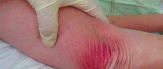
Rice. 12. Edema due to trichinosis.
Test for igg antibodies to Trichinella
As a rule, when infected with trichinosis, people do not even suspect that we are talking about a helminthic infestation, and attribute all unpleasant symptoms to gastrointestinal diseases or poisoning. The most reliable way to determine the true cause of painful health is to test for antibodies to Trichinella.
The presence of IgG antibodies to Trichonella is determined by ELISA, the sensitivity of which is 90-100%, and the specificity is 70-80%. The appearance of specific antibodies is usually recorded 2 weeks after infection, and their maximum number is observed at 4-12 weeks.
Even if the result is negative, it cannot be said that there was no infection, since this may simply be an early stage of infection, so the test is usually repeated after 2-3 weeks. The diagnosis of trichinosis is made if the antibody titer is increased fourfold. In people who have had trichinosis, antibodies can persist for more than 2 years.
Diagnosis
It is diagnosed intravitally using allergic methods that are applicable in breeding farms for high-breed animals. Immune methods (immunofluorescence assay and enzyme-linked immunosorbent assay) are also applicable.
In commercial farms, selective diagnostics are carried out followed by control slaughter.
The main diagnostics are carried out during veterinary and sanitary examination of meat. Trichinelloscopy is carried out by microscopy (compressor) of thin sections of the muscles of the favorite places of localization of the larvae (peduncles of the diaphragm, intercostal, cervical).
At processing plants, research is carried out by digesting prefabricated samples (from 10-50 carcasses) in artificial gastric juice, followed by microscopy.
It is necessary to differentiate the larvae by microscopy from cysticerci and sarcocysts (cylindrical or irregular shape). Treatment is carried out using the Yamshchikov method (using hydrochloric acid), in which the Trichinella capsule does not dissolve.
The diagnosis is considered established if at least one encapsulated or non-encapsulated larva is found in the sample.
Mechanism of infection
In all cases, infection occurs due to eating the meat of sick animals, i.e. the mechanism of infection is nutritional. It is especially dangerous to eat cured meats and undercooked meats. When salted, the larvae in the depths of the piece can survive for up to a year.
The larvae tolerate low and high temperatures quite well. At temperatures below -20 C they survive for up to three days, but the higher the temperature, the longer the larva survives. In the freezer (temperature about -10 C) the larva does not die for up to a year. When frying, if the temperature of the meat reaches +50 C, the larvae die within a few minutes. But if the animal has been sick for a long time, a calcified capsule could have formed around the larva, in which case the temperature is not capable of killing the larva.
It is possible to defeat parasites!
Antiparasitic Complex® - Reliable and safe removal of parasites in 21 days!
- The composition includes only natural ingredients;
- Does not cause side effects;
- Absolutely safe;
- Protects the liver, heart, lungs, stomach, skin from parasites;
- Removes waste products of parasites from the body.
- Effectively destroys most types of helminths in 21 days.
There is now a preferential program for free packaging. Read expert opinion.
Read further:
Roundworms: size, color, nature of movements, shape of eggs and larvae
Acanthocephalans: types and class of worms, life cycle of development
What can be the source of human infection with trichinosis, the causes of infection
Pork roundworm: description and structure, life cycle and ways to remove it
Pork tapeworm: symptoms, prevention, and treatment methods for pork tapeworm
Dysenteric amoeba: life cycle, structure, diagram, stages of development
Signs
Unfortunately, timely detection of trichinosis is not always possible, since in many pigs this disease occurs without severe symptoms. Only with a very severe infection can the waste products of parasites cause intoxication. In this case, the following symptoms are typical:
- increased muscle temperature;
- loose stools;
- intensive weight loss;
- severe vomiting;
- muscle pain (pigs are inactive, lie down and stretch their legs);
- difficulty breathing;
- severe swelling in the limbs and neck.
The disease does not always lead to death. But improving the well-being of pigs does not mean that they are no longer carriers. After a month and a half, the animal’s body produces antibodies that contribute to the formation of immunity.
The larvae that get from the gastrointestinal system into the blood, and with it into the muscle tissue of the animal, are encapsulated. Now they are ready to settle into the next host. After a few months, the capsules are covered with lime. Trichinella can remain in this form for about 25 years, which is why the likelihood of a new infection is still high.
Treatment
Since diagnosing the disease in a living animal is extremely difficult, treatment methods are considered by most modern veterinarians to be insufficient and poorly developed. Among the drugs that can destroy parasites localized in the intestines, as well as those going through the stage of capsule formation, there are the following: mebendazole, thiabendazole, parbendazole, albendazole.
There is a certain probability of stopping trichinosis at the initial stage of infection or clearing the muscle tissue of an already ill animal from encapsulated parasites.
Effective methods of combating the disease during the most active stage of the spread of Trichinella in muscle tissue (migration stage) do not yet exist.
Prevention measures
To prevent trichinosis, it is necessary to protect areas where pigs are kept from rodents. Rats and mice are carriers of this disease. Strong walls, floors, and the lack of holes in them will become an obstacle for them.
It is advisable to bury the carcasses of animals obtained during hunting to a depth of at least a meter, in places inaccessible to tearing by pigs, dogs, and other animals. Do not feed raw residues from the slaughter of domestic animals and fur-bearing animals to pigs. Follow the rules for slaughtering livestock on private farms and farms. To prevent infection of people, you should not purchase pork that has not passed a veterinary examination. Meat obtained from hunting must be taken to the nearest laboratory for testing. It is important to remember that any disease is easier to prevent than to treat.