- In which bronchi do malignant tumors occur?
- Classification of bronchial cancer
- Stages of bronchial cancer
- Causes of bronchial cancer
- Symptoms of bronchial cancer
- Methods for diagnosing bronchial cancer
- Treatment of bronchial cancer
- Surgical treatment of bronchopulmonary cancer
- Radiation therapy
- Chemotherapy
- Targeted therapy and immunotherapy
- Survival prognosis
“Bronchi cancer” is a term that is rarely used by doctors. In medicine, the more common concept is “bronchopulmonary cancer,” which combines malignant tumors of the lungs and bronchi. This approach to terminology arose due to the fact that the lung tissue and the bronchial tree are closely related, both in an anatomical and functional sense.
Some figures and facts regarding bronchial cancer:
- The disease most often occurs in older people. In more than half of cases, bronchial cancer is diagnosed after the age of 50 years. Another 25% of cases occur in the age group of 40–50 years.
- The main cause of malignant tumors of the bronchi is smoking. Up to 80% of patients are smokers.
- Most often, the tumor occurs in the upper parts of the bronchial tree, since they are better ventilated, and carcinogens contained in the inhaled air are more likely to lead to malignant transformation of cells.
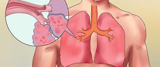
In which bronchi do malignant tumors occur?
The air that a person inhales passes through the nose, pharynx, larynx, and trachea. At the level of the upper edge of the fifth thoracic vertebra, the trachea ends and divides into two main bronchi . This location is called the tracheal bifurcation . The main bronchi are the first order bronchi , they are divided into lobar (second order) , then into segmental (third order), subsegmental (fourth order), lobular, and finally into terminal (end) bronchioles.
All these branches together make up the bronchial tree . The walls of large and small bronchi are structured in the same way: on the inside they are lined with mucous membrane , under it there is a framework - fibrocartilaginous membrane , on the outside - the adventitial membrane.
Malignant tumors that originate from the mucous membrane are called cancer. They can occur in any part of the lung, but are most often found in the hilum - where the main bronchus enters the lung. In two thirds of cases, cancer develops in the bronchi of the first, second and third order.
Pneumonia: features
The development of pneumonia is caused by a complication after an infectious process in the upper respiratory tract, bronchitis or a primary infection. Pneumonia affects the alveoli and terminal airways. Due to the fact that the disease manifests itself with symptoms identical to the common cold, its first signs are often neglected.
The cause of pneumonia often lies in an infection or an allergic reaction of the body. You can determine the difference between bronchitis and pneumonia with the help of an experienced diagnostician, since the inflammatory process in the alveoli of the lungs is accompanied by slightly different symptoms.
The presence of pneumonia can be assumed in the following conditions:
- high temperature (exceeding 38.5 degrees);
- active secretion of sputum;
- hemoptysis;
- pain in the chest area;
- difficulty breathing;
- increased heart rate (more than 100 beats per minute);
- increased breathing (more than 24 per minute);
- duration of the disease exceeding two to three weeks.
Complications of pneumonia can include:
- lung abscess;
- respiratory failure;
- exudative pleurisy;
- septic fever.
Damage to the alveoli of the lungs is detected using the same diagnostic tests that are carried out for bronchitis. The correct selection of antibacterial drugs and their timely administration guarantee effective treatment and elimination of infection in the lungs.
Classification of bronchial cancer
The histological (based on the appearance of tumor cells under a microscope) classification of bronchopulmonary cancer includes four main types of tumors:
- Small cell cancer occurs mainly in smokers, is highly aggressive, spreads quickly and is difficult to treat. It accounts for about 12% of all cases of bronchopulmonary cancer.
- Squamous cell carcinoma , together with the next two types, combines into a group of non-small cell tumors , which account for more than 80% of all cases of lung cancer. The risk of developing squamous cell carcinoma is strongly associated with smoking.
- Adenocarcinoma is most common among women and nonsmokers. Unlike squamous cell carcinomas, adenocarcinomas are usually smaller in size and tend to grow in the peripheral parts of the lung. A separate subgroup of bronchoalveolar adenocarcinomas is distinguished.
- Large cell carcinoma consists of large, undifferentiated cells.
Depending on how much the tumor tissue has lost its normal features, poorly differentiated and highly differentiated tumors are distinguished. The first ones are more aggressive.
When choosing treatment, the location of the tumor foci is of great importance. Depending on this indicator, bronchopulmonary cancer is divided into two types:
- Central - the tumor is located in the bronchi of the 1st–3rd order (main lobar, segmental).
- Peripheral - the tumor is located in the smaller bronchi.
Write to an oncologist
Visualization of bronchial diseases (part 2)
Transcript of the second part of the video lecture by Professor Igor Evgenievich Tyurin on the visualization of bronchial diseases, images from the second program of the radiology diagnostics series for therapists.
Igor Evgenievich Tyurin, professor, doctor of medical sciences:
— Since obstructive diseases themselves are considered a risk factor for the development of many socially significant, important, latent diseases, X-ray examination for such patients is of certain importance for identifying a malignant tumor, tuberculosis. It may be detected somewhat more often in this category of patients than in the general group of patients.
What changes can we detect with X-rays and CT scans? I tried to divide these pathological conditions into several component parts in order to make it more systematic, understandable, methodical, from the point of view of the perception of sometimes quite complex X-ray and tomographic symptoms.
X-ray changes. Signs of bronchitis, bronchial obstruction and emphysema.
Unfortunately, when it comes to a conventional X-ray examination or even a computed tomography examination, radiologists and attending physicians relatively rarely differentiate these symptoms and clinical radiological manifestations from each other. Although the manifestations, even with conventional x-ray examination, are completely different. We must be able to find these signs even on a regular x-ray in order to accurately characterize the pathological process.
What are x-ray changes in the form of signs of chronic bronchitis? Precisely chronic bronchitis, because acute inflammation of the bronchial mucosa, as a rule, does not find any reflection in X-ray and tomographic examination. In fact, neither one nor the other is intended for the diagnosis of acute respiratory viral infections with damage to the paraximal or middle caliber bronchi.
Common signs, such as enhancement and deformation of the pulmonary pattern, are added by radiologists to their protocols through the interstitial component. Thickening of the walls of the bronchi, which we can see on x-rays and tomograms, is defined as peribronchial couplings. At the same time, perivascular couplings.
Bronchial dilatation is possible. But with conventional X-ray studies, in contrast to tomographic studies, we see dilated bronchi much less often and not as confidently as with CT.
If we return to conventional X-ray examination, then in patients with obstructive diseases we see these changes in the large bronchi in the hilar region. This is due to thickening of the walls of the bronchi.
(Slide show).
The arrows here indicate peribronchial couplings, that is, thickening of the walls of the bronchi, which we can see in longitudinal or cross section. It quite accurately and adequately reflects the severity of the chronic inflammatory process in the bronchi themselves. Plus, there is a change in the pulmonary pattern due to the pulmonary interstitium, which we can observe in such patients.
03:22
But what I would like to draw your attention to. We will return to this more than once today. These changes immediately on the basis of one x-ray cannot be interpreted as irreversible changes, as manifestations of pneumosclerosis, or any specific morphological type of changes in the lung tissue.
Most often, we see such patients during an exacerbation of the disease, when it is not so much and not only about sclerotic changes in the walls of the bronchi or in the peribronchial lung tissue, but rather about swelling of the walls of the bronchi, incoming changes. In fact, this picture resembles interstitial edema with peribronchial changes.
As soon as these acute symptoms go away and the patients return to their stable state, the X-ray picture very often also indicates the disappearance of a significant part of these signs, which are sometimes not entirely correctly characterized as irreversible changes.
With a conventional X-ray examination, we rarely see signs of bronchi dilatation, bronchiectasis, or cystic cavities with relatively thick walls. Sometimes we even see fluid levels in them (during an exacerbation).
These are pronounced changes that can be observed in advanced stages. They need to be differentiated from other diseases that can lead to the formation of bronchiectasis in addition to obstructive pulmonary disease.
It is fundamentally important that on x-rays we see quite clearly signs of bronchial obstruction. This:
- flattening of the diaphragm;
- limitation of her mobility;
- vertical position of the heart;
- increase in retrosternal space;
- signs of pulmonary arterial hypertension that may occur in this type of patient.
05:14
It is no coincidence that the flattening of the diaphragm and the limitation of its mobility are written in the first two lines. It has long been known and well proven that these are the two most important, reliable, accurate signs that characterize a violation of the passage of air through the small bronchi and an increase in the residual volume in the lungs.
When we interpret or review x-rays of the chest cavity, then, first of all, we pay attention not to the degree of transparency of the lung tissue, which depends extremely variable on age, on the constitution, on the physical conditions of the x-ray, but on the position of the mediastinum, diaphragm, retrosternal space.
Flattening of the diaphragm and changes in the configuration of the chest are the two most important radiological signs that allow us to talk about the formation, the presence of obstructive changes in the lung tissue. Previously, fluoroscopic examination was much more often used for this purpose. Radiologists could evaluate the mobility of the diaphragm and the limitation of this mobility in patients with obstructive diseases.
Nowadays this is done much less frequently. But we see it well. An increase in the retrosternal space and a flattening of the dome of the diaphragm, which should have a dome shape as the main respiratory muscle. These are two of the most important signs of the presence of bronchial obstruction in this type of patient.
Where these changes reach a significant degree of severity, we see not only changes in the configuration of the chest, which takes on a typical beam-like shape. Not only a flat or even concave diaphragm towards the abdominal cavity, but also those signs that radiologists mention in their reports.
This is an increase in the airiness of the lung tissue, a depletion of the pulmonary pattern, which is practically indistinguishable against the background of the exceptionally high air content in the lung tissue. A classic example of such obstructive disease is on x-ray.
07:40
These changes can be traced over time if we observe such patients for quite a long time. This can lead to typical changes in the configuration of the chest cavity and changes in the position of the diaphragm and mediastinal organs.
Doubtful signs (that's how I would label them). I specifically draw your attention to this, because in the protocols these signs are often designated as characteristic manifestations of an obstructive state or manifestations of emphysema.
This is not entirely true, because the increase in airiness of the lung tissue, and the low location of the diaphragm, which we see on photographs in direct projection, and the expansion of the intercostal spaces are signs that to a very significant extent depend on the constitution, on many other technological factors. They do not always correctly reflect the condition of the lung tissue.
What signs characterize these same changes on CT. Everything that we just said for a regular X-ray examination can also be observed with a CT scan. But in addition to this, we observe several more very important and interesting signs, which we will talk about in more detail today.
These are expiratory air traps, deformation of the trachea, which is visible on X-ray examination, but is also accurately and clearly detected on CT. This is expiratory bronchial collapse. In some cases, this is a symptom of mosaic density, which characterizes the density and uneven airiness of the lung tissue.
Air traps are a classic sign of bronchial obstruction, which is detected on CT.
(Slide show).
In this patient, when examined during exhalation, the lower lobes of the lungs appear to be as airy as during inspiration. The upper ones naturally increase their density, or their airiness decreases due to the fact that the volume of air in the alveoli during exhalation becomes smaller.
Such large air traps, which occupy lobes of the lung or several segments or a segment of the lung, are indeed of great diagnostic value and allow us to judge the nature of the pathological process.
10:03
They are detected with high-resolution CT, at the height of deep delayed exhalation and occur when the passage of air through the small bronchi is delayed. They vary in volume. Only relatively large changes (segmental, lobar air traps) are of clinical significance.
Small areas of increased airiness during expiratory examination can also be observed in completely healthy people without any diseases in this area.
A saber-shaped trachea or trachea like a saber sheath is a radiological syndrome of bronchial obstruction, when the trachea is compressed by enlarged lungs on both sides of the sagittal plane and takes on a peculiar shape. In direct or frontal projection, it is small and narrow. In the lateral projection of the sagittal plane, the dimensions are quite wide.
Expiratory bronchial collapse can also be a manifestation of bronchial obstruction and create a very interesting clinical picture, which is not entirely typical for the usual clinical picture of obstructive pulmonary disease.
We can clearly see this with expiratory CT scans. The bronchi, segmental and subsegmental, clearly visible during inhalation examination, turn out to be collapsed and practically indistinguishable against the background of the lung tissue during exhalation examination. This slows down the release of air from the lung tissue and creates corresponding clinical symptoms.
11:36
Emphysema as one of the most important manifestations of obstructive pulmonary disease and chronic obstructive pulmonary disease. Standard, traditional definition that has existed for many years. Pathological irreversible increase in the air-containing spaces of the lung as a result of destruction of lung tissue in the absence of inflammatory changes.
What is fundamentally important in this definition is what is important for radiology diagnostics, for tomographic studies, for evaluating X-ray data. Emphysema is an irreversible pathological condition that is accompanied by pronounced morphological changes in the lung tissue. This is an increase in the amount of air in the lung tissue associated with the destruction of lung tissue.
This fundamentally distinguishes emphysema from many other functional disorders that can occur in the lung tissue. Functional changes as a result of hyperventilation, an increase in the volume of lung tissue, in which no disturbances in the structure of the lung tissue occur.
The task of X-ray examination and CT is precisely to try to identify these morphological signs, determine their localization, and degree of severity. On this basis, determine or propose some kind of possible treatment measures.
The types of emphysema are familiar to everyone. It differs in the nature of changes in the lung tissue. It can be localized in the upper and lower lobes and have a diffuse distribution.
From the point of view of morphological changes, well-known diagrams of the pathology of small airways and lung tissue clearly show why normal airways and the adjacent air-containing spaces, expanding, form panacinar, centriacinar and distal acinar emphysema.
13:39
These changes, if we are talking about radiographic studies, are not so easy to identify. With X-ray examination, we must determine morphological changes in the lung tissue. Not functional, associated with an increase in the amount of air remaining in the lung tissue, but morphological.
Conventional radiography is not very accurate in distinguishing between these two pathological conditions. We can detect direct radiological signs of emphysema. This is a local depletion of the pulmonary pattern (vascular zones). This is a violation of the architectonics of the pulmonary pattern (pushing, pushing apart, displacement of blood vessels, some individual areas due to the formation of individual cavities).
Direct visualization of the walls of the bullae, which we can detect on x-rays. This will be a direct indication for establishing the presence of emphysema as a pathological condition.
(Slide show).
This is one example of the fact that it is not always easy to navigate the nature of changes in lung tissue and reliably and accurately state that there really is emphysema. Signs of emphysema and increased lung volume in this patient are expressed completely differently than in previous patients.
However, if you look at the image in a lateral projection on the upper part of the lung field, you will notice an unusual pulmonary pattern - something similar to the cavities that form in this part of the lung.
If we return to the CT scan, in the left lung almost the entire upper lobe is replaced by a large number of air cavities with thin walls. Between them there is compressed lung tissue. This picture is the cause of the pathological changes that appear on the x-ray.
Identifying such vascular zones or areas lacking a pulmonary pattern, or the walls of the air cavities themselves, which we can sometimes see in such patients, helps us to correctly assess the nature of the pathological changes.
But when we see such a picture, when almost the entire right lung is replaced by huge air cavities, the remaining lung tissue is shifted towards the mediastinum, and most of the right lung and the entire upper lobe of the left lung are occupied by huge air cavities, in this case the radiologist’s conclusion about the presence of emphysema is correct, strict and completely objective.
16:27
Something to look at critically. When we do not see this kind of direct signs of emphysema, then talking about the presence of emphysema only on the basis of deformation of the pulmonary pattern or some asceological features of the pulmonary pattern (especially during fluorographic studies) is an element of overdiagnosis or an x-ray stamp. It is hardly reasonable to use it in protocols or in descriptions of radiological studies.
The types of emphysema that we can see on CT are centriacinar, panacinar, and distal acinar emphysema.
(Slide show).
This is what they look like on a CT scan. What is the advantage of this technology. The fact is that we can find direct morphological signs of this condition in situations where this is practically impossible with conventional x-ray examination.
This is especially true for small centrilobular or centriacinar changes, when small air-containing cavities are formed in the lung tissue, bordering the lung tissue. They can only be seen on CT when it comes to intravital examination of the lungs. Compared to morphological studies (inaudible, 17:42) this is very large.
Centrilobular emphysema is a common pathological condition that occurs in a significant proportion of people who abuse smoking, in most patients suffering from chronic obstructive pulmonary disease, which we see in almost all patients in the 3rd and 4th stages of this disease.
It is characterized by the predominance of changes in the upper lobes of the lungs, sometimes in the 6 bronchopulmonary segments, and the formation of relatively small air cavities in the lung tissue, the walls of which can be either the lung tissue itself or the interlobular septa.
This predominant localization in the upper lobes of the lung without pronounced subpleural localization is a typical picture for this type of patient. It is reliably detected by CT. Although it is completely unobvious with conventional x-ray studies.
18:43
Panlobular emphysema is a much rarer condition. It is characterized by diffuse expansion of the acini and fusion of intralobular structures into a single air space. This condition can often be observed in diffuse forms of emphysema and in cases of β-antitrypsin deficiency.
Congenital pathology, especially when it comes to young people. Especially in cases where these young people do not have obvious clinical symptoms for obstructive pulmonary disease, and there is no reason for its development.
In this case, performing a tomographic study and detecting a similar computed tomographic picture, when we see the predominance of these changes (increased airiness, destruction of lung tissue, formation of intrapulmonary cavities mainly in the lower parts of the lungs), suggests the possible nature of emphysema in these patients and forces carry out the necessary amount of additional research.
The most familiar pathological condition is paraseptal or bullous emphysema. Air cavities are localized along the visceral pleura, especially along the mediastinal pleura and costal pleura. This type of emphysema is and often serves as the cause for the formation of spontaneous pneumothorax. This is the clinical significance of this type of emphysema.
For patients with spontaneous pneumothorax, performing a CT scan after straightening the lung tissue is fundamentally important so that further tactics and the course of treatment of the patient can be determined.
Bullous emphysema is a condition that can almost always be seen with a routine x-ray. It is characterized by obvious changes on CT scan. Here, CT scans are performed not so much to diagnose this condition, but in most cases to those patients who are scheduled to undergo lung volume reduction surgery, to assess the exact location of the bullae and the possible type of surgery for the patient.
21:00
Such bullae can lead to compression of the lung tissue. This is clearly seen in the condition of the lower lobe of the right lung in this patient. As a consequence, surgical resection of these bullae results in straightening of the lung tissue. In many cases, it improves the quality of life of such a patient.
CT, in contrast to conventional x-ray examination, which is aimed more at identifying functional changes and signs of bronchial obstruction, is a very accurate, reliable tool for determining the phenotypes of chronic obstructive pulmonary disease and, in general, changes in lung tissue that occur in patients with any obstructive disease lungs.
The clinical presentation of these patients may vary significantly. The morphological changes in their lung tissue can also be completely different. Depending on whether in this patient changes predominate in the bronchial tree (in the middle, in the small bronchi) or in the lung tissue in the form of the development of emphysema and various types of air cavities of this localization.
Solving anatomical issues, distinguishing between types of obstructive pulmonary disease, and CT scans are of fundamental importance in differential diagnosis.
As a conclusion, a brief overview of what we see on X-rays and CT scans in patients with bronchial obstruction, I will repeat once again. In most patients, the presence of emphysema and bronchial obstruction can be confidently assumed based on conventional plain radiography.
To date, this is the original technique, research intended for such patients.
Why do you need high-resolution CT? Do you mean a conventional step-by-step study or a multi-slice CT scan? It allows you to identify emphysema, clarify the nature of changes in the lung tissue, the type of emphysema, and give it a quantitative description.
To this we add that multilayer CT today is a very accurate and powerful tool for assessing the bronchial tree in general. Not only in this category of patients, but also in patients with other pathological processes in the lung tissue (bronchiectasis, bronchiolitis).
Irina Alexandrovna will talk about this in detail. I'll end here.
Stages of bronchial cancer
Bronchopulmonary cancer is divided into stages depending on how far the tumor has spread in the body. In this case, they are guided by the generally accepted TNM classification. The letters in it mean:
- T—size of the primary lesion, degree of invasion into adjacent tissues.
- N - damage to regional (close to the primary tumor) lymph nodes.
- M—presence of distant metastases.
Depending on these indicators, the stage of the tumor is determined, which is designated by Roman numerals I–IV.
Causes of bronchial cancer
A normal cell turns into a tumor cell when certain changes occur in its genes. It is impossible to say exactly why, in one case or another, genetic defects arose that led to malignant transformation. But there are known factors that significantly increase the risk of the disease. They are called risk factors .
The main risk factor for bronchopulmonary cancer is smoking. The likelihood that a person will be diagnosed with a malignant tumor directly depends on the length of smoking, the daily number of cigarettes smoked, the age at which the person started smoking, and the brand of cigarettes (tobacco quality, carcinogen content). Not only active but also passive smoking is dangerous. If someone constantly smokes in an apartment, the risk of lung cancer is increased for all its residents.
Other risk factors:
- Occupational hazards: workers in mines, factories producing cement, glass and fiberglass.
- Contact with chemicals, some volatile substances, asbestos.
- Air pollution with radon , a radioactive gas that is naturally released from the soil when uranium decays.
- It can accumulate indoors.
Symptoms of bronchial cancer
In the early stages, there are most often no symptoms. The tumor is diagnosed accidentally during an x-ray. Regular fluorography helps in early diagnosis. Experts from the American Cancer Society recommend that long-term smokers consider regular CT scans. Such periodic studies designed to diagnose cancer in early, asymptomatic stages are called screening .
Possible symptoms of bronchopulmonary cancer:
- Persistent chronic cough.
- Sputum mixed with blood.
- Dyspnea.
- Chest pain.
Even when symptoms appear, it is not always possible to recognize a malignant tumor immediately. The picture may resemble sluggish pneumonia, pleurisy, or another disease. It is important to be attentive to your health. If you experience any unusual symptoms, if they persist long enough, you should contact your doctor and get tested.
Book a consultation 24 hours a day
+7+7+78
Cancer cells produce various substances that enter the bloodstream and can cause pathological changes in the body. This leads to the so-called paraneoplastic syndrome . In lung and bronchial cancer, the tumor often affects the nervous system. This manifests itself in the form of disturbances in gait, balance, coordination of movements, swallowing, speech, memory, vision, sleep, etc.
ACCESSORY TRACHEAL BRONCHUS
V. D. Stonogin, A. V. Bogdanov
2nd Department of Surgery (headed by Professor Timofey Pavlovich Makarenko) of the Central Institute for Advanced Medical Studies on the basis of the Central Clinical Hospital No. 1 of the Ministry of Railways (chief physician V.N. Zakharchenko), Moscow, Russia.
The publication is dedicated to the memory of Vasily Dmitrievich Stonogin (1933-2005).
ADDITIONAL TRACHEAL BRONCHIAL TUBE
VD Stonogin, AV Bogdanov
2-n faculty of surgery (managing professor Timofej Pavlovich Makarenko) Central institute of improvement of doctors on the basis of the Central clinical hospital No. 1 Ministries of Railways (head physician VN Zaharchenko), Moscow, Russia.
The publication is devoted Vasily Dmitrievich Stonogin to memory (1933-2005).
The tracheal bronchus, which can be truly accessory or displaced, is one of the rare malformations of the bronchopulmonary system. Thus, V.I. Struchkov et al. over 15 years, out of 1500 operated patients, only 5 (0.3%) patients with an accessory bronchus were observed, as well as 5 patients. F. G. Uglov and Yu. N. Levashov, individual observations are described by B. A. Alekseev and P. K. Nastepanin and others. Diagnosis of this defect is very important, since pathological processes can develop in the accessory bronchus and in the accessory lobe. There are no clinical symptoms specific to this malformation. Most often, the pathology manifests itself as hemoptysis, sometimes pulmonary hemorrhage or a chronic suppurative process.
In a relatively short period of time (3 years), we observed 4 patients with an accessory tracheal bronchus, 3 of whom were operated on. The diagnosis of accessory bronchus in 2 of them was made after bronchography, in 2 it was suspected during bronchoscopy and only then confirmed by bronchography and on the operating table. Of the 4 patients, 2 had the main complaint of hemoptysis, and 2 had predominantly complaints characteristic of a chronic suppurative process of the lungs.
It should be noted that from the beginning of complaints and the first visit to a doctor to the correct recognition of the disease, from 1 to 5 years passed. Moreover, in one of the patients, bilateral bronchography was performed, which revealed no signs of the presence of an accessory bronchus, and only a subsequent more thorough study with repeated bronchography helped establish the correct diagnosis.
We present this observation.
I am sick K., 24 years old, admitted to the clinic on October 20, 1971. with complaints of pain in the left half of the chest, cough with a small amount of purulent sputum, hemoptysis. Ill for 5 years. She was treated for chronic pneumonia. Bronchography was not performed.
As a result of an examination in the clinic, including bilateral bronchography, a diagnosis of chronic pneumonia with localization of the process in the region of the lingular segments was established. On January 19, 1972, resection of the lingual segments was performed. The operation and postoperative period proceeded without complications. 2 months after surgery, the patient’s hemoptysis resumed and she was re-admitted to the clinic. During bronchoscopy on October 5, 1972. on the right wall of the trachea, “at 4 o’clock”, 2 cm above the origin of the upper lobe bronchus, a diverticulum is identified, which looks like an underdeveloped bronchus with a diameter of 0.3-0.4 cm. There is blood at the mouth of the accessory bronchus. The presence of an accessory bronchus and bleeding from it was suspected, and bronchography was performed. On the right wall of the trachea at the level of the bifurcation, an accessory bronchus with small branches is identified (Fig. 1, 2). The upper lobe bronchus has a normal segmental division.
Figure 1 - Direct right-sided bronchogram of patient K. Above the origin of the upper lobe bronchus, an accessory tracheal bronchus arises from the right wall of the trachea.
Figure 2 - Lateral right-sided bronchogram of the same patient. Along with the 3 segmental bronchi of the upper lobe, an accessory bronchus is visible.
November 24, 1972 The additional lobe was removed with separate processing of its root elements. Histological examination of the removed part of the lung revealed no signs of bronchial malformation. The bronchial wall is edematous, densely infiltrated with plasma cells and leukocytes. The walls of the bronchioles are sclerotic. Reticular pneumosclerosis. Chronic diffuse productive panbronchitis, peribronchitis. The patient was discharged upon examination after 2.5 years; she had no complaints and was practically healthy. It should be noted that the correct diagnosis was made thanks to bronchoscopic examination, as well as targeted bronchography with the patient in the lateral position, on which side the pathology was expected, and the position of the end of the bronchography catheter not in the main bronchus, but in the trachea at the level of its bifurcation. In none of the patients, tomography revealed an accessory bronchus, which was apparently due to the fact that it did not extend from the trachea in the plane of the main bronchus.
As follows from our observations and literature data, most often the accessory lobe is functionally inferior, does not take part in the act of breathing or gas exchange, and a pathological process often develops in it.
LITERATURE
- Alekseev B.L., Nastepanin P.K. “Breast surgery,” 1968, No. 2, p. 113-115.
- Struchkov V.I., Vol-Epshtein G.L., Sakharov V.A. Malformations of the lung in humans. M., 1969.
- Uglov F. G., Levashov Yu. N. “Vestn. hir.”, 1972, No. 2, p. 14-20.
Author information:
| Vasily Dmitrievich Stonogin – Associate Professor of the 2nd Department of Surgery, CIU, Head of the Department’s academic department, Candidate of Medical Sciences. Email |
| Arkady Vasilievich Bogdanov - Associate Professor of the 2nd Department of Surgery of the Central Research University, Candidate of Medical Sciences |
Text restoration, computer graphics - Sergey Vasilyevich Stonogin.
Any copying of the material is prohibited without the written permission of the authors and editor.
The work is protected by the Federal Copyright Law of the Russian Federation.
Methods for diagnosing bronchial cancer
Usually the tumor is detected using x-rays. CT and MRI help to clarify its size, location, number of foci and their degree of invasion into surrounding tissues. Bronchoscopy is used - an endoscopic examination, during which a special instrument in the form of a long flexible hose - a bronchoscope - is inserted into the bronchial tree and the mucous membrane is examined.
During bronchoscopy, a biopsy can be performed: a fragment of a pathologically changed area of the mucous membrane can be obtained and sent to the laboratory for cytological and histological examination.
A biopsy can be performed in other ways: using a needle inserted into the lung through the chest wall, during thoracoscopy, or thoracentesis. You can also do a cytological examination of the sputum to look for cancer cells.
PET scanning helps detect small metastases. During this study, sugar with a special radioactive label is introduced into the body. The radiopharmaceutical accumulates in tumor cells and makes the lesions visible in pictures taken using a special device.
If necessary, the doctor may prescribe other diagnostic methods.
Clinical picture of acute bronchitis
The onset of bronchitis is similar to acute respiratory disease (acute respiratory disease). It manifests itself in the following conditions:
- runny nose and nasal congestion;
- dry cough without mucus at the beginning of the disease;
- the appearance after a few days of clear, yellowish or green sputum;
- the occurrence of headache, chills (in the absence of fever or the presence of slight hyperthermia);
- absence of pathologies in the lungs, confirmed by x-ray studies;
- recovery occurs in two to three weeks.
The symptoms of bronchitis and pneumonia are different, but both of these pathologies are accompanied by rapid breathing, wheezing and the appearance of a bluish tint on the skin associated with a lack of oxygen.
Treatment of the disease is aimed at preventing infection from entering the alveoli. To alleviate the condition, antihistamines, bronchodilators, steroids, and oxygen therapy are prescribed. With the help of properly selected medications, pulmonologists at the Yusupov Hospital Therapy Clinic are able to prevent the development of pneumonia and the chronic course of the disease, which may require longer treatment.
Surgical treatment of bronchopulmonary cancer
Surgical tumor removal is often the main treatment for local bronchial and lung cancer when there are no isolated metastases. The goal of radical surgery is to completely remove the tumor tissue. Depending on the size and location of the lesions, different types of surgical interventions are used:
- Removal of the entire lung - pneumonectomy .
- Removal of a lobe of the lung - lobectomy .
- Removal of a segment of the lung - segmental resection .
- Removal of the tumor with some surrounding healthy tissue - wedge resection .
- Removal of affected lymph nodes - dissection, lymphadenectomy .
The operation can be performed openly (through an incision) or endoscopically ( video-assisted thoracoscopic surgery , or VATS ).
Radiation therapy
Radiation therapy for bronchial and lung cancer can be combined with surgery. Before surgery, the doctor may prescribe neoadjuvant radiation therapy (including in combination with chemotherapy), this helps to shrink the tumor and make it operable. After surgery, adjuvant chemotherapy is used to kill any remaining cancer cells and reduce the risk of recurrence.
If the tumor is inoperable, radiation therapy becomes the main treatment. For metastatic cancer, it helps fight pain and other symptoms.
Chemotherapy
Chemotherapy drugs are drugs that destroy actively reproducing cells. Thus, their target in the body is primarily tumor cells, but healthy cells can also be affected, which is why chemotherapy is often accompanied by side effects. Modern doctors know how to reduce their risk and how to deal with them.
Like radiation therapy, chemotherapy for bronchial and lung cancer can be adjuvant, neoadjuvant, or can act as the main method of treatment. Different chemotherapy drugs with different mechanisms of action are used. Typically, your doctor will prescribe a combination of two or more drugs. Combinations are selected depending on the type and stage of the tumor.
Lung location
The lungs are located in the middle of the chest. They are protected and supported by a frame of ribs, 12 on the left and right. On the back side, the organs are protected by the spinal column.
To ensure the ability to breathe, there is muscle tissue between the ribs; the bones are fixed to the sternum by cartilage. From the back, the lungs are located 2-3 cm above the collarbones.
From below, the organ borders the diaphragm, which separates the peritoneum and chest. On the right side, under the lung, is the liver. On the left side, the heart is adjacent on top, and partly the stomach is below. The exact location of a person’s lungs can be clearly seen in the photo.
Targeted therapy and immunotherapy
Targeted therapy and immunotherapy are modern, youngest trends in cancer treatment, the emergence of which became possible thanks to the rapid development of molecular genetics in recent decades:
- Targeted drugs affect a specific target molecule that cancer cells use for survival, uncontrolled reproduction, and activation of blood vessel growth. They act more targeted and selective compared to classical chemotherapy drugs, and due to this they are safer, but still have some side effects.
- The most commonly used immunotherapy drugs checkpoint —molecules that cancer cells use to suppress the immune system.
Typically these treatments are used in later stages when there are metastases. Also, in the later stages, doctors have to deal with such a complication of bronchial and lung cancer as pleurisy.
Respiratory damage to the bronchi: features
With bronchitis, the bronchioles of the airways leading to the lungs become swollen and excess mucus accumulates. The disease is provoked by a viral pathogen, followed by the addition of a bacterial infection. Young children are most susceptible to developing bronchitis. This fact is due to the narrowness of their respiratory tract and the lack of maturity of the immune system, which is unable to fight numerous respiratory infections. Therefore, both bronchitis and pneumonia in children are quite common.
Bronchitis is considered a highly contagious disease, as it is easily transmitted by airborne droplets. You can easily become infected with a dangerous virus by sneezing, coughing, or by contact with infected secretions on the surface of objects.