What does it represent?
Keratopapilloma of the skin is a keratinized papilloma that has transformed into a keratoma. The neoplasm looks like a growth on the epidermis. At first it is simple pigmentation, which tends to grow rapidly. In a short time it becomes a full-fledged keratoma.
The disease usually appears in older people. Most often, a growth on the skin occurs in a person who has crossed the fifty-year threshold if he has a dry type of epidermis. Similar papillomas also occur in people over 40 years of age. Skin formations do not bother people younger than this age, since their immune system successfully copes with the pathology.
Places of localization of formations are open areas of the epidermis.
What is the difference between papilloma and keratoma?
Papilloma and keratoma are benign neoplasms. They differ in the following features:
- Keratoma is formed as a result of a violation of keratinization. The phenomenon of hyperkeratosis develops. The resulting structures have a dense consistency, and the keratinized epidermis peels off from the surface of the growth.
- Papilloma is formed as a result of active division of epithelial cells. As a result, the cells form cauliflower-like masses. The growth has a soft consistency, a developed network of capillaries and stromal elements.
- Difference in age group: keratomas are typical for older people, papillomas occur at any age.
- Papillomatosis is the result of exposure to human papillomavirus, unlike keratomas.
- The provoking factor for the appearance of keratomas is advanced age and excessive sun exposure. Locations are open areas of the body. Papillomatous growths appear anywhere.
Types of keratopapilloma
Keratomas are classified as follows:
- neoplasms that arise due to the penetration of various viruses into the body, which harden under the influence of ultraviolet radiation;
- seborrheic growths arising from keratosis.
Seborrheic growths have several stages of development:
- Dark spots . They are formed on the skin under the influence of ultraviolet radiation. They are brown spots that do not rise above the epidermis. Such formations are not accompanied by pain and do not cause discomfort.
- Papules . At this stage, the spots increase in size and rise above the level of the skin.
- Keratosis . White scales appear on the neoplasm, and the skin begins to peel off.
- Kerification of formation . The growth becomes hard and rises above the level of the skin. The neoplasm looks like a horn.
When a lump appears on the epidermis in older people, the doctor advises them to spend less time in the sun and not go outside without applying a protective cream. This will help avoid the development of a benign neoplasm into a malignant tumor.
Why are keratopapillomas dangerous?
Senile papillomas in themselves do not cause any harm to health. But some of them can degenerate into malignant formations, that is, oncology. The likelihood of cancer is especially high with the appearance of solar and horny keratoma. Therefore, if growths are detected, it is necessary to visit a dermatologist and get recommendations for treatment and skin care.
KK Adapt. 5 paragraph
Places where keratopapillomas have formed should be protected from the sun. Since exposure to ultraviolet rays increases the risk of cells degenerating into malignant ones.
Causes
Keratomas are not caused by the human papillomavirus. The appearance of a formation is associated with a malfunction of the entire body or internal organs, as well as any system. However, this is not the main reason for the development of the disease. Such skin papillomas occur due to exposure to sunlight. The reaction of the epidermis to ultraviolet radiation is a hereditary form of the disease.
The development of pathology is accelerated due to the following factors:
- taking certain medications for a long time;
- diseases of the gastrointestinal tract;
- disruption of the autonomic and endocrine systems;
- disruption of the process of intercellular metabolism;
- prolonged wearing of tight clothing made of synthetic materials;
- regular visits to the solarium;
- weakening of the body's defenses;
- unfavorable environment;
- deficiency of nutrients, in particular vitamin A.
Keratopapilloma, photos of the disease are presented on the Internet, usually develops in people with the following pathologies:
- autoimmune diseases, which include lupus erythematosus, multiple sclerosis and others;
- autosomal recessive diseases, in which a person’s growth slows down and there is a tendency to develop oncological pathologies;
- a rare genetic disease that causes nervous system disorders, growth retardation, problems with hearing, vision, epidermis, as well as premature aging;
- congenital absence of the pigment melanin, which is responsible for the color of hair, eyes and skin;
- an autosomal recessive disease in which skin lesions are observed.
Neoplasms of this type appear due to the death of the upper skin cells, which are called keratinocides. Dead cells gather together and form a compaction that rises above the epidermis.
Types of senile warts
The growth is similar to a wart, but its cause is different. Warts are caused by the human papillomavirus (HPV), and keratopapilloma is an age-related change.
Senile keratoma
Senile keratoma is known as senile keratoma. Characterized by gradual development. Initially, a small hyperpigmented spot appears, which is brown in color. Gradually, the surface of the spot begins to rise above the surface of the skin and acquires a papillary appearance (for which reason it can be confused with condyloma). On palpation it has a soft consistency. Later, the integumentary layer undergoes keratinization and falls off in the form of grayish plates.
It is considered a benign formation characteristic of old age. Located on the upper limbs, face, back and other closed areas of the body.
Follicular
Keratoma is located in the area of the hair follicle or nearby. It is a small flesh-colored nodule, sometimes pink or cream due to weak pigmentation, 1-1.5 cm in size. A hyperemic line outlines the growth around it. In the center there is a depression in which keratohyaline masses are located.
It is not dangerous, has a low probability of becoming malignant, but can reappear after removal. Favorite places of localization are nasolabial folds, upper lip, cheeks.
Seborrheic wart
Tumor of epithelial origin, benign. Develops from the basal layer of the epidermis. Typical for older people. It is formed over several decades. Can reach 4 cm in diameter. Having passed the stage of an inconspicuous yellowish spot, it gradually hypertrophies and grows. Throughout the entire period of formation, fatty scales peel off from the surface of the spot. Sebum imparts oiliness, which is why the tumor gets its name. It is most often localized in closed areas of the body. A seborrheic wart can be black in color and mushroom-shaped (or like a papilla). Senile (seborrheic) growths do not undergo malignant degeneration.
Possible complications
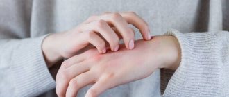
Keratopapilloma is a benign skin formation, so in itself it does not pose a threat to the body. Complications arise only due to mechanical damage to the growth. This may occur due to constant friction against the seams of clothing or due to the fact that the patient himself tries to tear off the formation.
As a result, the following complications arise:
- development of other skin pathologies;
- the appearance of fungus;
- the appearance of purulent formation;
- spread of keratopapillomas throughout the skin;
- penetration of various viruses and bacteria into the body.
If a keratopapilloma has been subjected to mechanical stress for a long time and begins to bleed, then there is a high risk of transforming the formation from benign to a malignant tumor. For this reason, such papilloma cannot be ignored. It is recommended to remove the keratoma to ensure that the formation does not become a malignant tumor.
Features of the treatment of seborrheic keratoma
Seborrheic keratoma can be treated with the following dermatological methods:
- Removal of the lesion by cryodestruction.
- Laser removal.
- Chemotherapy method.
- Use of aromatic retinoids.
A medical neodymium laser is used to remove keratoma. The principle of operation is similar to laser removal of other formations - layer-by-layer destruction of cells.
The chemotherapy method involves the use of 30% prospidin and 5% fluorouracil ointment, solcoderm. Ointments have an antitumor effect. Solcoderm causes mummification of the formation followed by self-elimination. Used only after checking for good quality. As a result, a reduction in keratotic elements is achieved.
Aromatic retinoids are synthetic analogues of vitamin A. They slow down cell division. There are a number of contraindications, which are prescribed individually.
Symptoms
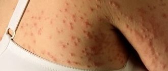
Papilloma of this type first appears as a light brown spot. After some time, it begins to grow and rises above the epidermis. The spot then acquires a dark brown or red tone. The surface of the formation has small depressions. Keratopapilloma does not stop there. Education continues to grow in size. This papilloma has a significant elevation above the skin. The shape of the formation is round, diameter is up to 6 cm. The surface of the growth is covered with horny scales. When subjected to mechanical stress, they tear off, and the damaged surface begins to bleed.
Typically, papillomas of this type appear where the epidermis is not protected by clothing. Formations rarely appear on other areas of the skin, but such cases have been recorded. Papillomas rarely occur in one copy. Multiple formations are more common.
Symptoms and diagnosis
It is possible to identify an age-related wart thanks to the symptoms:
- at the beginning, the pathological formation looks like a speck stuck to the skin;
- color: pink to black or dark brown;
- size and appearance: a small spot initially appears, which over time begins to grow, rises above the surface of the skin and takes on a warty appearance. Over time, it changes and takes on a mushroom-like appearance. Multiple formations that are closely located can merge together, then the size increases significantly;
- age-related keratomas are characterized by the development of hyperkeratosis, active keratinization of epithelial cells. As a result, a significant layer of exfoliated horny masses is formed, sometimes up to 2 cm thick;
- formations may differ in localization. Condylomas can occur on the mucous membranes, in the larynx (on the vocal cords), bladder, ureters, external auditory canal, and sometimes in the chest (intraductal);
- keratomas are never located on the mucous membranes, but can appear on the back, arms, chest, and head.
For such formations, malignancy is not typical, but in appearance they can resemble melanoma due to jagged edges, which is observed in some cases.
Diagnosis is carried out by a dermatologist (or dermatologist-oncologist). During the examination, the appearance, shape, edges, size, consistency are assessed, then a fragment (piece) of the warty growth is taken for histological examination. Only histology will make an accurate diagnosis.
Diagnostics
If papillomas of this type occur, it is recommended to consult a doctor. The specialist will diagnose the keratoma and choose the appropriate treatment method.
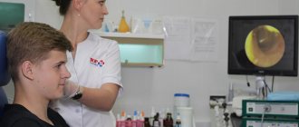
Any problems related to the skin are dealt with by a dermatologist. For this reason, if there is a suspicion of keratopapilloma, it is recommended to contact this specialist. The doctor will interview and examine the patient, and then perform a dermatoscopy. The essence of this study is to examine the tumor particles under a microscope. This allows you to see the characteristic cysts and cornea of this type of papilloma. Histology of the formation is carried out after its removal.
If a malignant papilloma is suspected, the patient is referred to a dermato-oncologist. The specialist will prescribe seascopy and ultrasound. Based on these studies, a diagnosis will be made.
Treatment
Keratopapilloma, what it is was discussed above, and how to treat the formation - the doctor will tell you after examination and relevant research.
One of the methods of treating papilloma of this type is the local use of medications. Usually the doctor prescribes:
- antitumor drugs to prevent the transition of a neoplasm from a benign papilloma to a malignant tumor. Most often, the specialist prescribes Bleomycin and Prospidin;
- agents that slow down the development of keratomas . These include Solcoderm and colchamine ointment;
- corticosteroid drugs . Such drugs are prescribed to relieve inflammation and itching, as well as reduce peeling.
It is recommended to take multivitamins in conjunction with local therapy. Such drugs will replenish the deficiency of nutrients in the body and improve its defenses, which will have a positive effect not only on the condition of the epidermis, but also on the activity of all organs and systems.
The drug treatment method is only a prevention of the transformation of keratopapilloma into a malignant formation.
Treatment methods
Need advice from an experienced doctor?
Get a doctor's consultation online. Ask your question right now.
Ask a free question
This pathology is typical for older people; classical methods of therapy may not be suitable due to the presence of many absolute and relative contraindications due to age characteristics and concomitant diseases.
Age-related (seborrheic) growths do not pose any danger or physical discomfort; doctors seek help for aesthetic reasons when the growths are located on the face.
Some warts are a symptom of other somatic disorders, which requires additional diagnosis.
Surgical removal
Surgical method is the traditional treatment option. In surgery, treatment is used only in the following cases:
- the likelihood of degeneration into a malignant tumor;
- inconvenient location when permanent damage occurs;
- when the process is pronounced and has a multiple character.
The essence of the operation:
- Inspection, selection of location and volume of surgery.
- Preparation of the surgical field. Treatment with an antiseptic solution (betadine).
- Conducting anesthesia (novocaine or lidocaine).
Individual intolerance to the anesthetic may occur.
- Tissue dissection, excision of the pathological area within healthy tissue.
- Antiseptic treatment.
- Skin suture with re-treatment with betadine.
- Applying an aseptic dressing.
Advantages of the operation:
- low probability of reappearance in the same place;
- acceptable price;
- get rid of pathological tissues as much as possible, which is important in the case of a malignant tumor.
Negative sides:
- a scar remains;
- the likelihood of infectious complications;
- relatively long healing.
Hardware procedures
Hardware procedures include:
- cryodestruction;
- radio wave method;
- laser removal.
Cryodestruction - the use of liquid nitrogen, low temperature allows you to destroy pathological tissue without damaging healthy tissue. The procedure is practically not felt, and no scars are formed. The pathological focus will not disappear immediately, but after a few weeks. This method is safe for older people
Radio wave - the use of high-frequency radio waves. The peculiarity of the method is the accuracy of execution, short procedure time and the likelihood of application in hard-to-reach places (on the eyelids).
Laser removal – layer-by-layer removal of cells with a special laser. It involves eliminating a cosmetic defect in several sessions; it will not be possible to remove everything at once. But the procedure has no age restrictions, is bloodless due to cauterization of blood vessels, and is short in duration.
Traditional methods of treatment
Traditional medicine allows you to treat keratopapillomas on the skin at home yourself. Treatment with folk remedies is varied.
| Onion | For the recipe you need onion peels, which it is advisable to chop, pour the dried peels into a jar and pour in table vinegar, leave for 14 days in a dark place. Then filter the tincture and apply externally (make compresses). First for half an hour, and then increase the time to 3 hours. Result: the wart should soften, which will reduce the likelihood of injury. |
| Propolis | The therapeutic effect of propolis slows down the growth of malformation. Propolis is kneaded until smooth and applied to the affected area for 5 days. You can secure it with a plaster or bandage. |
| Castor oil | This method requires warm oil. It must be rubbed into the malformation daily. As a result, education will decrease or growth will slow down. |
| Nuts | You will need to collect unripe nuts and remove the crust from them. Grind it and add it to your regular hand cream. Use the product twice a day. |
Removal methods
Removal of papillomas is carried out in the following cases:
- if the tumor appears on the face and causes aesthetic discomfort;
- keratopapilloma is constantly exposed to mechanical stress;
- the growth is accompanied by inflammation;
- there is a possibility of papilloma developing into a malignant formation;
- growths spread throughout the entire skin at high speed or combine into large formations that cause discomfort.
There are several methods for removing keratopapilloma:
- Surgically . This is one of the most reliable methods for removing growth. The essence of the procedure is cutting out the keratopapilloma using a scalpel. The disadvantage of this method of treatment is that scars remain at the site of the operation, which is unacceptable if the papilloma needs to be removed on the face.
- Laser removal of growth . The essence of the method is the impact on the affected areas of the epidermis with a laser. This method of treatment is effective, painless and safe. There are no traces left at the site where the wart was burned. After surgery, the risk of recurrence of the pathology is minimal. Laser removal can be performed on any part of the epidermis.
- Radiosurgery . Removal of papilloma is carried out without damaging the healthy epidermis. This method of treatment has proven itself to be positive. After removal of keratopapilloma, no traces remain on the skin. The risk of relapse of the pathology is minimal.
- Removal of keratopapilloma with liquid nitrogen . After this procedure, after seven days the growth disappears. The treatment does not go unnoticed. A small pink spot remains on the epidermis.
How are keratopapillomas treated?
Pharmaceutical preparations developed for the external treatment of papillomas do not help get rid of keratomas, since the nature of their occurrence does not have a viral etiology. This also applies to home remedies.
Keratopapilloma appears as a result of age-related changes. These are areas of the skin in which natural metabolic processes and cell functions are disrupted. Therefore, it is not possible to treat them using traditional methods. Such growth can only be removed in medical institutions using the following methods.
offer
- Surgical removal. This method is used to eliminate both benign and malignant tumors.
- Laser treatment. Using the beam, the DNA chain in the neoplasm cells is destroyed. This stops the growth of papilloma and reduces the risk of developing cancer.
- Cryodestruction is the effect of low temperature (freezing) on the skin. Removal can be performed using a special cryodestruction apparatus or by applying liquid nitrogen to the treatment area. Keratoma tissues freeze and destroy very quickly. After the procedure, the papilloma begins to gradually peel off and disappears completely.
- Radio wave therapy. This method is considered the most gentle and safe, therefore it is used on areas of thin sensitive skin.
- Electrocoagulation. This method is popularly called cauterization. Indeed, the removal of the growth takes place under the influence of a high-frequency current, which heats the nozzle of the device and, when cut, cauterizes the wound surface. This prevents the possibility of bleeding. But at the site of the removed keratoma, a crust forms, which subsequently separates on its own.
Folk remedies
If keratopapilloma is just beginning to grow, then you can use alternative medicine to get rid of the tumor. It is recommended to use such drugs only after consulting a specialist.
Aloe
Several of the fleshiest aloe leaves are cut from the bush, washed and put in the freezer for three days. After this, the plant is taken out and left to defrost. Then the leaves are cut and fixed on the affected epidermis with an adhesive plaster. The course of therapy is 21 days.
Potato
Peel fresh potatoes and grate them into a fine grater. Place the resulting puree on the affected area of the epidermis and leave for half an hour. Then remove the mixture and rinse the skin with warm water. The treatment period is 2-3 weeks.
Keratopapilloma is a neoplasm that causes aesthetic discomfort and can cause complications, including a malignant tumor. For this reason, the problem cannot be ignored and if it occurs, it is recommended to consult a doctor.