This oncopathology is usually registered in representatives of the stronger sex: men get sick 4 times more often than women. Seven out of ten cancer cases occur in people aged 65+.
Author:
- Sorokin Nikolai Ivanovich, head of the urological service of the university clinic of Moscow State University. M. V. Lomonosova
5.00 (Votes: 1)
Causes of the disease
There is no single and reliably proven cause of the tumor. Many risk factors contribute to the development of bladder cancer. Among them:
- Smoking. It is considered the main factor provoking bladder cancer. Approximately half of the detected cancer cases are associated with smoking; it is diagnosed in smokers 3 times more often than in non-smokers.
- Poor water quality and unfavorable environmental conditions. The chance of getting sick is increased by excess chlorine and fluorine in drinking water, and pollution of water and air by industrial waste.
- Abuse of alcohol, fried and fatty foods.
- Harmful working conditions – working with substances containing aromatic amines, phenols, phthalates. At risk are workers in the chemical, paint and varnish, rubber, oil, aluminum, leather and textile industries, artists, designers, and hairdressers. RMP often occurs in long-distance drivers due to prolonged sitting while driving, frequent hypothermia, and inability to urinate in a timely manner Source: Kulesh P.A. Study of clinical and morphological features of bladder cancer / P.A. Kulesh, A.A. Bagaeva // Eurasian Union of Scientists. — 2021..
- Non-infectious urogenital pathologies - chronic cystitis, urolithiasis, diverticulitis; genitourinary infections – chlamydia, mycoplasmosis, ureaplasmosis, trichomoniasis; parasitic diseases – schistosomiasis.
- Stagnation of urine or rare urination for various reasons - prostate adenoma, narrowing of the urethra, insufficient fluid intake.
- Radiation therapy for tumors of the cervix and ovaries, prostate gland, chemotherapy using Cyclophosphamide (increases the risk by 4.5 times), long-term use of phenacytin-containing analgesics (increases the risk by 2-6.5 times) Source: Kulesh P.A . Study of clinical and morphological features of bladder cancer / P.A. Kulesh, A.A. Bagaeva // Eurasian Union of Scientists. — 2021..
- In addition, the risk is significantly increased in people exposed to ionizing radiation Source: Kulesh P.A. Study of clinical and morphological features of bladder cancer / P.A. Kulesh, A.A. Bagaeva // Eurasian Union of Scientists. — 2021..
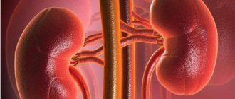
Risk group
As practice shows, the likelihood of tumors appearing in the genitourinary system increases in men:
- Those who abuse tobacco products. According to statistics, kidney cancer is diagnosed in smokers 2 times more often.
- Middle aged (after 45 years). This is due to a decrease in the level of male sex hormones along with an increase in female ones.
- People who work in hazardous industries (for example, when a person often comes into contact with hazardous chemicals).
- Those who have been on dialysis for a long time. This often leads to kidney cancer.
Symptoms of bladder cancer
The main symptom of bladder cancer is hematuria (blood in the urine), which is often transient in nature, its severity does not depend on the stage of the disease Source: Kulesh P.A. Study of clinical and morphological features of bladder cancer / P.A. Kulesh, A.A. Bagaeva // Eurasian Union of Scientists. - 2021.. Microhematuria is possible, when blood is not noticeable visually and is detected only during analysis, and macrohematuria - urine turns dark orange, pink, red, brown.
Early symptoms also include dysuric disorders: frequent or infrequent urination, accompanied by pain, stinging or burning, a weak, intermittent stream of urine.
All the first signs of bladder cancer are nonspecific, characteristic of other genitourinary diseases. It is important to seek medical help as early as possible in order to carry out a differential diagnosis, identify a tumor in time and begin treatment.
Pain syndrome in bladder cancer can have different characteristics depending on the nature of the spread of the tumor. With a locally advanced tumor process, pain initially most often occurs above the womb, provoked by the act of urination; subsequently, the pain becomes constant, pain appears in the perineum and in the sacral area. When the tumor obstructs the ureteral orifices, hydronephrosis develops, pain appears in the lumbar region, a positive symptom of effleurage in the lumbar region, and an enlarged kidney is palpated. Pain in the bones, as a rule, indicates metastatic damage to the skeleton Source: Kulesh P.A. Study of clinical and morphological features of bladder cancer / P.A. Kulesh, A.A. Bagaeva // Eurasian Union of Scientists. — 2021..
In the later stages of cancer, general weakness, pain in the lumbar and groin areas, in the lower abdomen, lack of appetite and weight loss, fever, swelling in the legs, and enlarged lymph nodes occur. Fistulas may develop between the bladder and rectum.
Rehabilitation
Following organ removal surgery for bladder cancer, clinical recommendations include:
- special care for the area where the urinary catheter is inserted;
- enhanced drinking regime to flush out toxins and bacteria;
- avoiding alcohol, smoking, drinking drinks containing caffeine, as well as spicy and salty foods;
- exclusion of heavy physical activity;
- attending prescribed physical procedures, taking medications.
Types of Bladder Cancer
By prevalence in the muscle layer:
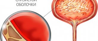
- muscular non-invasive – located only on the mucous and submucous membranes;
- muscle-invasive - grows into the thickness of the bladder wall.
By location:
- on the dome - the upper wall of the bladder;
- on the side, front or back walls;
- in Lieto's triangle - a space in the form of a triangle with vertices at the mouths of the ureters and the exit of the urethra;
- on the neck of the bladder;
- in the area of the ureteric orifices;
- in the area of the urachus - a normally closed rudimentary primary duct located above the bladder;
- the tumor covers several areas.
By type of growth:
- exophytic (papillary) – the formation grows upward from the mucous membrane, initially looks like a polyp;
- endophytic (infiltrative) – grows inside the wall of the organ with a transition to the muscle layer, has no clear boundaries;
- mixed form - the tumor grows simultaneously inward and outward;
- flat;
- nodular;
- intraepithelial.
According to histological structure:
- Epithelial cancer. In 98% of cases, bladder tumors develop from epithelial cells Source: Kulesh P.A. Study of clinical and morphological features of bladder cancer / P.A. Kulesh, A.A. Bagaeva // Eurasian Union of Scientists. — 2021.. In 90-95% of cases, bladder tumors are morphologically transitional cell carcinoma Source: Dolgikh D.V. Diagnosis of bladder cancer / D.V. Dolgikh [and others] // Siberian Medical Journal. - 2015. - No. 6. - P. 141-147.. It can be low- and highly differentiated, which determines the degree of malignancy of the formation. Verrucous carcinoma, adenocarcinoma and clear cell adenocarcinoma, urachus cancer, and small cell carcinoma are diagnosed much less frequently.
- Non-epithelial cancer. Tumors from muscle, fibrous vascular and other tissues: rhabdomyosarcoma, fibrous histiocytoma, leiomyoma, hemangioma, melanoma, germ cell tumors.
Symptoms of tumor diseases
Considering the symptoms of a bladder tumor, it is worth noting that at an early stage it does not manifest itself in any way. Signs occur when the tumor reaches such a size that it can irritate the walls of the organ. In such a situation, a person may have the following complaints:
- change in urine color;
- cramps and pain in the lower abdomen;
- feeling of a not completely emptied bladder;
- excretion of urine in small portions;
- frequent urge to urinate;
- disturbance of the menstrual cycle and discharge from the genital tract (in women);
- swelling in the legs, perineal area;
- disorders of the digestive system.
Bladder cancer grades
At the first stage
RMP tumor cells are found only in the epithelial layer. The process does not affect the wall of the bladder, nearby and distant organs.
For the second stage
The tumor is characterized by invasion of the muscle layer. Adjacent organs are not affected, there are no distant metastases.
At the third stage
, in addition to cancer growing into the muscle layer of the bladder, an increase in regional lymph nodes is observed.
Fourth stage
characterized by any tumor size, with regional lymph nodes affected and metastases observed in distant organs.
For international classification, the TNM system :
- T – indicates the extent of the tumor, indicated by numbers from 1 to 4.
- N – presence of metastases in lymph nodes. The number 0 indicates the absence of metastases in the lymph nodes, 1 - a single affected node, 2 - metastases in 2-5 lymph nodes.
- M – (x, 0 or 1) – impossibility of detection, absence or presence of distant metastases.
Diagnosis of bladder cancer
The first stage in diagnosing bladder cancer is examining the patient, collecting an anamnesis of life and illness, and conducting laboratory and instrumental studies. The doctor will ask the patient in detail about the symptoms, the time of their appearance, the frequency of occurrence, the presence of bad habits, and other factors that provoke the disease.
During a urological examination, the doctor will determine Pasternatsky's symptom
– the presence or absence of painful sensations when tapping on the lower back, and will also check the condition of the prostate gland using a digital examination through the rectum.
In addition to general and biochemical blood and urine tests, which are not informative in the diagnosis of cancer, the patient may be prescribed a microscopic examination of urine sediment
for the presence of pathological cells and determination
of window markers for bladder cancer
.
Ultrasound for bladder cancer allows you to visualize a tumor if its size exceeds 4 mm, determine the form of tumor growth (exophytic, endophytic or mixed) and the depth of its invasion into the wall Source: D.V. Dolgikh. Diagnosis of bladder cancer / D.V. Dolgikh [and others] // Siberian Medical Journal. - 2015. - No. 6. - P. 141-147..
Mandatory instrumental examination - cystoscopy
.
During cold cystoscopy, the bladder is examined and an area of affected tissue is excised for analysis. Another diagnostic and treatment method is TUR biopsy
, when the tumor is excised completely, capturing healthy tissue. During transurectal resection with photodynamic diagnostics, the doctor can see even the smallest areas of tumor damage in blue illumination.
To determine damage to the lymph nodes and metastasis to other organs, use:
- CT and MRI,
- scintigraphy,
- biopsy of sentinel lymph nodes,
- chest x-ray,
- ultrasound examination of the abdominal organs;
- tests for tumor markers.
Three markers have the greatest sensitivity and specificity in staging bladder cancer: UBC, TPS and VEGF. An increase in the level of UBC in urine characterizes the initial stages of bladder cancer and possible non-invasive tumor recurrence during organ-preserving treatment. In patients with non-muscle-invasive bladder cancer, an increased level of VEGF can serve as an additional criterion not only for relapse of the disease, but also for its progression. A simultaneous increase in the content of UBC in the urine and TPS, VEGF in the blood serum is associated with the progression of tumor growth and characterizes the initial stage of bladder cancer invasion into the muscle layer. A high level of VEGF is characteristic of a tumor extending beyond the organ, as well as with high tumor growth activity and a low degree of differentiation, which is a poor prognostic factor Source: D.V. Dolgikh. Diagnosis of bladder cancer / D.V. Dolgikh [and others] // Siberian Medical Journal. - 2015. - No. 6. - P. 141-147..
To determine the patency of the urinary tract, excretory urography . The study allows us to assess the patency of the ureters, identify neoplasms both in the upper urinary tract and in the bladder Source: Kulesh P.A. Study of clinical and morphological features of bladder cancer / P.A. Kulesh, A.A. Bagaeva // Eurasian Union of Scientists. — 2019..
The optimal method for diagnosing stage III and differentiating it from the stage of the disease, as well as for distinguishing between T2a and T2b degrees, is multiscan spiral computed tomography Source: D.V. Dolgikh. Diagnosis of bladder cancer / D.V. Dolgikh [and others] // Siberian Medical Journal. - 2015. - No. 6. - P. 141-147..
Chemotherapy
Chemotherapy is the use of drugs that kill cancer cells. For bladder cancer, several different ways of administering chemotherapy are used - intravesical chemotherapy, when the medicine is injected directly into the bladder, and systemic chemotherapy, when chemotherapy is administered intravenously:
- Intravesical chemotherapy targets cancer cells in the bladder and is used for superficial tumors to prevent tumor recurrence. When chemotherapy is injected into the bladder, side effects are usually minor. For several days after treatment, you may experience blood in your urine and more frequent and painful urination. These symptoms usually disappear after treatment.
- Systemic chemotherapy kills cancer cells throughout the body, not just the bladder, and is used for invasive or metastatic cancer. Chemotherapy is usually given in cycles. Each cycle has a treatment period and a rest period. Side effects depend on the chemotherapy drug and its individual tolerance.
Bladder cancer treatment methods
In the early stages of bladder cancer, when the spread of the tumor is limited to the epithelial layer, transurethral resection
followed by
intravesical chemotherapy
or
immunotherapy
- the introduction of drugs into the bladder cavity. The advantage of intravesical administration is the absence of side effects of systemic chemotherapy, such as nausea, vomiting, and hair loss.
The anti-tuberculosis vaccine BCG is used for intravesical immunotherapy. Weakened Koch bacilli activate immune system cells, which begin to attack the tumor.
A more modern method of immunotherapy is the use of checkpoint inhibitor drugs. Checkpoints are molecules that suppress the immune response, including to prevent the occurrence of autoimmune diseases. Inhibitors remove the blockage of these points and stimulate the immune system to fight cancer cells.
Another low-traumatic treatment method is en-bloc laser resection.
, which allows you to remove neoplasms in the mucous and submucosal membranes, the muscle layer as a single block. This method is not suitable for large formations without clear boundaries.
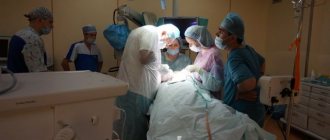
In late stages of bladder cancer, the process is very widespread, if the above methods do not help, they resort to complete removal of the organ - cystectomy
, followed by radiation or chemotherapy.
Isolated cystectomy (removal of only the bladder) for invasive cancer is rarely performed. More often it is combined with the removal of neighboring organs: the prostate gland and seminal vesicles in men, the appendages and uterus in women. At the same time, the entire urethra or part of it and the pelvic lymph nodes are excised.
After cystectomy, it is possible to perform a reconstructive operation - neocystoplasty, when the removed bladder is replaced with an isolated section of the intestine, to which the ureters and urethra are connected.
Prevention
To reduce the risk of developing a tumor in the bladder, you must:
- lead a healthy lifestyle, quit smoking;
- drink at least one and a half to two liters of water per day;
- promptly treat inflammatory pathologies of the urinary system;
- reduce contact with carcinogenic substances, constantly use PPE in production.
People at risk should undergo annual screening examinations to detect tumors at an early stage.
Disease prognosis
The survival prognosis depends on the stage at which the tumor is diagnosed and how timely and complete the treatment is. If a single epithelial lesion is detected and TUR is performed in conjunction with chemotherapy or immunotherapy, the chances of recovery are about 91%. In the second stage, without cancer spreading to other organs, timely transurethral resection and chemotherapy (less commonly, radiation) allow hope for recovery without relapse in 73-75% of patients. In the third stage and the process spreads to nearby organs, even after removal of the bladder and chemotherapy, there is a possibility of relapse; five-year survival is observed in 50% of patients. At the fourth stage, the process is considered inoperable, palliative methods are used, five-year survival rate is less than 7%.
What kind of observation is required after treatment?
After treatment, recurrence of the tumor (relapse) may occur, the probability of which in superficial forms reaches 70%. A new tumor may appear in the same place as the primary one, and often relapses occur in another area of the bladder mucosa. Therefore, patients undergoing treatment for bladder cancer need to be examined every 3-6 months. One of the most important components of the examination is cystoscopy and urine cytology. The examination also includes an examination, blood and urine tests, and an ultrasound or computed tomography may be prescribed.
Prevention of bladder cancer
Quitting smoking and alcohol and eating plenty of fresh fruits and vegetables, which contain antioxidants that prevent cell degeneration, will help reduce the likelihood of developing a tumor. Bladder cancer is less likely to occur in those who drink enough water per day. It is believed that the risk of neoplasm is reduced by 7% for every 240 ml of added fluid.
It is necessary to be attentive to your health and consult a doctor when the first, even minor, signs of illness appear. It is important to monitor the regularity of urination, protect against genitourinary infections, and promptly diagnose and treat inflammatory processes in the urinary tract. Workers in hazardous industries should not neglect medical examinations. People over 40 years of age are recommended to undergo annual medical examination.
Sources
- Dolgikh D.V. Diagnosis of bladder cancer / D.V. Dolgikh [and others] // Siberian Medical Journal. - 2015. - No. 6. - P. 141-147.
- Dolgikh D.V. Bladder cancer (issues of etiology and pathogenesis) / D.V. Dolgikh [and others] // Siberian Medical Journal (Irkutsk). - 2015. - No. 7. - P. 26-30.
- Dunaev M.G. MRI diagnosis of bladder cancer / M.G. Dunaev // Remedium Volga region. — 2021. — No. 6 (156). — P. 22.
- Karyakin O.B. Bladder cancer: what's new in 2019-2020. / ABOUT. Karyakin // Oncourology. - 2021. - No. 4. - P. 147-154.
- Kulesh P.A. Study of clinical and morphological features of bladder cancer / P.A. Kulesh, A.A. Bagaeva // Eurasian Union of Scientists. — 2021.
- Nemtsova M.V. Molecular pathogenesis of bladder cancer / M.V. Nemtsova, N.E. Kushlinsky // Almanac of Clinical Medicine. - 2015. - No. 41. - P. 79-88.
- Startsev V.Yu. Modern possibilities of organ-preserving treatment of patients with muscle-invasive bladder cancer / V.Yu. Startsev [et al.] // Urological Gazette. - 2021. - T.9. - No. 1. — P. 29-38.
The information in this article is provided for reference purposes and does not replace advice from a qualified professional. Don't self-medicate! At the first signs of illness, you should consult a doctor.
Where to contact
Time plays a key role in the treatment of any oncology.
It is the timing of tumor detection that largely determines the personal prognosis. You should not self-diagnose, prescribe treatment, or be afraid to hear a terrible diagnosis. Today, oncology is curable, and new methods of fighting cancer have emerged. The symptoms described above simply provide information so that you can consult a doctor in a timely manner and not delay the diagnosis. Clinic of Urology named after R. M. Fronshtein of the First Moscow State Medical University named after I.M. Sechenov (State Center for Urology) successfully diagnoses and treats urological pathologies, including oncological ones. Here patients are provided with medical care that meets modern standards. The clinic is equipped with the latest technology; robotic operations are performed here. Most patients of this public clinic return to a full life after treatment. July 28, 2020
Hakobyan Gagik Nersesovich - urologist, oncologist, MD, doctor of the highest category, professor
All the symptoms of the disease...