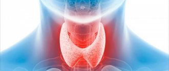
Ultrasound of the thyroid gland - (Thyroid Ultraschall) is the simplest and most highly informative diagnostic method, which provides a detailed assessment of the condition of the gland itself, as well as the anatomical formations adjacent to it - blood vessels and muscles. This is possible using modern ultrasonic sensors with additional capabilities. In particular, Doppler ultrasound allows you to examine the blood supply to the thyroid gland and nearby lymph nodes.
Make an appointment, ultrasound or tests
PRICE OF THYROID ULTRASOUND 1000 rub.
Indications for thyroid ultrasound
The content of the article
Before scheduling a procedure, please read the important information regarding indications and contraindications. A classic ultrasound examination of the thyroid gland is recommended to be done regularly as part of an annual medical examination, regardless of the patient’s age and the absence of characteristic symptoms. If the following symptoms appear, it is necessary to make an appointment with an endocrinologist as soon as possible and undergo an ultrasound examination of the thyroid gland:
- visible increase in the volume of the neck, the appearance of a compacted formation;
- redness of the neck area near the location of the gland;
- hoarseness not associated with colds;
- difficulty breathing, discomfort during swallowing;
- increase in body temperature within 37-37.5 degrees;
- the appearance of swelling;
- weakness, apathy, drowsiness, severe irritability;
- sweating, feeling hot or, conversely, cold;
- trembling of limbs;
- sudden change in body weight;
- cardiopalmus;
- baldness.
These symptoms may be an indicator of a malfunction of the thyroid gland, and therefore require examination by ultrasound. An endocrinologist may refer you for an ultrasound of the thyroid gland if palpation reveals lumps or the patient complains of pain when palpating the gland. A direct indication for ultrasound diagnostics is unsatisfactory results of hormone tests.
In addition, the following groups of people should sign up for a thyroid ultrasound :
- Pregnant women or those who are planning to conceive a child in the near future.
- People living in iodine-deficient areas.
- Patients diagnosed with obesity.
- For those taking hormone-containing medications.
Ultrasound of the thyroid gland is also prescribed to patients to monitor the treatment. In some cases, the frequency of such a procedure reaches several times a week, which is necessary for dynamic monitoring of the gland’s reaction to drug correction.
Ultrasound examination is completely safe for humans, has no contraindications, and if a pregnant woman undergoes it, it does not have any effect on the fetus. Therefore, ultrasound can be done as many times as needed.
How to prepare for a thyroid ultrasound
This gland is absolutely accessible for research due to its convenient location: ultrasonic waves easily reach the thyroid gland and are fully reflected from its tissues. Therefore, this procedure does not require special preparation. But there are a couple of recommendations that should be considered when planning an ultrasound of this gland:
- Patients who take medications that affect cardiac output and blood pressure should stop taking them the day before ultrasound diagnostics.
- Do not drink alcohol during the day.
- Elderly people are advised to come for the procedure on an empty stomach. This recommendation is associated with the appearance of a gag reflex at this age when pressing on the neck area.
When coming for an ultrasound, you can take a personal towel with you to wipe off any remaining gel from your neck after the procedure.
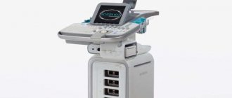
How to do an ultrasound of the thyroid gland
The procedure uses a linear sensor with a high scanning frequency. With its help, you can obtain a high-quality image that shows the structure of the organ and focal changes. During the procedure, the size of the gland, its structure, configuration, and blood flow condition are determined. In parallel, the parathyroid glands and regional lymph nodes are examined.
No preparation is required for diagnosis, since nothing interferes with the examination of the organ. Ultrasound diagnosis of thyroid pathologies is an accessible, non-traumatic and painless diagnostic method. Complications after ultrasound are not observed. During the study, the patient is not administered drugs, and the organs are not irradiated, but the diagnostic efficiency is high. There are no contraindications to the ultrasound procedure. It can be done many times even during pregnancy.
The examination is carried out in a lying position. A special gel is applied to the patient’s neck to increase the conductivity of ultrasound. The results are displayed on the screen. Modern ultrasound machines determine not only the presence, but also the size of thyroid nodules, which is important for choosing treatment tactics.
The patient is given an image and a description, based on which the endocrinologist will prescribe treatment for the identified pathologies. To make a final diagnosis, the patient needs to undergo blood and urine tests and consult with a therapist and oncologist.
In what cases does the doctor prescribe an examination?
Studies of the thyroid gland are carried out if the attending physician or endocrinologist suspects diseases of the gland, and when palpation reveals formations that are not characteristic of a healthy organ.
Diagnosis of pathologies is based on the following symptoms:
- feeling of a lump or sore throat;
- cough of unknown etiology;
- sudden loss or gain of body weight;
- difficulty swallowing;
- emotional instability;
- hair loss;
- hoarseness of voice;
- swelling;
- hand tremors;
- rapid pulse, arrhythmia;
- insomnia, other sleep disorders;
- fluctuations in blood pressure;
- enlargement of regional lymph nodes;
- profuse sweating.
Emotional lability in diseases of the thyroid gland can manifest itself with symptoms such as irritability, anxiety, nervousness, and sudden changes in mood.
Ultrasound examination is prescribed for preventive purposes to people working with harmful production factors or a hereditary predisposition to the disease. The procedure is necessary for patients after a course of hormonal medications, patients with diabetes, as well as men and women after 40 years.
Endocrinologists and gynecologists recommend that women undergo ultrasound diagnostics during pregnancy and when planning it. Sometimes hormonal disorders and pathology of the thyroid gland are one of the reasons for the inability to get pregnant.
The study is indicated for children who have experienced stress, as it can become a trigger in the development of thyroid disease.
Interpretation of ultrasound of the thyroid gland
To obtain comprehensive information that reflects the condition of the thyroid gland, the diagnostician during the ultrasound procedure evaluates and enters into the protocol the following parameters:
- Location . The typical location of the gland is considered standard when it is within the anatomical norm. If there is a pathology, a record of its aberrant location is entered into the protocol, when the thyroid gland may be located behind the sternum, in the area of the root of the tongue, etc.
- Size . Determining this parameter allows one to judge the presence or absence of hypo- or hyperplasia of the thyroid gland.
- Structure . Normally, the thyroid gland consists of two symmetrical lobes, which are connected by a small isthmus. In the case of abnormal intrauterine development of the gland or its pathological acquired modification, tissue outgrowths, an additional lobe, and bifurcation may be absent.
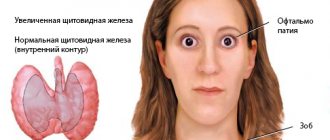
- Form . Normally, the thyroid gland is flat, but may be slightly elongated. If it is greatly increased in volume, swollen, with thyroid tissue, diffuse goiter, viral or bacterial pathology is suspected.
- Outlines . The clear contours of the thyroid gland are taken as normal. Otherwise, if its contours are not clearly visualized, one can judge the presence of a tumor or inflammatory focus in the gland.
- Structure . If there is no pathological process in the gland, its structure is homogeneous and has a specific granularity.
- Echogenicity . Visually, this parameter can be defined as a gradation of gray shades over the entire area of the study area. Normally, the echogenicity of thyroid tissue should be visible in light gray shades.
- Focal formations . During ultrasound diagnostics, the doctor must examine the gland for the presence of pathological formations - nodes, calcifications, cysts or tumors.
- Blood supply to tissues . Impaired blood supply to the thyroid gland can lead to the death of its tissue. Normally, the multi-colored signals visualized on the ultrasound monitor screen should be uniform over the entire area of the thyroid gland. An increase in color and “pulsation” of shades indicates an increase in blood flow, which may be associated with an inflammatory process inside the gland.
- The structure of the cervical lymph nodes . To determine their condition, the following indicators are determined: structure, size, presence of pathological inclusions, cystic transformation, presence of the “gate” of the lymph node, assessment of blood flow.
History of ultrasound
Ultrasonic vibrations were discovered by Italian scientist Lazzaro Spallanzani back in 1794. While watching the bat, he wondered how it was possible to navigate in space at night while flying. It turned out that this is carried out using high-frequency sound vibrations, inaudible to humans. But animals, particularly bats, perceive them very well. The principles of echolocation are also used by marine animals - killer whales, dolphins, whales, etc. Over time, they became known as ultrasonic waves.
In 1942, for the first time, the German doctor Theodor Dussick and his brother, scientist and physicist Friedrich Dussick, tried to use ultrasound to diagnose a tumor in a patient’s brain. The first medical ultrasound device was created in 1949 by the American scientist Douglas Haury.
Doppler Christian Anders made a great contribution to the development of ultrasound diagnostics. While working on his scientific work “On the collometric characteristics of the study of double stars and some other stars in the sky,” he noticed that the frequency of the signal reflected from an object depends on its speed and direction of movement. This property is called the Doppler effect, which is widely used in Dopplerography to examine blood flow parameters.
How much does a thyroid ultrasound cost?
The cost of the procedure depends on the medical center where the study is carried out and on the quality of the device used to perform the procedure. In St. Petersburg, the price of an ultrasound scan of the thyroid gland varies from 1000 rubles to 2000 rubles. At the Diana Clinic, you can undergo a study on a new device for 1 thousand rubles.
ADDITIONAL RESEARCH
Interpretation of ultrasound of the thyroid gland: norm and deviations
To understand whether a pathological process is present in the thyroid gland, the following points must be carefully assessed:
Structure, contours
The homogeneous structure of the gland is considered standard; its granularity is clearly visible. Normal variants include a moderately heterogeneous structure in patients in whom laboratory tests have revealed an increase in the titer of antibodies to thyroid peroxidase and thyroglobulin. If the gland is visualized as a “honeycomb,” it means that there is an abnormal degeneration of its tissues. The contours of the gland should be clear. If they are blurred and difficult to read, it means that an inflammatory process or tumor is developing in the gland. If the structural outlines of the thyroid gland are poorly visualized, there is reason to believe that malignant tumors have grown into nearby organs.
Main settings
To determine the presence of edema, tumor or thyroid tissue, the sizes of the lobes must be assessed: the length should not go beyond the range of 2.5-4 cm, width - 1.5-2 cm, and the range of 1-1.5 cm is taken as the norm for the thickness of the gland During the study, the volume of the thyroid gland is determined. Its norms may vary based on the patient’s weight:
| Patient weight (kg) | Maximum gland volume (cm cube) |
| Up to 55 | 15,5 |
| 56-65 | 18,5 |
| 66-75 | 22 |
| 76-85 | 25 |
| 86-95 | 28,5 |
| More than 96 | 32 |
In case of non-compliance with the standards, a record is made in the study protocol - a diffuse increase, which may indicate the presence of bacterial or viral damage to the gland, diffuse goiter, an oncological process, etc.
Echogenicity
The density of the echostructure may vary depending on the age and gender of the patient, but should be visualized in light gray tones. Inflammatory foci are visualized in a dark gray color, closer to black. Black echogenicity is a sign of a malignant process in the thyroid tissue. The following echogenicity characteristics may be included in the study protocol:
- isoechoic area – a sign of the absence of pathological processes in the tissues of the gland;
- hyperechogenicity - light areas that indicate the absence of diffuse changes in the area under study;
- hypoechogenicity - the presence of dark areas in the examined area, which indicate the presence of inflammation;
- anechogenic zone - black areas indicating the presence of cysts or tumors in the thyroid gland.
Focal changes
The study protocol must indicate the absence or presence of focal formations - neoplasms of a benign or oncological nature. If cysts or nodes are identified, the protocol indicates a focal lesion with a description of their location and nature.
Small cyst-shaped inclusions up to 4 mm are considered an acceptable variant of the norm.
Benign neoplasms are visible on ultrasound as clearly isolated objects, while a cancerous tumor has blurred boundaries and is black in color.
Cysts are fluid-filled blisters and can be congenital or acquired. If the doctor diagnoses the presence of nodes, then they were formed from the growing tissue of the gland itself. The presence of calcifications in the thyroid gland suggests that these formations consist of potassium salts, and therefore can contribute to the death of thyroid cells and their further degeneration into a tumor.
Blood supply to tissues
The intensity of this parameter is determined using Doppler ultrasound. Normally, single signals of pulsating blood flow can be seen on the surface of the thyroid gland. When its speed increases, one can judge the inflammatory process or tumor inside the gland.
Regional lymph nodes
The norm is smooth, clearly visualized contours, the absence of any neoplasms, cysts, the presence of a “gate” of the lymph node, moderate blood flow, length greater than width. Any deviation from the norm indicates a developing pathology.
The protocol has a conclusion, which indicates all the information about the study performed and the presence or absence of identified pathologies. Based on the ultrasound examination, the endocrinologist can clarify the diagnosis and select the appropriate treatment.
What diseases can be detected by ultrasound of the thyroid gland?
Based on the results of ultrasound diagnostics, an endocrinologist can diagnose the following diseases:
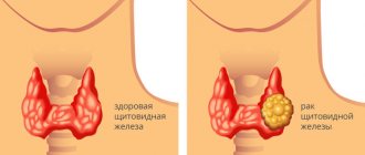
- Nodular goiter : a disease in which compactions are clearly palpable in the area of the gland. Ultrasound reveals an increased density of formations bordering unchanged tissues.
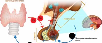
- Diffuse toxic goiter : a pathology in which the diffuse toxic state of the thyroid gland is within normal limits, but the volume of the gland is increased, and laboratory diagnostics show increased synthesis of thyroid-stimulating hormone (TSH) and T3.
- Hashimogo's thyroiditis : an inflammatory disease of an autoimmune nature, in which part of the thyroid tissue dies, therefore the function of the gland is impaired.
- Hypothyroidism : an endocrine disease characterized by the presence of diffuse degenerative changes in the thyroid tissues. In this case, the gland is small in size compared to the norm; thyroid hormones are produced in insufficient quantities.
- Thyroiditis : a disease in which inflammation develops in the gland. The main symptoms of this condition are an increase in the volume of the neck, pain in the thyroid gland, swelling of adjacent tissues.
- Cyst : a formation that resembles a bubble with fluid inside. On ultrasound it is visible in the form of formations with clear contours.
- Follicular adenoma : a nodular formation of a benign nature that may contain colloid.
- Papillary adenoma : a benign formation with a cystic structure and papillary growths inside.
- Cancerous tumor : a malignant neoplasm that is a mass accumulation of cancer cells.
In some cases, to clarify the diagnosis, additional tests for thyroid hormones are required, which reflect the balance of the production of T3 and TSH hormones.
Read also
Hyperparathyroidism
Physiology of the parathyroid glands The parathyroid glands produce parathyroid hormone, the main hormone that regulates the metabolism of calcium and vitamin D (under the influence of parathyroid hormone, vitamin D becomes active)…
Read more
Thyrotoxicosis (hyperthyroidism)
Physiology of the thyroid gland: The function of the gland is to synthesize and secrete thyroid hormones. An increase in their number leads to more intense metabolism and vice versa. Thyroid function is regulated...
More details
Complications of diabetes
Diabetes mellitus (DM) is a systemic disease caused by absolute (type 1) insulin deficiency or insulin resistance (type 2), which causes impaired carbohydrate metabolism, manifested by chronic...
More details
Itsenko–Cushing syndrome
Itsenko-Cushing syndrome is a disease that occurs against the background of chronic hypercortisolism, i.e., an increased level of the hormone cortisol in the blood secreted by the adrenal cortex, or long-term treatment...
More details
Pheochromocytoma
Pheochromocytoma is a tumor of the adrenal medulla that produces catecholamines (adrenaline, norepinephrine, dopamine). Pheochromocytoma consists of chromaffin cells. Pheochromocytoma may have...
More details
Advantages of the method - ultrasound examination of the thyroid gland
If there is a suspicion of a malfunction in the endocrine system, the doctor will refer the patient for an ultrasound scan of the thyroid gland. This study is informative enough to determine the presence of pathologies in the area under study, while being absolutely safe for humans. MRI and CT are indicated only in cases where it is necessary to detail the structure of the pathological formation inside the gland.
The main advantages of ultrasound examination of the thyroid gland include:
- lack of special training;
- simplicity and speed of the procedure;
- absence of ionizing radiation;
- harmlessness, including for pregnant women and the fetus;
- no risk of infection.
Such diagnostics have no contraindications, so they can be performed on children of any age, pregnant and lactating women and the elderly.
Contraindications
Ultrasound screening is a publicly available examination. There are no contraindications to it. It can be performed an unlimited number of times at any time interval, for any category of patients, including pregnant women. Sometimes patients with scars in the neck area may experience difficulty during the examination. Due to the roughness of the skin, the doctor may not be able to achieve close contact with the surface, and the intensity of the ultrasound signal will decrease.
Author: Telegina Natalya Dmitrievna
Therapist with 25 years of experience
>