- Ultrasound machine
Ultrasound machine
SAMSUNG SONOACE R7
Class:
Expert (installation year 2019)
What is included in the cost of ultrasound:
Ultrasound diagnostics, interpretation of images, written diagnostic report
What is important to know about making an ultrasound appointment?
Do I need to make an appointment: ultrasound diagnostics are carried out by appointment
Appointment schedule: you can make an appointment 24 hours a day
Form of payment for diagnostics: cash, credit cards
Doctor's referral: not required
Ultrasound screening during pregnancy: we do not provide
Minimum patient age: 18 years.
Content:
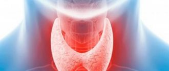
- Why do you need to do an ultrasound of the thyroid gland?
- How to do an ultrasound of the thyroid gland correctly?
- Ultrasound of the thyroid gland: interpretation
Hypoechoic formations
If a specialist (sonologist) speaks about decreased echogenicity of the thyroid gland, then this may indirectly indicate the presence of the following conditions:
- the appearance of a “nodule” in the organ, which often indicates an iodine deficiency in the patient;
- hypoplasia;
- diffuse goiter;
- tumor process.
In some situations, doctors do not reject the possibility of the influence of a genetic predisposition to the formation of hypoechogenicity in the thyroid gland.
Risk factors:
- living in an ecologically unfavorable area where background radiation is high, there is iodine deficiency, etc.;
- insufficient and poor nutrition (fasting, long-term strict diets, unbalanced diet);
- constant stressful situations. As is known, they are the cause of most pathologies;
- taking certain medications;
- bad habits, chronic alcoholism and smoking.
As a rule, a person accidentally learns about existing thyroid pathologies during a routine ultrasound examination. Even relatively small nodes can be painless and not cause discomfort in the cervical area.
With careful palpation in the projection area of the thyroid gland, you can sometimes detect a slippery and dense nodule. Large formations, the size of which exceed 30 mm in diameter, become noticeable even with the naked eye.
The first symptoms of hypoechoic formation in the form of nodules are observed only with their persistent increase:
- foreign body sensation;
- dryness and sore throat;
- hoarseness, difficulty in voice reproduction;
- pain in the anterior neck.
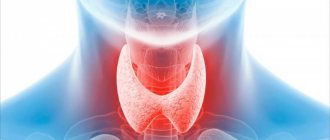
What is thyroid ultrasound?
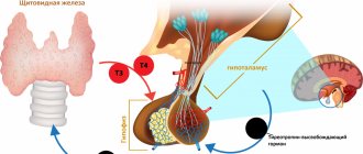
The tissues of the human body have different structures, different densities and, accordingly, will reflect ultrasonic waves differently. Ultrasound diagnostics are based on this physical phenomenon. The diagnostic apparatus is equipped with sensors of various frequencies that supply and receive the return signal. During the examination, the scanner sends a signal of ultrasonic waves into the soft tissues of the neck. Some of the waves, reflected, return to the sensor, and some continue on their way. All data on the behavior of waves is sent to the device’s processor, and based on them, the computer constructs an image that is sent to the monitor. The computer of a modern ultrasound machine is capable of making an accurate calculation of tissue thickness, distance from other nearby organs or vessels, and tissue density. During the examination, the diagnostician has the ability to change the frequency and intensity of sound waves using a control panel, which is located just below the monitor, thereby obtaining the necessary information.
Echogenicity
This parameter shows the density of a substance, how strongly tissues reflect or do not reflect ultrasound.
There are 4 types of echogenicity:
▪ Anaechogenic
The tissues on the monitor are black and absorb ultrasound (blood vessels, benign formations)
▪ Isoechoic
The tissues on the monitor are light gray and partially reflect ultrasound (healthy tissues)
▪ Hypoechoic
Dark area that reflects little ultrasound (cysts)
▪ Hyperechoic
Very light parts on the monitor, fully reflect ultrasound (connective tissue)
Increased echogenicity on ultrasound may be a sign of immune diseases.
Why do you need to do an ultrasound of the thyroid gland?
Ultrasound of the thyroid gland allows you to timely detect the disease in the early stages of its development or prevent it. Using this type of diagnostics you can determine:
- size and structure of the organ, size of the lobes;
- benign and malignant tumors, sites of metastases;
- cysts and fibrous tissue, inflammation;
- local lymphatic drainage;
- anomalies and malformations.
An ultrasound of the thyroid gland with interpretation of the results will allow us to determine the true condition of the patient and the nature of his complaints. Otherwise, with a lack of hormones in the body, you can get, for example, a violation of the natural bone density and osteoporosis. An excess of hormones leads to increased blood pressure, increased cholesterol, poor daily well-being and new problems with the functioning of other organs.
Causes of thyroid diseases
Many factors lead to organ pathologies, among which the following play the most important role:
- hereditary predisposition;
- dysfunction of the endocrine system;
- infectious and chronic diseases;
- disruptions in the functioning of the immune system;
- taking certain medications;
- stress factor, regular psycho-emotional overload;
- unbalanced diet associated with a lack of essential vitamins and iodine deficiency;
- unfavorable ecological environment (in particular, increased background radiation).
Many provoking factors can become a trigger, which over time will lead to system failure. A wide variety of reasons affect the state of the endocrine gland and cause it to both increase and decrease the production of hormones. Over time, the organ wears out, and failures lead to hypo- or hyperthyroidism or the appearance of goiter and tumors.
It is important to remember that only timely diagnosis is the key to health and longevity.
Nodular goiter on ultrasound of the thyroid gland
Using an ultrasound of the thyroid gland, the doctor will be able to quickly see all the signs of nodular or diffuse goiter. Nodular goiter is a limited compaction in the tissues of the thyroid gland, which can be detected even by palpation. This is a dense node, which on ultrasound of the gland is significantly different from healthy tissue of the organ. Diffuse goiter is a disease associated with an increase in the volume of the thyroid gland and the hormones it produces. The increase in volume in women can reach more than 19 ml, and in men - more than 25 ml. The cause of the formation and development of the disease is autoantibodies that cause active stimulation of the thyroid gland. Signs of diffuse goiter include:
- irritability;
- increased excitability;
- sudden weight loss with good appetite;
- rapid heartbeat (attacks);
- slight increase in body temperature.
Ultrasound diagnostics for diffuse goiter reveals an increase in the size of the scale insect with a constant homogeneous structure.
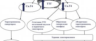
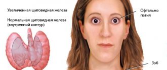
Hyperthyroidism on thyroid ultrasound
Hyperthyroidism is a disease of the thyroid gland associated with excessive production of the hormones triiodothyronine and thyroxine. There are primary, secondary and tertiary hyperthyroidism (pathology of the thyroid gland, pituitary gland and hypothalamus, respectively). It is difficult to make this diagnosis based only on ultrasound results. In addition to the scan results, the patient needs to donate blood for hormones. Severe hyperthyroidism can actively affect the functioning of the brain and heart, thereby threatening the patient’s life.
Hypothyroidism on thyroid ultrasound
Hypothyroidism is a disease associated with decreased activity of the thyroid gland, low production of hormones, and a decrease in the volume and size of the thyroid gland. The main causes of hypothyroidism are: surgery on the thyroid gland, Hashimoto's goiter (replacement of healthy gland tissue with connective or fibrous tissue). With this disease, an ultrasound scan of the thyroid gland may reveal some diffuse changes.
Thyroiditis on ultrasound of the thyroid gland
Thyroiditis is inflammation in the tissues of the thyroid gland. Symptoms of the disease may include pain in the neck and head, a slight increase in body temperature and an increase in the size of the gland, although not in all cases. When carrying out ultrasound diagnostics in case of thyroiditis, small cavities with liquid (pus), swelling and an increase in the volume of the organ are detected. In the case of autoimmune thyroiditis, changes in the structure and size of the organ (decrease or increase) are detected. Previous Next
Cyst on ultrasound of the thyroid gland
A thyroid cyst is a cavity filled with fluid. The cyst can be either congenital or acquired. It can be detected by ultrasound examination. In the case of an inflammatory process or suppuration of cysts, the patient may notice a sore throat and a slight increase in body temperature. Ultrasound of the thyroid gland notes the formation of cysts in the form of round, clear cavities with fluid inside. The rest of the organ tissue remains healthy. In some cases, in order to exclude malignant formations, fluid is taken from these cavities (puncture) directly during ultrasound diagnostics using a needle, or the patient is sent for further examination using MRI of the thyroid gland with contrast.
Tumor on ultrasound of the thyroid gland
In addition to cysts, benign or malignant tumors can form in the thyroid tissue. Ultrasound of the thyroid gland diagnoses tumors in the form of limited (for benign tumors) or invasive compactions. In most cases, when a tumor is suspected, special attention is paid to the lymph nodes. In some situations, evidence of cancer is enlarged lymph nodes and associated diseases - lymphadenitis. However, there is no direct relationship between cancer and lymph nodes, that is, lymphadenitis does not always become a determining factor in cancer. If an ultrasound of the thyroid gland shows signs of a tumor, the patient is necessarily referred for either a biopsy or an MRI of the soft tissues of the neck and lymph nodes with contrast.
How the research works
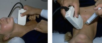
The procedure for ultrasound diagnosis of the thyroid gland is absolutely painless and does not cause any discomfort to the patient. First, the subject lies on his back on a special couch and exposes the neck area. In order to increase the visual area of examination, an additional pillow is usually placed under the patient’s shoulders. After this, a gel is applied to the sensor, which is used to establish contact and improve the passage of ultrasonic signals.
At the next stage of the procedure, the doctor presses the device to the patient’s neck and begins to examine the thyroid gland area from different angles. During the study, a specific ultrasound signal is reflected from the tissue structures of the organ. It is processed using computer equipment and then visualized on a monitor screen. The specialist records the results, alternately examining the isthmus and thyroid lobes, assesses the size and structural condition of the gland, after which an appropriate conclusion is made.
The duration of the session varies from ten minutes to half an hour. It is important to know that an ultrasound specialist only issues a conclusion about the condition of the thyroid gland, and an endocrinologist is responsible for making a diagnosis and determining the feasibility of additional diagnostic procedures.
How to prepare for the procedure
Ultrasound examination of the thyroid gland does not require special preparation. It is necessary to free your neck from jewelry and remove your scarf or neckerchief. It is recommended to wear clothing with an open collar or neckline. You may want to bring a towel with you if the scan is being done at a public health facility. It is recommended to place it under the head, and then use it to remove the remaining gel from the neck.
There are no restrictions on meal time, foods and spices. The recommendations apply only to older people and children. They do not need to eat shortly before the test to avoid nausea that can be caused by pressing on the thyroid gland with the sensor.
All preparation of the child for the procedure takes place in conversation, his parents prepare him psychologically: they tell him that it doesn’t hurt at all, what the doctors do, why it is necessary.
If a scan is performed to determine the effectiveness of treatment, it is necessary to bring the results of a previous ultrasound so that the doctor can assess the dynamics of changes in the gland.
Contraindications
The procedure has no strict contraindications. Diagnosis can be carried out for all people.
A limitation may be damage to the skin at the site of application of the gel:
- pustular formations;
- painful rash;
- burn;
- eczema.
Once the problem has been treated and resolved, an examination can be performed. The procedure is not recommended to be performed in the first days of the menstrual cycle, since during this period the gland is enlarged, which can distort the results.