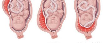
This week the baby's growth ranges from 0.36 mm to 1 mm. The period of development that began at 4 weeks is called “embryonic” and will last until the beginning of the eleventh week of your pregnancy. The fourth week is extremely important for the baby’s future; now all its organs are beginning to form, and some of them will even begin to function! Germinal petals begin to form, which will subsequently develop into various tissues and organs. The inner layer, or endoderm, serves as the basis for the formation of the lungs, digestive system, liver and pancreas. The middle leaf, or mesoderm, will be transformed into the bone, muscle and circulatory systems, heart and kidneys. The outer layer, or ectoderm, will further develop into hair, skin, nervous system, eye lenses, and tooth enamel. The head of the embryo also begins to form. Now it is a small droplet, delicate and transparent.
In addition, at the 4th week there is an active development of extra-embryonic organs - amnion, chorion, yolk sac. It is these organs that will provide breathing, nutrition, protection and support to your baby. The amnion will become the basis for the development of the fetal bladder, and the chorion - the placenta. In general, the most important weeks in the development of the unborn child are from 1 to 12; during this time, not only internal organs are formed, but also the placenta, which plays a huge role in providing the baby with nutrition, oxygen, and also in the production of hormones.
Indications for ultrasound in the 4th week
The content of the article
There are a lot of indications for early ultrasound, because the fourth obstetric week is an important period of fetal development, since the transformation of the fertilized egg into an embryo occurs there. Also, ultrasound during this period is prescribed for suspected ectopic pregnancy, fetal fading, etc. There is no need for early research in the normal course of pregnancy, if the woman has not previously had cases associated with pregnancy pathologies, if there is no need to confirm the date of conception, etc.
Indications for ultrasound at 4 weeks of pregnancy:
- Confirmation of pregnancy if it is unwanted
. This is due to the need to comply with the deadlines for a mini-abortion. Medical abortion is only possible up to 6 weeks of pregnancy. Complications are guaranteed at later stages. To initially confirm pregnancy, you need to do a blood test for hCG. Then you can do an ultrasound. - Ectopic pregnancy
. Most women are still just guessing about an interesting situation at this stage, unless the pregnancy was planned or resulted from IVF. Therefore, an ultrasound is performed at 4 weeks to confirm the fact of pregnancy. During this period, you can see not only the fetal sac itself, but also its location. This is why it is possible to diagnose an ectopic pregnancy. - Threat of miscarriage
(severe nagging pain in the lower abdomen, bleeding, myometrial hypertonicity). If a woman is confident that she is pregnant, which was confirmed by a test and analysis for hCG, but she begins to be bothered by bloody vaginal discharge and periodic pain in the lower abdomen, an ultrasound scan is performed at such an early stage in order to exclude the possibility of termination of pregnancy - miscarriage. - Frozen pregnancy
. To diagnose a frozen fetus in the uterus, an ultrasound is performed vaginally - through the vagina. - Suspicion of oncology (cancer)
of the reproductive organs.
What other changes occur in the mother's body?
- During implantation of the embryo into the lining of the uterus, substances are released into the body that reduce the mother's immunity to prevent the production of large numbers of white blood cells.
- During a healthy pregnancy, discharge from a woman's genitals is white or clear and often has a sour odor. The amount of secretions may increase, which is considered normal, taking into account hormonal changes.
- It is also possible that spotting and spotting may occur. This is the so-called implantation bleeding. If the discharge is scanty and brief, then there is no need to worry.
Prenetics is a way to find out the risk of chromosomal abnormalities
already from the 10th obstetric week. Recommended for every pregnant woman. Duration of two days!
More details
What can a fourth week ultrasound show?
Ultrasound at the 4th week of pregnancy allows you to determine the expected date of birth, assess the course of pregnancy and identify possible abnormalities in the development of the fetus. Early diagnosis of pathologies allows either to begin treatment, or, if it is impossible, to terminate the pregnancy with medication without complications for the woman.
- Presence of pregnancy.
At this stage, the fetal sac is clearly visualized in the uterine cavity. With a delay of 1 or 2 days, the size of the fertilized egg is 1-3 mm. - Accurate determination of gestational age
. In the early period, you can determine the exact gestational age (plus or minus 2-3 days) by measuring the coccygeal-parietal size of the embryo. But this can only be done at 6 weeks. At 4 weeks, the doctor can find out the gestational age by measuring the size of the fetal sac. - Localization of the fertilized egg
. If the egg is in the uterus, the pregnancy is normal. Outside the uterus, for example, in the tubes, an ectopic pregnancy. However, the presence of a fetal sac in the uterus does not exclude an ectopic pregnancy. Every 10,000th patient experiences both a normal pregnancy and an ectopic pregnancy. - Number of fruits
. Ultrasound reveals multiple pregnancy at the beginning of the 5th week. Multiple pregnancies cause complications 10 times more often than singleton pregnancies. Therefore, ultrasound should be performed more often. - Embryo viability.
Having counted 3 weeks and 4 days from the moment of conception, you can already see the heartbeat. But the embryo begins to move at 5-6 weeks. - Early pathologies
. For example, you can see a hydatidiform mole. - False pregnancy
. This is the name for formations in the pelvis that resemble pregnancy. Ovarian cysts, tumors and large uterine fibroids can be mistaken for pregnancy. These formations are hormonally active, so they can give positive results when tested for hCG. Such formations can develop in parallel with pregnancy, interfering with the development of the fetus in the early stages. - Risk of miscarriage.
If there is discharge or spotting, you need to immediately evaluate the heartbeat of the embryo. An ultrasound will also show placental abruption. Timely consultation with a gynecologist will help maintain pregnancy. - Location of the chorion.
The placenta develops from it.
Methods for calculating gestational age
The main method for determining the duration of pregnancy is the method used by the gynecologist at the antenatal clinic, or the obstetric method. He calculates the gestational age from the first day of the last menstruation and prescribes routine instrumental and laboratory tests based on this date.
The embryonic counting method takes the date of ovulation as a starting point and lags behind the obstetric period by about 2 weeks. Women may believe that their pregnancy began on the day when the expected menstruation did not arrive. To avoid confusion when communicating with your doctor, it is recommended to adhere to the obstetric method.
How is an ultrasound performed at 4 weeks of pregnancy?
At this stage of pregnancy, the most accurate and informative method of examination will be transvaginal ultrasound.
– by inserting a special sensor into the vagina. It is carried out as follows: a woman bares her body below the waist, lies on the couch with her back, bending her knees. The doctor inserts an ultrasonic sensor into the vagina, onto which a condom was previously put on and lubricant was applied. On the screen of the ultrasound machine, the doctor can examine the uterus, appendages, embryo and its size. The doctor may move the sensor slightly from side to side, but this should not cause any painful sensations; only slight discomfort is possible at the time of insertion of the sensor.
If the test is positive and the woman has complaints of bloody vaginal discharge, then a transabdominal ultrasound
– through the anterior abdominal wall. This is necessary to reduce the risk of miscarriage. The woman should lie with her back on the couch and expose her stomach, after which the doctor applies a special gel to its lower part, which removes air and improves contact with the sensor, and scans with the device. This type of ultrasound at 4 weeks is not very informative in terms of determining the size of the embryo, but can detect an ectopic pregnancy or a pathological source.
Lifestyle, nutrition and vitamins
Week 4 is, as a rule, the last week when a woman cannot yet reliably know that pregnancy has occurred. If you were planning a child and did everything to conceive, lead a healthy lifestyle, give up alcohol, smoking, taking antibiotics, avoid contact with sick people, try to be calmer, picky about food and drinks. And don't forget about vitamins. But remember: only a doctor can give you a detailed answer to the question of what vitamins to take and in what quantity.
How to prepare for the procedure
The quality of ultrasound results at 4 weeks of pregnancy directly depends on the quality of preparation for the study. So, if a transvaginal ultrasound is prescribed, then the woman should only empty her bladder immediately before the procedure.
Transabdominal ultrasound is performed on an empty stomach and with a full bladder. To do this, in the morning before the procedure, the woman should not eat anything, and an hour before the ultrasound, drink 1 - 1.5 liters of non-carbonated and non-dairy liquid. When the urge to urinate occurs, you can conduct an examination. Some clinics recommend bringing a towel or diaper to cover the couch, and napkins to wipe off the gel from the skin of the abdomen.
When to see a doctor?
If you feel normally, you can still postpone a visit to the doctor. If the test result is positive and there is nagging pain, signs of early toxicosis, or atypical discharge, you should consult a doctor.
Possible actions of a gynecologist if a woman feels well at the 4th obstetric month:
- Taking anamnesis;
- Visual examination, examination on a gynecological chair;
- Prescription of vitamin-mineral complexes containing, among other components, folic acid.
Signs of disease and a history of miscarriages require specific treatment. Women who have problems conceiving and become pregnant using IVF or ICSI are under the supervision of a doctor from the first days of pregnancy.
Watch a video that will tell you why you need to do a genetic analysis, what happens in the female body, about hereditary diseases - cystic fibrosis, hemophilia, Down syndrome and screening for chromosomal pathologies:
Fetus at 4 weeks of gestation
At this time, the fertilized egg turns into an embryo, which is already fixed in the uterus. The formation of embryonic tissue occurs, represented by three main leaves:
- ectoderm (outer sheet)
- the basis for the formation of skin, hair, nails, tooth enamel and the nervous system; - endoderm (inner layer)
- the digestive system and lungs are formed from it; - mesoderm (middle layer)
is the foundation for the formation of the heart, blood vessels, bones and muscle tissue.
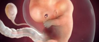
In this period, the embryo resembles a tadpole - it already has eyes and a head. It not only firmly attaches to the wall of the uterus, but also grows and develops rapidly. On the ultrasound machine monitor it is depicted as a small round formation.
At 4 weeks of pregnancy, a small bubble forms around the fetus, which will soon turn into an amniotic membrane containing amniotic fluid. Formation of the umbilical cord and placenta is also observed.
What's happening to mom
In the fourth week, external signs of pregnancy are not yet obvious. But the body is already actively restructuring. A woman may not be aware of her situation at this time, since the symptoms resemble the precursors of menstruation. By the end of the fourth week, pharmacy tests can already determine pregnancy.
Feel.
In the fourth week, signs of pregnancy become clearer. Increased production of progesterone provokes constipation and bloating. Frequent urge to urinate also causes discomfort. Early toxicosis may appear. Physiological sensations and emotional mood change:
- Mood swings.
A sharp change in mood from euphoria to apathy, causeless tearfulness, and sensitivity to words and reactions appear. Sometimes unreasonable fears appear. - Drowsiness and fatigue.
The body retains energy for restructuring, so even after rest and full sleep, the desire to sleep does not go away. - Changes in taste.
Habitual food can be disgusting or tasteless. At the same time, new unexpected addictions may appear. - Odor intolerance.
The urge to vomit from the aroma of your favorite perfume or the smell of familiar dishes is already a reason to buy a pregnancy test and check your suspicions. - Appetite.
A change in appetite can either increase or manifest as a complete refusal of food. - Chest pain.
In the fourth week, the mammary glands enlarge, causing pain. Nipples become especially sensitive. - Stomach ache.
The uterus stretches and increases in size, which can cause pain.
Changes in the body.
In the fourth week, active production of hormones and restructuring of the entire body continues. The fetus is already attached to the wall of the uterus and receives all the necessary nutrients from it. The formation of the placenta begins, which will take on the burden of fetal development from the twelfth week of pregnancy until the birth itself. Blood circulation in the mammary glands increases, the breasts prepare for the upcoming lactation.
External changes.
Significant visible changes occur in the breasts in the fourth week. Breast volume increases (sometimes by a size) within a week. A vascular network appears. The halo darkens and pigmentation may appear in other areas of the skin. The volume of the abdomen does not change and does not outwardly indicate an “interesting position.”
If the fetus is not detected on ultrasound
In some cases, the embryo may not be detected in the uterine cavity, while the pregnancy test is positive. Possible reasons for this are the following:
- bend or other abnormality of the uterus;
- inflammatory or neoplastic process in the uterine cavity;
- ectopic pregnancy, which can be localized on the ovary, in the fallopian tube, on the wall of the bladder or abdominal cavity;
- frozen pregnancy;
- incorrectly calculated period;
- insufficient qualifications of the doctor or the resolution of the device (the ultrasound machine simply does not see the fertilized egg).
The course of action in this case is as follows: repeat ultrasound in a week and a blood test for the content of human chorionic gonadotropin. It is important to establish a diagnosis in a timely manner in order to exclude dangerous causes - frozen or ectopic pregnancy.
Restrictions in the life of a pregnant woman
Restrictions on the use of medications during pregnancy are not accidental.
Substances included in medications remain in the body for some time after taking them, and therefore can affect both the quality of sperm and eggs before conception, and the formation and development of the Baby after conception has occurred. If it is absolutely impossible to avoid taking medications, then it is better to consult a doctor and choose the safest medications. Every time you are prescribed medication, remind your doctor that you are planning to conceive a child, especially if its development has already begun.
Although homeopathy, aromatherapy, and herbal medicine are safer than traditional medicines, their use should also be cautious - and only after consultation with a doctor.
But the health and life of a small creature can be threatened not only by diseases and medications, but also by:
- X-ray examinations of the abdominal cavity, pelvis, back (lower back) in the second half of a woman’s menstrual cycle (since she may be pregnant without knowing it) and even more so during the period of “non-occurrence” of the next menstruation. X-ray radiation has oncogenic and mutagenic effects;
- harmful working conditions for future parents. The most dangerous in this regard are professions in which you have to come into contact with chemicals (this includes, for example, such a seemingly harmless profession as a hairdresser). Radioactive radiation, chemicals, metals (mercury, lead), drugs (for example, tetracycline) cause abnormalities in the chromosomal apparatus at the level of eggs and sperm.
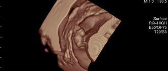
What can be seen on an ultrasound
At 4 weeks of pregnancy, a tiny fertilized egg (blastocyst) will appear on the monitor in the form of a small anechoic round formation up to 5 mm in the uterine cavity.
A detailed examination allows you to see the yolk sac in the form of a ring-shaped hyperechoic formation up to 3 mm in diameter. It is he who, during the development process, will be the supplier of nutrients to the growing fetus. In the uterus, the doctor observes diffuse dilatation of blood vessels. This is a physiological response to the progressive increase in the embryo's needs for nutrients and oxygen.
The ovary contains a functioning corpus luteum, which produces the pregnancy hormone progesterone. Duplex mapping shows a decrease in the resistance index in the arterial vessels of the corpus luteum. There is an increase in the size of the uterus and the intensity of the echostructure.
Data decryption
At 4 weeks of pregnancy, the normal indicators are as follows:
| Index | Norm |
| Uterus size | Slightly enlarged |
| Uterine tone | Not promoted |
| Uterine vessels | Expanded |
| Cervix | From 3 cm |
| Corpus luteum | Should be visualized in the ovary. Size – from 1 to 3 cm |
| Yolk sac | 2 – 3 mm in diameter |
| Embryo | 0.4 – 1 mm. Its localization is established |
If the length of the cervix is shortened, isthmic-cervical insufficiency will be diagnosed, requiring observation and, if necessary, treatment.
Subsequent ultrasounds will allow you to determine the size of the fetus, compare them with gestational age norms and more accurately indicate the gestational age. The gender of the child can be determined in the second trimester after the 20th week, since at this time the sexual characteristics will be practically formed and visualized quite well.
Good to know
Diabetes and pregnancy
Pregnancy by week: how the baby develops in the belly
Is it possible to self-medicate during pregnancy?
Boy or girl? Determining the gender of a child based on characteristics
Signs of pregnant women - should you believe in them?
5 ways to find out the gender of your unborn child - scientific and not so scientific
All texts for pages about mother and baby were kindly provided by RAMA Publishing - these are chapters from the book by Svetlana Klaas “Your Favorite Little Man from Conception to Birth”, reviewer Irina Nikolaevna Kononova, Candidate of Medical Sciences, Associate Professor of the Department of Obstetrics and Gynecology of the Ural State Medical Academy (Ekaterinburg).
Where to do an ultrasound in the 4th week of pregnancy in St. Petersburg, price
At the Diana clinic on Zanevsky, 10, a new expert ultrasound machine with Doppler has been installed, so here you can do an ultrasound during pregnancy at any time. Not every clinic in St. Petersburg has such ultrasound machines. The cost of the examination is only 1000 rubles. Here you can take a blood test for hCG. If the pregnancy is unwanted, a medical abortion can be performed at the medical center. The cost of abortion with pills is 3,500 rubles. The price includes everything you need - ultrasound, doctor’s consultation, a set of abortion pills.