We continue to talk about how mother and baby feel at different stages of pregnancy. Our permanent expert, candidate of medical sciences, obstetrician-gynecologist, gynecologist-endocrinologist Maria Prokhorova the questions of the FAN .
The time has come for the 12th week of pregnancy, which ends the difficult and exciting first trimester. The greatest vulnerability of the embryo to external environmental conditions is behind us, all associated fears and anxieties are behind us, toxicosis is slowly receding, and ahead of the expectant mother is a pleasant and calm state, which lasts from approximately 12 to 24 weeks and is called the “golden” period of pregnancy.
The interesting position remains a mystery to others, and the barely noticeable growth of the belly is still easy to hide under looser clothing. It is, of course, too early for the first movements, but the hormonal storm has calmed down a little, the woman has already gotten used to her new state and is preparing every day for the responsible role of a future mother.
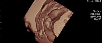
At this stage, the baby is still too small to show any noticeable presence inside the abdomen, and the woman can’t wait to see what the baby looks like, at least in an ultrasound photo. Well, soon she will be given such an opportunity, because the 12th obstetric week of pregnancy is the optimal period for carrying out the all-important first screening.
We will tell you what interesting things happen to the baby and mother during this period, what examinations need to be completed to make sure that everything is going as it should.
pixabay.com/raman bhardwaj
What is placenta
During the development of the embryo, a chorion is formed on its outer shell - specific outgrowths. It gradually grows into the woman’s uterus and increases in size. As a result, the so-called "children's place"
The process comes to an end at 12-16 weeks after conception. At this point, the organ becomes flat, resembling a disk in shape (placenta in Latin). The connection between the mother and child is provided by the umbilical cord. It has a length of 5-55 cm, and functions as follows:
- the artery delivers nutrients and oxygen;
- the vein is necessary to remove processed substances.
The formed placenta consists of decidual tissues, embryoblast and trophoblast, and the main element is called the villous tree. This organ is designed to prevent Rh conflict. Its thickness is approximately 3.5 cm, and its diameter is 20 cm; weight – about 600 g.
The development of the placenta until the 4th month occurs faster than the growth rate of the embryo. At this point, the villi and blood vessels are completely formed. If a child dies at any stage, degenerative phenomena increase.
The placenta consists of two sides:
- rough maternal surface, formed from the basal component of the decidural layer, facing the uterus;
- the fruit side, covered with the amniotic layer, turned towards the embryo.
If a woman is pregnant with twins, there are three options:
- Dizygotic twins implanted separately. The placenta is divided into two parts, each of which has its own membranes and blood vessels.
- Dichorionic twins separated from each other by a septum. The main part of nutrition comes from amniotic fluid, and there are practically no blood vessels in the placenta membrane. In this case, pregnancy management under the supervision of a specialist in our center is mandatory, since the risk of transfusion syndrome is high.
- Monoamniotic monochorionic twins not separated by a septum.
The placenta begins to form from conception. Starting from the 2nd week of pregnancy, its growth is activated, by the 13th the structure is fully developed, and maximum activity is observed by the 18th week. Complete cessation of growth and changes occurs only after delivery.
Monitoring the development of the placenta
Changes occurring in the placenta are monitored using ultrasound. To do this, use the classification of organ maturity:
- Stage 0 – up to the 30th week;
- Stage 1 – from 30th to 34th;
- Stage 2 – from 34th to 37th;
- Stage 3 – from 37th to 39th;
- Stage 4 – before delivery.
By monitoring the rate of organ maturation, our doctors can promptly determine the pathology:
- too early development indicates disturbances in the placental blood supply (if the woman does not have a genetic predisposition to this phenomenon);
- delayed maturation does not affect the child’s condition and does not pose a threat to him.
Research is carried out not only using ultrasound, but also by determining the level of lactogen, a hormone that indicates normal maturation. Normally, it should be at least 4 mcg/ml.
Pregnancy management in Nizhny Novgorod also includes daily monitoring of estrogen concentrations. A low level may indicate renal failure of the expectant mother, severe liver pathologies, or be a consequence of taking antibiotics.
What examinations need to be completed at 12 weeks?
Despite the fact that the most dangerous time for the fetus is already over, the risks of a frozen pregnancy exist at any stage, so the expectant mother must register with the antenatal clinic, if she has not already done so, be regularly monitored and undergo all examinations prescribed by the doctor. .
Maria Prokhorova warns that it is not possible to independently determine the child’s condition, since the woman does not yet feel fetal movements, and heartbeat parameters cannot be determined at home, even with the help of special sensors.
“There may be a slight deterioration in health,” explains our expert. “But this is not a direct sign of a missed abortion, so women undergo a screening examination at this stage, and if they feel unwell, they should consult a doctor.”
pixabay.com/No-longer-here
Therefore, expectant mothers are advised to register for pregnancy as early as possible. By the way, for registration up to 12 weeks you even receive some benefits from the state. The payout is small, but still.
Important! At 12–13 weeks, the first prenatal screening is carried out, which makes it possible to identify possible fetal malformations at an early stage, if any.
This most important examination includes a highly professional ultrasound and a special blood test. Diagnosis should be performed by a doctor who specializes in performing such screenings. Using the device, the doctor will see the general condition of the baby, as well as a number of important parameters, such as the thickness of the nuchal translucency (NVP), the length of the nasal bone, the coccygeal-parietal size, head circumference and some others.
Many parents can't wait to find out the gender of their unborn child. Maria Prokhorova explains that at week 12 one can only guess whether a boy or a girl will be born, but the gender can only be reliably determined by a blood test. Or you will have to wait for the next scheduled ultrasound, which is done at 20–22 weeks.
On the day of the ultrasound examination, the patient simultaneously donates blood, which is sent to the laboratory, where a special program calculates the genetic risks of the fetus.
Sometimes it happens that bad results come. In this case, Maria Prokhorova urges, first of all, not to panic, since the screening parameters are influenced by many factors, including, for example, age calculation. If the future parents are over 35 years old, then the program automatically includes an age parameter and the results will be slightly worse than for younger patients. In any case, the decision always remains with the woman, and the doctor has no right to incline her in one direction or another. He can only reassure, support and inform about possible options for the development of events.
If a woman has not yet had time to be examined when registering, then the first screening can be combined with a classic examination and a general blood and urine test. In addition, now is the time to visit specialists. On the mandatory list:
- therapist;
- dentist;
- otolaryngologist;
- ophthalmologist;
- other doctors - if indicated.
Possible pathologies
The most common violations:
- discrepancy between the pace of development of the placenta and the baby;
- the appearance of blood clots that obstruct blood flow;
- incorrect location of the organ;
- excessive thickening;
- heart attack - cessation of blood flow through the arteries;
- various inflammations, tumors.
The reasons for these phenomena may be:
- toxicosis;
- diabetes;
- atherosclerosis;
- infectious phenomena in the mother's body;
- Rhesus conflict;
- excessive or insufficient weight of a woman;
- emotional stress;
- bad habits;
- woman's age over 35 years.
It is important that the expectant mother monitors her own well-being throughout the entire period of carrying the child. The appearance of any unusual symptoms - vaginal discharge, pain, cramps, increased swelling should be a signal for urgent contact to our center.
The most common problems in the 1st trimester of pregnancy
Bleeding : due to a lack of progesterone, spasm and relaxation of the muscles of the uterus occurs and, accordingly, detachment of the ovum and bleeding (in severe cases, miscarriage). Also, with hypothyroidism, there is a violation of the implantation of the fertilized egg, which also leads to bleeding.
Thrombosis . During pregnancy, blood clotting increases (this is normal - the body is preparing for blood loss). To prevent thrombosis of the veins of the lower extremities, it is necessary to stop taking oral contraceptives 6 months before planning a pregnancy, and during pregnancy, maintain sufficient physical activity (long walks, swimming) and be observed by a doctor. If pain, swelling, or “cyanosis” occurs in the lower extremities, you should immediately consult a doctor.
Developmental anomalies and genetic pathologies . In the 1st trimester, abnormalities in the development of the fetus most often develop and manifest themselves, which can occur as a result of ionizing radiation, infectious diseases suffered during pregnancy (rubella, influenza, etc.), taking drugs prohibited during pregnancy (fluoroquinolones, macrolides, tetracyclines, tranquilizers , etc.), genetic pathologies and hereditary diseases.
Anemia . More often it happens due to iron deficiency, because. the fetus consumes a huge amount of it, taking away iron reserves from the mother, less often due to bleeding. B12 and folate deficiency anemia also occur, which is also associated with increased consumption and insufficient consumption by the mother. Anemia is dangerous due to chronic lack of oxygen, causing fetal growth restriction syndrome. In severe cases, it can lead to intrauterine fetal death.
Chronic diseases may worsen during pregnancy . This is due to the fact that the fertilized egg already carries part of the DNA that is foreign to the mother. To prevent egg rejection, the mother’s body suppresses its immunity throughout pregnancy, which affects the body as a whole. Therefore, all diseases that were not detected at the stage of preparation for pregnancy should be detected and treated. This applies mainly to infectious diseases. For example, exacerbation of chronic pyelonephritis, or herpes virus infection.
Also, during pregnancy there is a sharp restructuring of hormonal levels , which, in turn, affects somatic (non-infectious) diseases. For example, due to thickening of bile and compression of the gallbladder by the uterus, chronic cholecystitis may worsen. As the fetus develops, the islets of Langerhans (cells that produce insulin in the pancreas) sometimes do not develop quickly enough and the baby begins to consume the mother's insulin. If the mother has diabetes or impaired glucose tolerance (including hidden), both the child and the mother lack insulin, which leads to tissue damage to both the child and the mother (early aging of the placenta, delayed fetal development, premature birth, polyhydramnios, increased blood pressure in a pregnant woman, the formation of a large fetus, the risk of injury to the woman and child during childbirth and the most dangerous complication - intrauterine fetal death).
In addition, the fetal liver does not yet function as a detoxification organ, and all decay products produced by the child are forced to be processed by the mother’s liver. 2% of pregnant women develop benign cholestatic hepatosis of pregnancy - a violation of the formation and outflow of bile, which causes:
- Itchy skin, especially at night
- Digestive disorders (as bile is involved in the emulsification of fats)
- Inflammation of liver tissue due to toxic effects on cells and bile ducts