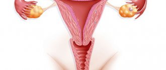
Leiomyoma (myoma, fibromyoma) of the uterus is a hormonal-dependent, benign, tumor-like formation of the uterus, the development of which begins in smooth muscle tissue. Uterine leiomyoma can be single or multiple, localized in different parts of the organ. Most often, the neoplasm develops in the body of the uterus, in rare cases - in the cervix of the organ. The size of the tumor varies from a few millimeters to a kilogram. With small-sized leiomyoma, clinical manifestations are usually absent, as a result of which the disease is often discovered accidentally during a gynecological examination.
Differences between uterine leiomyoma and fibroids
Many women are interested in the differences between fibroids, fibroids, fibromyomas and uterine leiomyomas. The medical history of patients often contains both one and the other diagnosis. Essentially, uterine fibroids are the common name for fibroids, fibroids, and leiomyomas. In turn, some differences between leiomyoma, fibroma and fibromyoma still exist and they relate to the structure of the tumors. Leiomyomas contain predominantly smooth muscle cells, whereas fibroids and fibroids are composed of fibrous tissue cells.
Varieties
In accordance with the direction of growth of the myomatous node, the structure and number of neoplasms in the uterus, the following types of leiomyoma are distinguished:
- subserous;
- submucosal;
- intramural (interstitial);
- unspecified;
- cellular;
- multiple.
Intramural leiomyoma of the uterus
Intramural uterine leiomyoma is characterized by localization of the tumor node in the thickness of the muscular walls of the uterus. Treatment of this form of leiomyoma involves both dynamic observation and surgical intervention, up to radical removal of the uterus, which depends on the size of the node and the intensity of its growth.
Subserous leiomyoma of the uterus
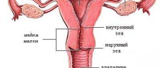
Subserous uterine leiomyoma is characterized by subperitoneal localization, under the serous membrane of the uterus. The myomatous node has a wide base or a long stalk.
Submucosal leiomyoma of the uterus
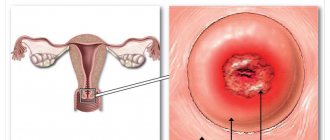
Submucosal (submucosal) leiomyoma is most often localized in the body of the uterus, sometimes in its cervix, its growth is directed into the organ cavity.
Leiomyoma of the uterus, unspecified
Uterine leiomyoma of unspecified form is essentially a latent form of tumor formation that cannot be confirmed diagnostically, which is associated with the small size of the myomatous node or its slow growth.
Cellular leiomyoma of the uterus
Externally, cellular (high cell) leiomyoma looks the same as regular uterine leiomyoma, but it is more “fleshy” and soft. In section, cellular leiomyoma of the uterus has a reddish-brown color; foci of hemorrhage and necrosis are visualized. According to the results of histological examination, cellular leiomyoma is a tumor with an extremely developed cellular structure, relatively uniform nuclei and the absence of nuclear atypia.
Multiple uterine leiomyoma
Multiple leiomyoma is diagnosed when there are several tumors, which may have different volumes, tissue composition and location in the uterus.
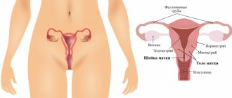
Myomatous nodes. Histological structure
Myomatous nodes are heterogeneous in their histological structure. They are divided into myomas, fibromas and angiomyomas, as well as adenomyomas.
There are three main forms of uterine fibroids (depending on morphogenetic characteristics):
- simple fibroids, which develop as benign muscle hyperplasia;
- proliferating fibroids, which have the morphogenetic criteria of a true benign tumor, are found in every fourth patient;
- presarcomas are a stage on the path to true malignancy.
Depending on where the myomatous node is localized and how it progresses, the following types are distinguished:
- Submucosal submucosal myomatous node. It grows into the uterine cavity and deforms it.
- Subserous, or subperitoneal myomatous node, which grows towards the abdominal cavity.
- The intraligamentary myomatous node is located between the leaves of the broad ligament of the uterus and pushes it apart.
- The interstitial (intermuscular) myomatous node grows from the middle layer of the myometrium and is located in the thickness of the myometrium.
Causes
There is currently no clear answer about the reasons for the development of uterine leiomyoma. Experts agree that this disease often develops in women with impaired ovarian function, which is accompanied by excessive production of estrogen. However, in contrast to this theory, there are cases of the development of uterine leiomyoma in patients whose hormonal levels are within normal limits.
In addition, the occurrence of uterine leiomyoma can be provoked by other negative factors:
- surgical termination of pregnancy;
- complicated pregnancy and childbirth;
- endometriosis (adenomyosis) of the uterus;
- inflammatory diseases of the fallopian tubes and ovaries;
- absence of pregnancies and childbirth in women over 30 years of age;
- obesity;
- hereditary predisposition;
- immune and endocrine disorders;
- long-term insolation.
Why did you get fibroids?
Fibroids most often develop due to hormonal imbalances. The development of tumors is caused by:
- disruption of estrogen production;
- imbalance in the mechanisms of estriol and estrone secretion;
- influence of progesterone.
Unwanted nodules can also appear due to chronic infections, untreated diseases, inflammatory processes, trauma to the myometrium during curettage, abortion, and manual manipulation during childbirth. It is believed that a hereditary predisposition can also lead to the development of fibroids. There is even an opinion that the uterine cells from which the tumor grows are formed incorrectly even at the stage of human intrauterine development.
Uterine fibroids are a signal that there is a malfunction in the body’s functioning
Some researchers claim that taking hormonal birth control reduces the risk of unwanted inflammation by a quarter. When prescribing medications for uterine fibroids, the doctor may ask how exactly you protect yourself.
Symptoms
In women with small uterine leiomyoma, clinical manifestations may be absent for a long time, so it is often accidentally diagnosed during a gynecological examination. Malignancy of leiomyoma occurs in extremely rare cases.
As the myomatous node grows, symptoms appear, the most common of which include the following:
- menstrual bleeding intensifies and lengthens (menorrhagia develops);
- blood clots are found in menstrual discharge;
- acyclic uterine bleeding occurs (metrorrhagia develops);
- As a result of metrorrhagia, iron deficiency anemia develops.
Uterine leiomyoma is characterized by pain, which depends on the location and size of the myomatous node. Most often, pain appears in the lower abdomen or lower back. The nature of pain with slow growth of leiomyoma can be constant and aching.
Submucosal leiomyoma is characterized by sudden, cramping pain. The severity of the pain syndrome increases as the uterine leiomyoma increases in size; in the initial stages it is practically absent.
The intensive development of leiomyoma can lead to the fact that the tumor begins to compress nearby organs, as a result of which their functions are disrupted: a woman often experiences constipation and the process of urination becomes difficult.
With large leiomyomas, compression syndrome of the inferior vena cava may develop, resulting in shortness of breath (usually in a horizontal position), and increased heart rate.
How to get rid of fibroids without surgery?
When the diagnosis is beyond doubt, it is important to first think about how to cure uterine fibroids without surgery. The fact is that after surgery, unwanted nodules may appear again. Therefore, the following methods are used to treat fibroids:
- drug therapy. The non-hormonal drug Esmya is used, the active ingredient of which is ulipristal acetate. Esmya is prescribed before surgery or during a course of treatment of moderate and severe symptoms of the disease for women of reproductive age. Reviews indicate that in almost 80% of cases, patients stop menstruation while taking the drug. The maximum duration of one course of treatment is 3 months. If the course needs to be repeated, this should be done during the second menstrual cycle, which will occur after the end of the previous course. The best results are achieved in the early stages of the disease.
- hormonal treatment. Reviews indicate that this type of therapy is most effective when uterine fibroids are still small. In this case, derivatives of 19-norsteroids (for example, Norethisterone, Norkolut) are used, which reduce blood loss and normalize hemoglobin levels. Antagonists of gonadotropin-releasing hormones (for example, Buserelin, Diferelin) help to significantly reduce the size of the nodes due to the fact that they contribute to the onset of menopause. Therefore, they are not recommended for use in the treatment of women of childbearing age. Of the antigestagens (drugs that inhibit ovulation), Ginestril, the active ingredient of which is mifepristone, is often used. You must take tablets for uterine fibroids strictly according to the regimen prescribed by your doctor - the effectiveness of the treatment depends on this.
- embolization of the uterine arteries. Special substances are injected into the uterine arteries to block the flow of blood to the cells that make up the tumor. In this case, emboli are introduced so that the nutrition of healthy myometrial cells is maintained. Without receiving nutrition, fibroid cells are replaced by connective tissue. There is no need to be afraid that the doctor will block the “wrong” vessels - the arteries that feed the tumor practically do not connect with other vessels, and they are also several times thicker than “healthy” ones. The doctor decides whether to prescribe auxiliary drugs for uterine fibroids that have undergone embolization or not.
- FUS ablation. During the procedure, focused ultrasound is directed to the node. This heats up the tissue and destroys it. The progress and results of the process are monitored using MRI. Large units are heated in parts. There are no incisions made and the patient is sent home the same day.
- physiotherapeutic methods. This is an auxiliary set of measures that cannot replace primary treatment. It is aimed at increasing the tone of the body, stimulating recovery processes, and stabilizing hormonal levels. To treat uterine fibroids, patients, in addition to taking medications, are recommended to undergo electrophoresis, radon and iodine-bromine baths, and magnetic therapy. Most often, a wellness course begins on the 5-7th day of the menstrual cycle.
But experts do not recommend getting carried away with traditional medicine. Even if herbs and herbs contain the same active ingredients and vitamins as pharmaceutical drugs, it is impossible in this case to select an effective dosage and maintain it throughout the entire course of treatment.
Medicines for uterine fibroids should only be prescribed by a doctor
Sometimes, as an auxiliary method of treating uterine fibroids without surgery, hirudotherapy is called. Expert opinions are divided on this matter.
- Proponents of the method claim that the use of leeches evens out hormonal imbalances. However, no one can guarantee that the tumor will not increase due to exposure to worms. It is reported that hirudin contained in the saliva of leeches normalizes the fluidity and coagulability of blood and helps eliminate blood stagnation in the pelvic organs.
- Opponents of the method point out that the use of leeches causes blood loss, which is unacceptable in case of anemia, which is usually observed with fibroids.
Diagnostics
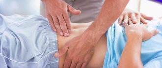
Uterine leiomyoma can be diagnosed during an initial gynecological examination. Using a two-manual vaginal examination, the doctor palpates a dense, enlarged uterus with a lumpy, nodular surface. To more reliably determine the size of uterine leiomyoma, its localization and classification, an ultrasound examination of the pelvic organs is prescribed.
One of the most informative diagnostic methods is hysteroscopy, during which a specialist examines the cavity and walls of the uterus using an optical device - a hysteroscope. Hysteroscopy is performed for both diagnostic and therapeutic purposes.
Additionally, hysterosalpingoscopy (ultrasound examination of the uterus and fallopian tubes) and diagnostic tests for sexually transmitted infections may be prescribed.
FAQ
What do gentle surgeries give a woman?
Thanks to the possibility of preserving the uterus, the woman will not experience psychological discomfort. If it is impossible to preserve the uterus, we strive to use gentle techniques. Preservation of the cervix allows not to disturb the erogenous sensitivity of this zone, and the ovaries - to avoid hormonal disorders. Thanks to organ-preserving techniques, the risk of prolapse of internal organs after surgery is also minimal.
What is the essence of the author’s technique for removing multinodular fibroids?
During laparoscopy, a temporary occlusion of the uterine arteries is performed, blood flow to the uterus is suspended, the surgical area remains dry, and visualization is not impaired. There is no need to use coagulation. After removal of the nodes, blood flow is restored. The bloodless technique not only serves to prevent intraoperative complications, but also reduces the duration of the operation.
Treatment
The treatment tactics for uterine leiomyoma are chosen by the doctor in accordance with the size of the myomatous nodes, the severity of its clinical symptoms and the age of the patient. Both conservative and surgical methods can be used for treatment.
All patients with uterine leiomyoma are advised to undergo dynamic observation by a gynecologist, who must be visited at least once every three months.
Asymptomatic small uterine leiomyoma is subject to conservative treatment with hormonal drugs - progesterone derivatives, which help normalize ovarian function and prevent tumor growth.
In addition, to suppress the secretion of gonadotropins and create pseudomenopause, injections of GnRH agonists are prescribed, which have a prolonged effect. It must be taken into account that long-term use of these drugs can lead to the development of osteoporosis.
Conservative treatment helps to restrain further growth of myomatous nodes, but is not able to completely eliminate the disease.
It is possible to radically get rid of uterine leiomyomas with the help of surgical treatment, but certain indications are necessary for its implementation:
- large size of the myomatous node;
- rapid rate of increase in the size of uterine leiomyomas;
- severe pain syndrome;
- concomitant diseases: ovarian tumors or endometriosis;
- torsion of the leg of the node and its necrosis;
- impaired function of adjacent organs (rectum, bladder);
- infertility;
- submucous uterine leiomyoma (surgical treatment is prescribed if conservative therapy is ineffective);
- suspicion of malignancy of uterine leiomyoma.
The nature of the surgical intervention and its volume are determined taking into account the patient’s age, state of reproductive and general health, and the degree of expected risks.
The obtained objective data allows the doctor to make the right choice of the type of surgical intervention:
- conservative – the uterus is preserved;
- radical - the uterus is completely removed.
Conservative surgical treatment is preferable for women planning pregnancy, since this method does not interfere with their reproductive function.
In addition, myomectomy is an organ-preserving method, during which myomatous nodes are removed from the uterus.
The least traumatic method of performing myomectomy is hysteroscopy, which involves excision of the myomatous node using a laser. The manipulation requires the use of local anesthesia and visual supervision by a physician.
During radical surgical interventions, the uterus with myomatous nodes is completely removed, as a result of which the woman loses her reproductive function. Similar methods include hysterectomy, supravaginal amputation, panhysterectomy.
The use of laparoscopic techniques for conservative myomectomy and supravaginal amputation of the uterus provides a significant reduction in surgical trauma to tissues, the severity of adhesions in the future and significantly shortens the rehabilitation period.
How to cure fibroids: cut them or take pills?
Previously, surgery was considered the main method of treating uterine fibroids. According to some data, removal of nodules accounted for about 80% of gynecological operations. Gynecologists sought to get rid of them as quickly as possible, since they considered them to be harbingers of a malignant disease - leiomyosarcoma. However, after the “benign quality” of fibroids was proven, doctors began to give preference to non-surgical methods.
After excision of the tumor, a scar remains on the uterus. Therefore, it is recommended to wait 6-12 months after the operation, and only after this period plan a pregnancy. To reduce the risk of childbirth, the patient may be advised to have a caesarean section.
Laparoscopy is a minimally traumatic method of surgical intervention
In each specific case, the final word remains with the doctor. In general, surgery is necessary when the tumor:
- located under the mucous membrane of the uterus or in its cervix;
- has large dimensions;
- causes bleeding (especially with anemia);
- grows quickly;
- compresses neighboring organs - bladder, rectum;
- increases during postmenopause.
If the above symptoms are absent, the doctor will most likely allow the patient to be treated with medications, or suggest minimally invasive or physical therapy methods. Doctors are in no hurry to perform operations if:
- the shape of the uterus has not changed, or the organ has already changed shape, but is still small in size;
- the tumor is between the muscles;
- a woman is still of childbearing age.
To avoid suffering from fibroids, you need to take treatment seriously
It is important to remember that whether uterine fibroids can be treated without surgery depends in each case on the patient herself. To fully recover, she will have to significantly adjust her lifestyle:
- observe a natural sleep schedule - avoid night shifts, do not stay up late;
- have regular sex life;
- give up smoking, alcohol, junk food;
- control your weight;
- give yourself regular physical activity, regularly spend time in the fresh air;
- take vitamins (for example, Aevit).
If the patient becomes pregnant during treatment, the pregnancy is usually maintained. At this point, the use of hormonal drugs is stopped. The fact is that during pregnancy, childbirth and breastfeeding, fibroids often stop growing, decrease in size and may even disappear altogether.
In some cases, fibroids do not interfere with pregnancy
Treatment with Mirena coil
The Mirena intrauterine device for uterine leiomyoma is a hormonal IUD of the latest generation and is used for contraception and treatment. Thanks to the main active ingredient - levonorgestrel, the endometrium is thinned, the blood supply to the tumor is reduced, as a result of which many symptoms of uterine leiomyoma disappear.
The use of Mirena for uterine leiomyoma allows you to achieve the following results:
- cessation of intermenstrual bleeding;
- reduction in the volume of menstrual flow;
- elimination of painful sensations in the lower abdomen characteristic of leiomyoma;
- reducing pressure on nearby organs (with subserous leiomyoma);
- improvement of general well-being.
However, the main advantage of the Mirena spiral is that its use helps to reduce the myomatous node itself. Thanks to the normalization of hormonal balance, the tumor loses its stimulus for development. In addition, Mirena helps prevent the development of endometriosis, hyperplastic processes of the endometrium, often accompanying leiomyoma.
Advantages of treating multinodular fibroids at the Swiss University Hospital
- Our Center performs more than 1,500 surgical interventions per year, with an average of 5 operations daily.
- Having our own laboratory and endoscopic operating rooms allows patients to undergo a full examination and treatment in the shortest possible time.
- Equipping with modern equipment, using proprietary techniques and advanced technologies allows complex operations to be performed without the risk of complications.
- The clinic employs specialists of the highest category, some of them are the authors of patented techniques that are also recognized abroad.
- We offer simultaneous operations, when a patient can get rid of 3-5 pathologies requiring surgical treatment during one intervention.
Reviews, prognosis and prevention
According to doctors, if detected early and treated correctly, leiomyoma has a favorable prognosis. Organ-conserving surgeries performed on young women allow them to preserve their reproductive function. Radical surgery may be required only if the fibroids grow rapidly and some other factors, when removal of the uterus becomes inevitable.
In order to prevent the development of this pathology, it is imperative for every woman to regularly visit a gynecologist and perform ultrasound diagnostics, which will allow timely detection of a tumor in the uterus.
The Yusupov Hospital offers a comprehensive examination using modern high-tech equipment. Thanks to the modern equipment of the hospital, the experience and high professionalism of our specialists, we guarantee the most accurate results in the shortest possible time.
Doctor's comment
If a multinodular fibroid is detected, it is necessary to begin treatment immediately, since there is a risk of severe complications. In our clinic, preference is given to organ-preserving techniques, which make it possible to do without removing the uterus. If this is not possible, we strive to preserve the cervix, appendages and ligamentous apparatus, which is important for the patient’s future quality of life. The specialists of our clinic are fluent in all methods of surgical treatment, so the most effective method will be selected for each patient. Moreover, to achieve a better result, it is possible to combine different techniques, for example, for large multinodular uterine fibroids and submucous nodes, laparoscopy and hysteroresectoscopy can be performed, without incisions of the uterus itself. But it is important to remember: the possibility of organ-preserving surgery depends on the timeliness of treatment. Therefore, sign up for a consultation without delaying until later.
Head of the surgical service at SwissClinic Konstantin Viktorovich Puchkov