The postpartum period is the period of time during which the female body returns to normal after childbirth. It begins immediately after the birth of the child and ends after 6-8 weeks. Usually this period proceeds normally, but sometimes complications arise that require medical attention.
At MedicCity, qualified obstetricians-gynecologists, gynecologists-endocrinologists and gynecologists-mammologists, who are distinguished by their professionalism and delicacy, receive daily consultations. The latest Voluson 10 ultrasound scanner allows you to identify problems in the female body at the earliest stages. We know how to solve any problems that a patient may encounter during the postpartum period!
1 Observation in the postpartum period
2 Observation in the postpartum period
3 Observation in the postpartum period
Postpartum stages
Changes in the postpartum period
caused by pregnancy and childbirth, regress in a woman.
This is associated with postpartum ailments, weakening of the body, as well as with the feeling of new, strong and as yet unknown emotions associated with the birth of a child.
The postpartum period is divided into three stages: direct puerperium, which is the first day
after birth, early puerperium
, which lasts up to 7 days after birth, and late puerperium, which lasts
up to 6 weeks after birth.
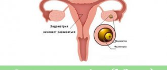
The most important changes
occurring in a woman’s body during this period are: contraction of the uterine muscle,
healing of the perineal wound after childbirth
and postoperative wound after cesarean section, as well as the beginning of milk secretion or lactation.
Immediately after childbirth, the process of involution begins, that is, contraction of the uterine muscle
.
Before pregnancy, the uterus is approximately the size of a fist; before childbirth, its bottom reaches the height of the ribs, and immediately after childbirth - at the level of the navel. From the moment of birth there is a gradual
return to the state in which it was before pregnancy. This is accompanied by cramping pain in the abdomen with various pain sensations. This is a rather unpleasant phenomenon, but every woman has to go through it.
There is also the so-called postpartum discharge.
At first, that is, in the first week after birth, they are bloody, sometimes with clots, then the discharge becomes pink, brown and
approximately 5-6 weeks
after birth. Hygiene is very important during this time to avoid infection.
It is worth remembering that uterine contractions and bleeding from the genital tract are very similar in women after cesarean section and in those who gave birth naturally.
During this period, you should refrain from physical activity and sexual intercourse. After giving birth you should wait at least 2 months
. In practice, this is an individual issue that should be discussed with your doctor.
Observation plan
The doctors of our IVF Center are convinced that the first day after childbirth, a woman should be in the clinic under close supervision, since it is during this time that most complications arise. If factors were identified during pregnancy that increase the likelihood of complications, the period of hospital stay may be increased. It is important to monitor the dynamics of basic blood parameters, the tone and degree of uterine contraction, and ensure that bleeding does not develop. For these purposes, regular examinations are carried out, instrumental examinations and laboratory tests are prescribed. Once the doctor is satisfied that there is no serious threat to the woman's health, she can be discharged.
Throughout the entire postpartum period, the likelihood of developing some complications remains, so our specialists tell the basic rules of behavior during this time and explain in what situations it is necessary to seek medical help. The general list of recommendations is as follows:
- Limiting physical activity.
- Refusal of sexual activity.
- Careful adherence to personal hygiene rules.
- Proper nutrition.
Antibiotics, painkillers, anti-inflammatory and other medications are taken only as prescribed by a doctor. Based on the individual characteristics of each woman, this list is adjusted individually. Strict adherence to these rules will help reduce the risk of complications, but cannot completely eliminate this risk. Therefore, you need to remember about the symptoms that may indicate the development of pathological conditions:
- Increased body temperature.
- Vaginal discharge with an unpleasant odor, with copious amounts of blood or pus.
- Severe pain in the lower abdomen.
- Redness, swelling and tenderness of the mammary glands.
Our doctors are ready to help women in the most difficult situations. Objective observation during the succession period and individual recommendations will help to avoid serious problems, as well as quickly eliminate many diseases at their very beginning. The best specialists and the entire range of necessary diagnostic equipment are at your service.
Proper wound care
Depending on the type of delivery, the woman
may have difficulty healing painful wounds in the perineum or abdomen after a cesarean section.
In both cases, care should be taken to ensure that the wounds heal properly. The most important thing in this case is hygiene and disinfection with drugs that accelerate healing.
In case of a cut wound to the perineum, be careful when sitting down.
For better healing of the perineal wound, you need to ventilate it more often. Bathing should be done in the shower, not in the bath
.
Especially in the first weeks, both after childbirth and after cesarean section. All these measures will prevent infection of the uterus.
Surgical treatment of postpartum hemorrhage
During vaginal birth, after taking conservative measures to stop bleeding, performing manual exploration and curettage of the uterus, if they are ineffective, the patient is transferred to the operating room for laparotomy and surgical stopping of bleeding.
During laparotomy, the presence of hemoperitoneum is assessed, which may indicate uterine rupture. In the absence of coagulopathy and the patient's stable condition, the first stage of surgical treatment is bilateral ligation of the uterine arteries. The second step is to ligate the hypogastric or internal iliac arteries. If the cause of bleeding is uterine atony, hemostatic compression circular sutures are applied to the body of the uterus to achieve hemostasis. If these measures are ineffective, a hysterectomy (postpartum hysterectomy) is performed.
If placenta accreta is found during cesarean section, the first step (after separation of the placenta) is to place hemostatic sutures on the placenta site. If the bleeding does not stop and there are no other causes of bleeding, the second step for an open uterus is to place circular sutures on the body of the uterus. If ineffective, the next step will be suturing the uterus (with or without tamponade) and ligation of the hypogastric arteries. If bleeding continues, a hysterectomy is performed.
If the bleeding is not massive, there is a reserve of time; if the patient’s condition is stable and there is a desire to preserve reproductive function, temporary uterine tamponade and further embolization of the uterine arteries under angiographic control can be performed.
With the development of consumption coagulopathy (DIC syndrome), a hysterectomy is performed with simultaneous rapid restoration of BCC and coagulation factors (fresh frozen plasma, cryoprecipitate, platelets, red blood cells, refortan, albumin, colloidal and isotonic solutions) under the control of hemostasis and coagulogram parameters.
Psychological aspect
A big problem for women
In addition to the physical changes occurring in the body, there are mental changes.
Stress, pain, and fatigue burden the psyche
of every young mother, often causing mood swings, nervousness and low self-esteem.
Therefore, she needs support and assistance both in caring for the newborn and in caring for her psychological comfort. These emotions are caused by hormonal changes
in the body, and this is completely normal.
However, if mood disorders
and discomfort persist for a long time, you should consult your doctor as they may lead to postpartum depression.
During the postpartum period, it is worth freeing the woman from household responsibilities
and providing additional support. Allow yourself to rest more, eat right and get enough sleep.
The postpartum period is definitely
, a very difficult time for a woman.
But if she has the right support and enough knowledge, she can go through it calmly and mindfully. Only then is it possible to fully enjoy motherhood.
That's why it's so important that every pregnant woman is well prepared for this wonderful, but often difficult time.
Ruptures of the genital tract
Vaginal lacerations and hematomas
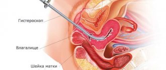
Immediately after birth, the mother's birth canal (perineum, labia, periurethral area, vagina, cervix) is examined in the speculum; Any tears found are sutured. Deep vaginal tears (up to the fornix) may be difficult to visualize, impinge on arterial vessels, and may cause noticeable bleeding or hematoma. To ensure adequate restoration of injuries to the birth canal, suturing is performed under adequate anesthesia (regional anesthesia).
Large hematomas are opened, injured vessels are found, stitched, and damaged vaginal tissue is restored. In some cases, extensive hematomas may form in the retroperitoneal space.
Clinical signs of such hematomas include back pain, anemia, and decreased hematocrit. The diagnosis is confirmed using ultrasound and, if necessary, computed tomography (CT). For small hematomas, a wait-and-see approach is chosen and anemia is treated. If the patient’s condition is unstable, surgical evacuation of the hematoma and ligation of injured vessels are performed.
Cervical ruptures . Cervical ruptures can lead to significant postpartum bleeding. The cause of these ruptures may be the rapid dilatation of the cervix in the first stage of labor or the beginning of the second stage of labor before the cervix is fully dilated. Immediately after birth, the cervix is examined in speculums using sequential application of fenestrated forceps following the movement of the clock hand. Sutures are repaired under adequate anesthesia (epidural, spinal or pudendal) with a continuous or interrupted suture using suture materials that are absorbable (absorbable).
Atony (hypotony) of the uterus
Uterine atony is the leading cause of postpartum hemorrhage. Increased risk of postpartum hemorrhage in patients with chorioamnionitis, when using magnesium sulfate, in multiple pregnancies, fetal macrosomia, a history of postpartum hemorrhage, in multiparous women (> 5 births). Developmental abnormalities and uterine fibroids can also affect the contractile function of the uterus and lead to atony and bleeding.
The diagnosis of uterine atony is determined by palpation of the uterus, which is soft, enlarged and weak. The fundus of the uterus may be contracted, and the lower segment may be relaxed. If uterine atony (hypotony) is suspected, Asos recommendations include the following:
1. Massage the uterus to stimulate its contractions and introduce an infusion of oxytocin (10-20 IU/l), which is usually also administered prophylactically; if ineffective, 20 IU of oxytocin is added intramuscularly. You should make sure that the bladder is stool (a full bladder prevents full contraction of the lower uterine segment). If bleeding continues, administer methylergonovine 0.2 mg IV or IM.
Maternal hypertension is a contraindication for the administration of methylergonovine. If bleeding continues, 250 mcg of PCP2a (contraindicated in bronchial asthma) is injected into the myometrium (transabdominal or transcervical) or administered no more than after 15 minutes with a maximum dose of 2 g. If ineffective, 1000 mg of misoprostol is administered rectally.
2. Examine the mother’s birth canal. If there are no visible ruptures, a careful manual examination of the uterine cavity is performed to remove retained products of the concept or to detect a uterine rupture (by the scar from a previous cesarean section). At the same time, an intensive infusion of colloidal (refortan) and isotonic solutions is carried out, and tests are performed before transfusion of fresh frozen plasma and blood.
3. If the bleeding continues, but is not rapid and massive, as an alternative to hysterectomy, if appropriate conditions are available, uterine artery embolization under radiological control can be performed. If delay is impossible or embolization is ineffective, laparotomy, ligation of the pelvic vessels (hypogastric, uterine, ovarian arteries) or hysterectomy are performed.
Remains of placenta and membranes
Immediately after birth, the placenta and membranes are thoroughly examined (integrity, presence of broken blood vessels, which may indicate an additional placental lobe). But with vaginal birth, it is often difficult to assess the retention of small parts of the placenta and membranes in the uterus. Typically, scraps of placental tissue and membranes emerge from the uterine cavity during postpartum contractions along with lochia. But the remains of concept products in some cases can lead to the development of endomyometritis and postpartum hemorrhage.
If remnants of the placenta and membranes are suspected in the postpartum period, a manual (if the cervix has not contracted) or, more often, instrumental inspection of the uterine cavity is performed. If after instrumental revision (curettage of the mucous membrane) of the uterus bleeding continues, placenta accreta is suspected.
Placenta accreta
Placenta accreta, as well as placenta accreta and placenta accreta, occur due to abnormal attachment of the placenta to the uterine wall, which can extend into the myometrium, leading to incomplete separation of the placenta from the uterine wall and the development of postpartum hemorrhage. Risk factors for placenta accreta include placenta previa and previous uterine surgery (cesarean section or myomectomy).
Clinical signs of placenta accreta may include a slowdown in the third stage of labor and fragmented separation of the placenta. If the duration of the third stage of labor exceeds 30 minutes, and there are no signs of separation of the placenta, perform manual separation and release of the placenta under adequate anesthesia. If the placenta is separated in fragments, a diagnosis of “placenta accreta” is made.
With placenta accreta, bleeding does not stop after uterine massage, the use of oxytocin, ergonovine and prostaglandins. If placenta accreta is suspected, treatment includes explorative laparotomy and surgical cessation of bleeding, which usually involves a hysterectomy. There are reports of cases of preservation of the uterus when fragments of the placenta are left in the uterus and subsequent successful treatment with methotrexate.
Uterine rupture
Uterine rupture may occur in 0.5-1% of patients with a previous uterine scar and in 1: (15,000-20,000) of women with an intact uterus. Uterine rupture can be traumatic (complicated childbirth, surgical vaginal delivery) and spontaneous (along a scar). This complication occurs during childbirth, but bleeding can develop in the postpartum period.
Uterine rupture is rare in nulliparous women (the uterus of primiparous women is “resistant” to rupture). Risk factors for uterine rupture include prior uterine surgery, fetal extraction in breech presentations, clinically narrow pelvis (disproportion between the fetal head and maternal pelvis), and an increased number of births in history. The classic clinical symptoms of uterine rupture are acute abdominal pain and a feeling of a “tear in the abdomen.” Treatment consists of urgent laparotomy, repair of the rupture, and, if surgical correction is not possible, hysterectomy.
Inversion of the uterus
Uterine inversion occurs when the fundus of the uterus is “born” through the cervix. Postpartum uterine inversion is rare (1:2000-1:2500 births). Risk factors for the underside of the uterus may include attachment of the placenta to the fundus of the uterus, uterine atony, placenta accreta, excessive traction on the umbilical cord in the third stage of labor. The diagnosis is determined by identifying the underside of the uterine fundus through the cervix, possibly with the placenta attached, at the birth of the placenta. Urgently perform manual separation of the placenta. In response to uterine inversion, the patient may experience a vasovagal reflex.
The doctor’s algorithm of actions after separation of the placenta in case of uterine incision includes stabilizing the patient’s condition, administering adequate anesthesia and restoring the position of the uterus (uterine reduction). To facilitate the reduction of the uterus, its relaxation is carried out using an infusion of beta-adrenergic agonists (terbutaline, ritodrine), magnesium sulfate or nitroglycerin. If it is impossible to manually reduce the uterus, a laparotomy is performed to surgically restore the position of the uterus using traction on the round ligaments. Sometimes, to restore the position of the uterine fundus, it is necessary to make a vertical incision of the myometrium.
How long does the postpartum period last?
The postpartum period for women begins after the birth of the child and lasts 6-8 weeks. This time is considered the final stage of pregnancy. The beginning of the period can be determined accurately, but there are no signs of its end, because the process of reverse restoration of all organs and systems occurs in women at different speeds and depends on a number of factors associated with:
- the constitution of the body;
- external conditions (rest, care, nutrition, sleep, etc.).