Pulpitis is an inflammation of the internal structures of the tooth, consisting of a neurovascular bundle and connective tissue. Develops as a complication of infectious, traumatic or other pathogenic processes. It manifests itself as severe throbbing pain, local and general increase in temperature, and pronounced tooth sensitivity. Most often it occurs with advanced caries with deep damage to dentin. Treatment is carried out by a dental therapist; in severe cases, the help of a dental surgeon is required.
Etiology of pulpitis and its symptoms
All provoking factors are conventionally divided into groups:
- mechanical - damage to the pulp due to acute or long-standing chronic injury;
- physical – irritation due to high temperatures;
- chemical or toxic - pathogenic effects of medications, filling materials and other irritating substances;
- biochemical - metabolic or age-related disorders with the deposition of calcium crystals in the pulp (the process of petrification, calcification with the formation of denticles);
- infectious - local (oral microflora) and general infections (brought in by blood).
Reference! According to statistics, the percentage of patients with pulpitis in dentistry reaches 14-20% of the total number of visitors. Among them, the most common form of inflammation is infectious; traumatic and iatrogenic are less common.
Infectious pulpitis
The entry of microorganisms into the dental cavity leads to the development of an infectious process. Routes of infection:
- caries - bacteria penetrate through the thinned layer of dentin;
- trauma – infection through cracks and chips in the area of the crown and root of the tooth;
- chronic periodontitis and periodontal disease - infection penetrates from deep gum pockets through the root canal opening;
- purulent inflammatory infections of the maxillofacial area (sinusitis, abscess);
- general infectious processes in the body - bacteria with the blood and lymph flow are able to penetrate any tissues and organs.
More than 19 types of microorganisms, both obligate and pathogenic, can act as pathogens. These are various types of staphylococcus and streptococcus, micrococci, Escherichia coli and Pseudomonas aeruginosa, diplococci, as well as fungi of the genus Candida, actinomycetes, and molds.
Traumatic pulpitis
Mechanical damage to the crown and root of a tooth occurs as a result of sports, household or industrial trauma, and less often - due to unsuccessful crown preparation. One of the reasons is exposure to high temperatures due to improper tooth preparation, when abrasive instruments cause tissue overheating and pulp burns.
Causes of traumatic pulpitis:
- fracture of the crown or root part of a tooth exposing the internal cavity;
- dislocation;
- tooth indentation;
- influence of denticles;
- opening of the dental cavity during preparation;
- the appearance of microcracks due to excessive vibration of the dental bur;
- bruxism against the background of increased tooth wear (can lead to exposure of the pulp horn).
Cracks and chips themselves do not always lead to inflammation, but they allow bacteria into the pulp cavity, which causes an infectious process. When the pulp is opened, the situation is more serious - acute inflammation develops with a high risk of post-traumatic necrosis, which, if left untreated, can occur within a week.
Iatrogenic pulpitis
Iatrogenic, or toxic form of pulpitis occurs as a response to irritation of the pulp by chemicals. Among them:
- acid pickles;
- aggressive antiseptics;
- adding medications for the treatment of deep caries;
- irritating filling materials when the backing is too thin or missing.
Causes of acute pulpitis
The following reasons lead to the occurrence of this disease:
- Untreated caries in time, as a result of which pathogenic microorganisms penetrate the tooth pulp and cause its inflammation.
- Poor treatment of caries can also lead to pulpitis. In this case, it can be caused by a thermal burn of the pulp, poor-quality filling materials, overdrying of the pulp tissue, or incomplete preparation of the carious cavity.
- Tooth injury. A fracture of the dental crown or its root, for example, due to injury or bruise, can cause a disruption of the blood supply to the pulp and an inflammatory process in it.
- Periodontitis. As a result of this disease, infection can penetrate into the pulp and cause retrograde pulpitis.
- In children, acute pulpitis can occur as a result of diseases such as scarlet fever, tonsillitis or measles.
Differential diagnosis of pulpitis
The main sign of pulp inflammation is severe toothache. It occurs spontaneously and is not associated with eating, brushing teeth, cold or mechanical stress. Often worries at night. At first, painful attacks appear several times a day for 10–20 seconds. Over time, their frequency increases and the pain becomes continuous. In an acute process, it radiates to other parts of the craniofacial zone, which complicates diagnosis.
Unlike pulpitis, with deep caries pain occurs only under the influence of external factors - when brushing teeth, while eating, when drinking cold or hot drinks. With periodontitis, the main difference is the localization of the source of inflammation outside the tooth itself and damage to adjacent tissues.
Symptoms
In addition to pain, you may experience:
- hyperemia;
- edema;
- bleeding of the pulp (at autopsy);
- suppuration;
- local and/or general increase in temperature;
- bad breath;
- darkening of the tooth due to pulp necrosis.
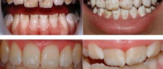
What does pulpitis look like in the photo?
In this section, clinic patients can see what tooth pulpitis looks like in the photo:
It is important to remember that dental pulpitis is a serious and dangerous disease that can cause a lot of discomfort and harm to health. However, with timely treatment of the disease, it is possible not only to save the tooth, but also to leave the pulp completely viable. Every patient can count on high-quality and effective treatment by making an appointment with a dentist. "Dent Academy", where the best specialists work, there is no place for negligence and mistakes.
Forms of pulpitis
There are 2 main forms of pathology:
- Acute – has pronounced symptoms of inflammation, lasts from 3 to 5 days. Inflammation is accompanied by circulatory disorders with hyperemia of blood vessels and expansion of their lumen. This increases blood flow and the accumulation of exudate in the area of inflammation - first serous, then purulent. If left untreated, it leads to the formation of first a pulp abscess, and then empyema of the entire tooth.
- Chronic – occurs during an advanced acute process, is characterized by sluggish symptoms and gradually leads to pulp necrosis.
Types of acute pulpitis
By area of damage there are:
- Focal - individual areas of the pulp chamber and its structures become inflamed. It is formed at the initial stage of the process and is a response to a local source of infection or injury.
- Diffuse - the entire coronal and root pulp is involved in inflammation. It becomes a continuation of untreated focal pulpitis or develops independently with generalized infections.
Depending on the course of the disease, there are 3 forms of the disease:
- Serous – the initial stage of inflammation with the accumulation of serous exudate.
- Purulent - pus begins to accumulate in the pulp chamber.
- Purulent-necrotic - against the background of hyperemia and suppuration, areas of tissue necrosis appear. In the case of putrefactive processes, wet gangrene develops, the pulp becomes black, and the nerve fibers disintegrate. As a result, the neurovascular bundle completely dies, the tooth acquires a dark shade, and the purulent inflammation itself can extend beyond the boundaries of the tooth.
On a note! Serous inflammation of the pulp usually turns into a purulent stage within 24 hours, so many authors identify a general serous-purulent form, which reflects the natural clinical picture of the development of the pathology.
Classification of chronic pulpitis
- Fibrous - there is no constant pain syndrome, but the tooth reacts to cold or hot. When tapping the crown, bleeding of the pulp is observed.
- Hypertrophic - the tooth does not hurt, does not react to hot/cold, even with severe destruction of the coronal part. Inside the cavity, granulation tissue grows to form a polyp.
- Gangrenous - the pulp begins to rot and die. The affected tooth darkens, and an unpleasant putrid odor appears from the mouth.
Pulpitis of wisdom tooth
The peculiarities of the location of the “eights” and the eruption pathologies characteristic of them provoke many problems. Among them are difficulties with care and a high risk of developing caries. If in the treatment of ordinary teeth the emphasis is placed on conservative methods with the preservation of the tooth and its pulp, then in the case of rudimentary wisdom teeth, dentists advise simply getting rid of the potential source of problems. The exception is cases when the “eight” has erupted completely and without damage, and the adjacent tooth is missing or has significant damage with a recommendation for removal.
Attention! An additional problem is the difficulty in diagnosing pulpitis. Against the background of painful eruption, the pain from pulpitis may go unnoticed. The patient (and often the dentist) may suspect something is wrong only if there are obvious complications.
Reviews about our doctors
I am very grateful to Evgeniy Borisovich Antiukhin for removing my three eights.
Especially considering that the lower tooth was not the simplest (it was located in an embrace with a nerve). The removal took place in 2 stages, one tooth under local anesthesia, two under general anesthesia. I had no idea that wisdom teeth could be... Read full review Sofia
28.12.2020
Words cannot express my gratitude to Elena Nikolaevna Kiseleva. This is the best doctor in the world. I got an appointment after many years of being ignored by the dentist’s office and with a bitter experience of treatment in another paid clinic, the mistakes of which had to be corrected in the first visits. Thank you for this... Read full review
Roman Stanislavovich Sh
25.07.2020
Complications
If pulpitis is neglected or treated incorrectly, concomitant pathologies arise:
- necrosis, or gangrene of neurovascular tissue;
- pulp degeneration with the formation of solid inclusions in the cavity - denticles, pulp stones;
- improper formation of hard structures - secondary or irregular dentin is actively formed.
If the infection extends beyond the dental cavity, the adjacent tissues of the maxillofacial region are involved in the pathological process. Diagnose:
- inflammation of the periosteum;
- abscess;
- osteomyelitis – purulent softening of bone tissue;
- phlegmon - purulent melting of soft tissues.
Treatment of pulpitis
2 methods are used:
- conservative (drug treatment, physiotherapy) – aimed at preserving the viability of the pulp;
- surgical – removal or amputation (partial removal) of the pulp, as well as complete removal of the entire tooth.
To preserve the pulp, conservative treatment methods are used. They are aimed at eliminating pain, eliminating the source of inflammation and restoring the functionality of the neurovascular bundle. In dentistry, this method is called biological and includes:
- drug treatment - antibacterial and anti-inflammatory drugs are prescribed;
- physiotherapy – electrophoresis, amplipulse, laser therapy, UHF therapy.
Important! The pulp nourishes the tooth tissue, maintaining its vitality. After it dies or is removed, the tooth becomes “dead”, its tissues lose strength and collapse faster (on average, such a tooth lasts no more than 5-8 years). Therefore, if possible, the pulp can be removed not completely, but partially.
If pulp removal (complete or partial) cannot be avoided, endodontic treatment is performed. Main stages:
- Anesthesia is given - usually local injection.
- The hard tissues of the tooth are prepared, removing all damaged structures and potential sources of infection.
- The pulp or its section is removed using vital or devital methods. In the first case, this is done “live”, without pre-treatment with destructive compounds, in the second, special means are used to pre-kill the tissue.
- Anti-inflammatory drugs are applied under the temporary filling to stop the further infectious process.
- The root canals are filled and a permanent filling is placed.
Surgical removal is resorted to if it is not possible to preserve the neurovascular structures. Vital amputation (partial removal) is performed only when treating multi-rooted teeth with a clear boundary between individual sections of the pulp.
Is it possible not to perform tooth depulpation?
Yes, in the early stages of the development of pulpitis, it is quite possible to treat it without removing the nerve bundle. But to do this, you should go to the doctor at the first signs of the disease, and not wait for it to go away on its own.
If possible, one should try to avoid pulp removal, since teeth without a nerve become more fragile and vulnerable to the action of bacteria and negative external factors. Moreover, in teeth depulped during the treatment of pulpitis, the enamel may lose its natural shine and whiteness.
If a person goes to the clinic for treatment of pulpitis, at a stage when the inflammation has already completely affected the nerve bundle, the nerve of the tooth will have to be removed. The dentist will remove the pulp from the tooth with a special tool, after which he will thoroughly rinse the tooth canals. We will describe in detail how this process works below.
Take a short test and calculate the cost of treatment!
Take a short test
- Which teeth have caries?
- Visual assessment
- Reaction to stimuli
- Cost calculation
×
Manukyan Artavazd Genrikovich
Chief physician of the clinic
Pulpitis of baby teeth
On baby teeth, inflammation of the neurovascular bundle has some features. This is due to the anatomical and physiological nuances of the structure of children’s teeth and the pronounced reactivity of the child’s body to various infections.
The pulp cavity of a baby tooth follows the shape of the tooth itself - there are pronounced pulp horns and a relatively thin layer of hard tissue on the chewing surface. This leads to the fact that with deep caries, pulpitis develops much faster than in adults. Additional complexity is created by the looser structure of dentin.
Attention! Parents often treat caries of baby teeth superficially, justifying it by saying that new teeth will grow anyway. But the danger of deep caries (and pulpitis in particular) lies in the high risk of damage to the rudiments of permanent teeth, which can become infected from the underlying tissues.
Prevention
Pulpitis can only be prevented by careful hygiene and timely treatment of any dental damage, from cracks, chips and caries to periodontitis. Basic preventive measures:
- Maintain good oral hygiene - in addition to brushing your teeth at home twice a day, use an irrigator and have your teeth professionally cleaned at a dental clinic.
- Eat right - a lack of calcium, phosphorus, vitamins C, D, group B in the diet, as well as microelements such as fluoride, leads to weakening of enamel and loosening of dentin, opening the way for the penetration of carious infection.
- Drink more water - a lack of this “universal solvent” in our body can cause a disruption in the acid-base balance, increased acidity of saliva and destruction of the enamel layer.
- Monitor the general condition of the body - monitor the course of chronic diseases. Control your hormonal levels and psycho-emotional state - most of the biochemical processes inside your body depend on them.
- Visit your dentist twice a year for preventive examinations.
All these efforts will help not only avoid tooth decay, but also contribute to the overall improvement of your health. Remember, a damaged tooth cannot be restored, and a new one cannot be grown in place of the lost one.
Symptoms
Inflammation of the pulp is not difficult to differentiate already at the stage of examining the oral cavity and interviewing the patient. This disease is characterized by:
- sudden onset of pain and its intensification at night;
- a gradual increase in pain intensity and duration in the first 3-5 days from the onset of the disease;
- irradiation (spread) of pain to the ear, eye, chin, depending on the location of the diseased tooth.
A characteristic feature of pulpitis, which makes it possible to distinguish it from caries and other dental pathologies, is a sporadic increase in pain, which is not associated with thermal, mechanical or other effects on the tooth.