Listeriosis is an infectious disease caused by gram-positive bacteria Listeria monocytogenes in humans and animals, Listeria ivanovii in animals.
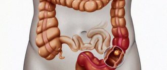
Listeriosis affects humans and animals (rodents, domestic cats and dogs, cows, sheep). Listeria can be infected through food by consuming meat, dairy products, soft cheeses, semi-finished products, seafood, vegetables and fruits, and can multiply in refrigerator conditions at a temperature of +4 - +6ₒ C and remain viable under vacuum packaging. They are stable in the environment: in water, soil. Capable of penetrating into the human body through the mucous membranes of the eyes and respiratory tract when inhaling dust that contains the pathogen. When Listeria crosses the placenta, it can infect the fetus.
Listeriosis is an opportunistic infection that affects people with a weakened immune system (mature people with chronic diseases, HIV-infected people, pregnant women, newborns), and those with congenital immune defects.
Listeriosis as an occupational disease can occur among veterinarians, meat processing and dairy workers, and livestock farms.
Classification and clinical picture of listeriosis
From the moment of infection with Listeria until the appearance of clinical symptoms, it can take from 2 to 4 weeks, sometimes up to two months.
Clinical symptoms are varied: there can be either an asymptomatic course or a severe septic condition.
The following forms of the disease are distinguished:
- Glandular form. Characterized by fever, headache, weakness, and enlarged lymph nodes. There may be plaque on the tonsils, unilateral purulent conjunctivitis.
- The nervous form most often develops in children and the elderly and is manifested by meningitis and meningoencephalitis. Against the background of high temperature, severe headache and impaired consciousness appear.
- Septic form. Muscle and joint soreness, hepatitis, and pneumonia are also present.
- Listeriosis in pregnancy can develop at any stage of pregnancy. It is usually asymptomatic or mild with a favorable outcome. The fetus becomes infected with listeriosis through the placenta with the development of intrauterine infection. If infected in the early stages of pregnancy, developmental defects and intrauterine fetal death may occur.
- Listeriosis in newborns occurs unfavorably, in a severe septic form, the mortality rate reaches 20%.
Causes of listeriosis in pregnant women
- The main cause of listeriosis in pregnant women is the consumption of foods infected with listeria - unboiled milk and products made from it, including soft cheeses, butter and ice cream, semi-finished meat products, sausages and sausages, cold cuts and other delicacies in vacuum packaging, seafood and salmon grown in artificial conditions, contaminated fruits and vegetables, including frozen ones, fast food products that have not undergone proper heat treatment before consumption.
- The high incidence of listeriosis is due to a decrease in the level of cellular immunity in women during pregnancy.
- A feature of the disease is that bacteria can hide inside the host cells for a long time, which makes them inaccessible for diagnosis and treatment.
- Listeria infection in a woman’s body can persist in the kidneys for quite a long time and become more active during pregnancy against the background of decreased immunity.
- Pregnant women with listeriosis often have a burdened obstetric and gynecological history: chronic adnexitis, cervical erosion, spontaneous and induced abortions. This category of patients is subject to mandatory screening for listeriosis.
Rice. 2. Products that can be harmful to humans.
Laboratory diagnosis of listeriosis
Laboratory diagnosis is based on identifying listeria from biological material: blood, cerebrospinal fluid, eye discharge, oropharynx, nasopharynx, feces. For diagnosis, the bacteriological method and the polymerase chain reaction (PCR) method are used.
But it must be taken into account that listeria can be detected in stool in healthy individuals, so detection of it in stool by PCR without clinical symptoms does not serve as a basis for diagnosis. But pregnant women who are found to have listeria in their stool and urine, even in the absence of symptoms of disease, are subject to treatment.
- Microbiological (cultural) study on Listeria (Listeria monocytogenes) with determination of sensitivity to antimicrobial drugs.
- Listeria (Listeria monocytogenes), qualitative determination of DNA (CSF, AF) - allows you to detect listeria in the cerebrospinal fluid or amniotic fluid.
- Listeria (Listeria monocytogenes), qualitative determination of DNA (whole blood).
- Listeria (Listeria monocytogenes), qualitative determination of DNA (feces)
Congenital listeriosis
• significant place of IUI in the structure of the causes of the pathological course of pregnancy and its unfavorable outcomes (miscarriages, stillbirths, premature births); • pathological influence of IUI on embryophetogenesis, leading to the development of congenital defects and/or infectious and inflammatory lesions of the central nervous system (CNS), heart, liver, kidneys and other organs and systems in the fetus; • high mortality rate among newborns and infants with manifest forms of IUI; • adverse impact of IUI on the health of children in subsequent periods of postnatal development, in some cases leading to disability and a decrease in the quality of life in general [1–4,7–9,11]. The true frequency of congenital infections has not yet been established, but, according to a number of authors, the prevalence of this pathology in the human population can reach more than 10%. IUIs develop as a result of intrauterine infection. It should be especially emphasized that in the vast majority of cases, the source of infection in IUI is the mother, from whom pathogens penetrate to the embryo and/or fetus in the ante- or intranatal periods [1,4,9,11]. The range of etiological agents of IUI is very wide. Most often, IUIs are caused by viruses (cytomegalovirus, herpes simplex viruses, rubella, enteroviruses, etc.) and other intracellular pathogens (toxoplasma, chlamydia, mycoplasma, etc.). In addition, it has been convincingly shown that IUIs can also be of a bacterial nature. Thus, in cases where a woman suffers diseases such as syphilis or gonococcal infection during pregnancy, there is a very high risk of intrauterine infection with Treponema pallidum and Neisseria gonorrhoeae, respectively, with the development of a corresponding congenital infection [1,8,9]. The potential danger of intrauterine infection of the fetus with a bacterial pathogen such as Listeria monocytogenes has also been known for a long time. It should be especially emphasized that congenital listeriosis has been studied quite well and completely. Its clinical, epidemiological and pathomorphological features have been determined, principles of diagnosis, treatment and prevention have been developed [1,3,5,8,9,12,14]. However, at present, alertness regarding congenital listeriosis among practicing obstetricians, gynecologists and neonatologists (especially those serving the urban population) has decreased significantly. This is likely due to a number of factors, the main ones of which are presented below. Firstly, there is no reliable information about the spread of listeriosis in Russia, despite the mandatory registration of cases of this infection, regulated in the Russian Federation since 2002. Secondly, ideas about the exclusive connection of listeria infection with living in rural areas and mandatory established contact of the sick person with domestic and/or wild animals are still dominant, although it has been convincingly shown that infection with Listeria monocytogenes can also occur in urban conditions if infected animals are consumed products (cheese, meat, milk, etc.) that have not undergone adequate heat treatment. It is also impossible not to pay attention to the fact that listeria is not detected during routine bacteriological examination, because This requires special environments. At the same time, the low demand from clinicians for tests for listeriosis has led to the fact that many hospital bacteriological laboratories are no longer equipped with special media for the detection of Listeria monocytogenes. A vicious circle arises - there is no true information about the frequency of listeria infection, because Research is not carried out in the proper amount to verify it, and research, in turn, is not carried out, because Practitioners do not prescribe them due to a lack of alertness regarding listeriosis. As a result, it turns out that the etiological significance of Listeria monocytogenes in the genesis of TORCH syndrome is rarely discussed, even in cases where there are typical manifestations of congenital listeriosis (exanthema, fever, meningitis). All of the above determined the need for this publication. Congenital listeriosis is IUI caused by Listeria monocytogenes. Intrauterine infection with Listeria often leads to miscarriages, stillbirths, the development of fetal defects, as well as serious infectious and inflammatory lesions of various organs and systems. After birth, such children in the neonatal period experience severe meningitis, pneumonia, and sepsis, which are characterized by a high mortality rate. In general, it should be especially noted that congenital listeriosis is a serious disease with a serious prognosis, which determines significant perinatal losses [1,5,8,9,11]. The causative agent of listeriosis, Listeria monocytogenes, is a short, gram-positive, non-spore-forming rod of regular shape, which is a facultative anaerobe. Listeria is highly stable in the external environment, growing in a wide temperature range (from +1 to +45°C) and high salt concentration. This explains the fact that listeria can grow both in foods that are refrigerated and remain viable in brine. When heated to 70°C, listeria dies in 29–30 minutes, and when the temperature reaches 100°C, in 3–5 minutes. The growth of Listeria is suppressed by ampicillin, gentamicin, chloramphenicol and erythromycin, while the pathogen exhibits varying degrees of resistance to cephalosporins, sulfonamides and polymyxin B [5,6,12,14]. If previously listeriosis was considered a typical zoonosis, now it is classified as a sapronotic disease, and the main source and reservoir of the infectious agent are environmental objects and natural substrates in which listeria is capable of multiplying [6]. The main route of human infection with listeria is through food. People become infected by consuming contaminated food that has not been adequately processed. Particularly noteworthy is the importance of heat treatment for products such as meat, milk, and cheeses. Fast food products (hamburgers, hot dogs, etc.) also pose an increased risk. In addition to nutrition, other ways of infecting a person with listeria are also possible. Among them, the most significant are contact (from infected animals) and aerogenic (when working with contaminated skins and wool) [5,6,12,14]. Of particular importance is the possibility of transmission of Listeria from an infected pregnant woman to her fetus. It should be noted that listeriosis in pregnant women can occur not only manifestly (both with a typical clinical picture and in the form of an undifferentiated influenza-like syndrome), but also as an asymptomatic carrier of the bacilli. In all these cases, there is a risk of transplacental transmission of infection. When Listeria penetrates the placenta and fetal membranes, conditions are created for intrauterine contamination. Subsequently, hematogenous and lymphogenous spread of Listeria monocytogenes occurs in the fetus, affecting various internal organs and the central nervous system. In the tissues where the pathogen accumulates, specific granulomas are formed - listeriomas [3,8]. The formation of granulomas is due to the peculiarities of the immune response to Listeria monocytogenes [13]. It has been established that due to the fact that cellular immunity cannot ensure adequate elimination of listeria that has penetrated into tissues, T-lymphocytes rush to areas where the pathogen accumulates. Stimulation of T lymphocytes is accompanied by active production and release of cytokines. Under the influence of cytokines, the attraction of other lymphocytes to the affected area increases (migration of both CD4+ and CD8+ cells is noted) and macrophages, which subsequently determines the formation of granulomas. Chronic stimulation of macrophages and hyperproduction of cytokines in the area of granulomas leads to pronounced vascular and dystrophic changes in the affected organs and tissues. It is assumed that the maximum development of granulomas is associated with T-helper-1 cytokines [13]. In congenital listeriosis, the granulomatous process is generalized and is considered as granulomatous sepsis [3,8]. At the autopsy of those who died from congenital listeriosis, all organs on the surface or in the section are as if “sprinkled with millet” due to the abundance of granulomas. In this case, whitish-grayish, yellowish-grayish granulomas are found under the pleura, in the lungs, under the liver capsule, in the kidneys, under the pia mater, in the substance of the brain, in the spleen, lymph nodes, intestines, stomach, adrenal glands, thymus. Microscopically, productive vasculitis is observed in the skin, and in the liver - multiple submiliary foci of hepatocyte necrosis with severe hyperplasia and proliferation of stellate endothelial cells, in place of which granulomas are formed [3,8]. Transplacental infection of the fetus with Listeria is possible throughout pregnancy. It has been noted that when infected in the early stages of gestation, spontaneous abortions occur or developmental defects are formed. When infected in the second half of pregnancy, as a rule, a specific infectious-inflammatory process develops in utero. In this case, the child is born with clinical manifestations of congenital listeriosis, or the manifestation of the disease occurs during the first or second day of life. At the same time, attention is paid to the serious condition of the newborn, the appearance of fever and exanthema, represented by papular elements with a hemorrhagic component. Less commonly, the rash has a roseolous character. The child's condition progressively worsens over several hours, anxiety, shortness of breath, cyanosis appear, respiratory failure increases, and convulsions develop. The disease becomes septic in nature and is characterized by a high (more than 50%) mortality rate [1,9,11,12]. In addition to transplacental transmission, transmission of infection during the intrapartum period is also possible. In this case, infection occurs due to the child’s contact with the mucous membranes of the mother’s birth canal contaminated with Listeria monocytogenes. Infection through aspiration of infected amniotic fluid is also possible. For intrapartum infection, the development of the disease in a later period after birth is typical. Thus, in most cases, the clinical picture manifests itself on the 10th–12th day of life and usually occurs in the form of meningitis. Less commonly, the onset of the disease is manifested by symptoms of pneumonia. In general, listeriosis caused by intrapartum infection, as well as by transplacental transmission of infection, is characterized by a severe course and has a high mortality rate (20–25%). At the same time, it was noted that with intrapartum infection with Listeria monocytogenes, transient colonization is also possible, which does not lead to the development of the disease. It should be noted that it is quite difficult to establish a diagnosis of congenital listeriosis based on clinical and anamnestic data. Laboratory research methods are of decisive importance in this regard. At the same time, bacteriological examination is traditionally considered the “gold standard” for diagnosing congenital listeriosis. In this case, a preliminary conclusion can be obtained quickly - based on bacterioscopy of Gram-stained smears of cerebrospinal fluid sediment, amniotic fluid, tracheobronchial aspirate and other biological fluids. However, it is necessary to differentiate between Listeria, Streptococcus and Corynebacteria. The definitive bacteriological conclusion confirming listeriosis is based on the isolation of Listeria monocytogenes from crops. It should be emphasized that Listeria colonizes weakly on ordinary media. Therefore, to isolate Listeria it is necessary to use special media. Thus, it has been established that Listeria grows well on meat-peptone liver media with the addition of glucose, serum, glycerol and blood. However, as practice shows, in recent years, due to the low demand for tests for listeriosis (especially in urban areas), hospital bacteriological laboratories are rarely equipped with media for verifying listeriosis. In addition, one cannot fail to note the fairly long period (3–7 days) required for a full bacteriological examination. This significantly limits the ability of clinicians to quickly establish a diagnosis and timely prescribe etiotropic therapy. These disadvantages can be eliminated if molecular biological methods are used to diagnose congenital listeriosis, in particular polymerase chain reaction (PCR) [10]. It should also be noted that it is possible to use serological methods for identifying listeria infection. However, in recent years, traditional serological tests such as RPHA, RSK, RA are increasingly used to verify congenital listeriosis, while ELISA with the determination of specific antibodies in the IgM and IgG classes in “paired sera” is considered a promising diagnostic method [12] . Treatment of children with congenital listeriosis is very difficult and should begin as early as possible. Therefore, the deterioration of the child’s condition on days 1–2 of life, accompanied by the appearance of fever, exanthema in the form of papular or roseolous rashes, anxiety, shortness of breath, cyanosis, hepatomegaly, and convulsions requires an urgent differential diagnosis and clarification of the genesis of the disease. At the same time, among the probable reasons for the deterioration of the child’s condition, it is necessary to take into account the possibility of developing congenital listeriosis. For emergency verification of the genesis of the disease, it is advisable to use PCR, which has a high level of diagnostic specificity and sensitivity and allows you to quickly (within a few hours) obtain the final results of the etiological decoding [10]. At the same time, it is considered justified, without waiting for laboratory results, to empirically prescribe starting antibiotics if clinical and epidemiological data indicate possible listeriosis. In this case, the antibacterial therapy of choice for congenital listeriosis is a combination of ampicillin and gentamicin (Table 1). It should be noted that the ampicillin dosage regimen depends not only on the age of the child, but also on the characteristics of the clinical form of the disease. Thus, for congenital listeriosis, which occurs with damage to the meninges, ampicillin is prescribed: in the early neonatal period - 100–200 mg/kg/day. in 2 injections with an interval of 12 hours, and for children over 7 days old - at the rate of 300–400 mg/kg/day. in 3-4 doses. In cases where congenital listeriosis occurs without meningitis, ampicillin is prescribed: in the early neonatal period - 100 mg/kg/day. in 2 injections, and for children over 7 days old - at a dose of 100–200 mg/kg/day, which is administered in 3–4 doses (Table 1). The duration of ampicillin use for congenital listeriosis occurring with meningitis is 14–21 days. If the disease occurs without damage to the meninges, then ampicillin is prescribed for 14 days [1,11,12]. For congenital listeriosis, gentamicin is prescribed together with ampicillin to potentiate the antibacterial effect aimed at eliminating Listeria monocytogenes. At the same time, a single dose of gentamicin does not depend on the form of the disease and the age of the child and is 2.5 mg/kg, while the frequency of administration of the drug has a clear age dependence. So, if children during the first 7 days of life are given gentamicin at a dose of 2.5 mg/kg with an interval of 12 hours, then for children over 7 days old - 2.5 mg/kg with an interval of 8 hours (Table 1). It should be especially noted that the duration of gentamicin therapy should not exceed 7 days [1,11,12]. Considering that congenital listeriosis, even with timely diagnosis and early initiation of therapy, is characterized by a high mortality rate, the main efforts should be aimed at preventing this disease. At the same time, the basis for the prevention of congenital listeriosis is to prevent women from becoming infected with Listeria monocytogenes during pregnancy. Considering that the nutritional route is the main route for Listeria to enter the human body, it is necessary to use in food only those products that have been subjected to adequate processing that strictly meets sanitary and hygienic requirements. In this case, special attention should be paid to the inadmissibility of using meat and dairy products that have not undergone adequate heat treatment. For women living in rural areas, having animals in the household, engaged in animal husbandry and processing of hides, wool, etc., compliance with basic sanitary and hygienic requirements is very important. The prevention of infectious diseases in general and listeriosis in particular largely depends on the sanitary culture of society. However, it is obvious that at the present stage, sanitary education work carried out by traditional methods does not allow actively promoting hygienic knowledge among the population and therefore must be revised. The promising areas in this regard should be considered sanitary -hygienic inserts (as social advertising) in the programs of central and local television and radio channels, popular publications in magazines for adolescents, young parents, on the Internet, etc. In cases where, during pregnancy, a woman with anamnestic risk factors for infection Listeria Monocytogenes develops, the clinical picture of which resembles a leaferiosa infection or there is a flu -like syndrome, it is necessary to conduct a targeted examination for listeriosis. In case of confirmation of the diagnosis, the immediate prescription of pregnant etiotropic therapy is shown (at the same time, ampicillin is more often used), which can significantly reduce the risk of intrauterine infection and development of congenital leatherosis.
Literature 1. Congenital, perinatal and neonatal infections: Transl. from English / Ed. A. Greenough, J. Osborne, S. Sutherland. - M.: Medicine, 2000. 2. Zaplatnikov A.L., Korovina N.A., Korneva M.Yu., Cheburkin A.V. Intrauterine infections: diagnosis, treatment, prevention. Attending doctor. – 2005. – No. 8. – pp. 54–62. 3. Ivanovskaya T.E., Leonova L.V. Pathological anatomy of diseases of the fetus and child. – M.: Medicine, 1989. 4. Protocols for diagnosis, treatment and prevention of intrauterine infections in newborns of the Russian Association of Perinatologists (methodological recommendations). – M.: GOU VUNMC Ministry of Health of the Russian Federation, 2001. 5. Tartakovsky I.S., Maleev V.V., Ermolaeva S.A. Listeria: role in human infectious pathology and laboratory diagnostics. – M., 2002. 6. Uchaikin V.F., Nisevich N.I., Shamsheva O.V. Guide to infectious diseases in children. – M., 2007. 7. Tsaregorodtsev A.D., Ryumina I.I. Morbidity of newborns with intrauterine infections and tasks to reduce it in the Russian Federation // Ros. Vestn. perinatol and pediatrician. – 2001. – No. 2. – P. 4 – 7. 8. Tsinzerling V.A., Melnikova V.F. Perinatal infections. Questions of pathogenesis, morphological diagnosis and clinical and morphological comparisons. Guide for doctors. – St. Petersburg: Elbi-St. Petersburg, 2002. 9. Shabalov N. P. Neonatology. – St. Petersburg, 2006. 10. Shipulina O.Yu., Piksasova O.V., Sadova N.V. and others. The importance of molecular biological methods in the diagnosis of listeriosis in pregnant women and newborns. – M., 2008 11. Infections Diseases of the Fetus & Newborn (Ed. JS Remington, JO Klein). – WBSaunders Company, 1995. 12. Red Book: Report of the Committee on Infectious Diseases. 27h ed. Elk Grove Village, IL: American Academy of Pediatrics, 2006; 992. 13. Roitt I., Brostoff J., Male D. Immunology. 5th ed. – Mosby International Ltd., 1998. 14. Wing EG, Gregory SH Listeria monocytogenes Clinical and experimental update. J. Inf. Dis. 2002, 185 (suppl. 1): 18–24.
How does listeriosis spread?
Listeriosis infection occurs:
- when eating raw or insufficiently heat-treated contaminated products of animal origin (dairy products, meat and poultry), vegetables, fruits and seafood;
- through inhalation of dust containing the pathogen;
- through contact with a sick animal (or with an animal carrying listeria);
- through the placenta or through contact of the newborn with the birth canal of the woman in labor;
- through contact of newborn children with infected care items and medical instruments in maternity hospitals.
Symptoms of listeriosis in pregnant women
Listeriosis in pregnant women most often develops in the third trimester. The insidiousness of listeriosis lies in the fact that the disease occurs easily - in the form of an acute respiratory viral infection with symptoms of mild intoxication, symptoms of inflammation of the urinary tract and/or symptoms of gastroenteritis, sometimes tonsillitis or purulent conjunctivitis. Rarely, the infection affects the central nervous system.
Pregnant women with symptoms suspicious for listeriosis and with a complicated obstetric-gynecological diagnosis should be examined for the presence of this infection.
In some cases, listeriosis in pregnant women is asymptomatic and remains unrecognized throughout pregnancy. The disease is diagnosed after spontaneous abortion or premature birth.
Most often, listeriosis occurs in a chronic form in pregnant women. During exacerbations, short-term fever and mild catarrhal symptoms may occur. Sometimes exacerbations occur with symptoms of chronic pyelonephritis or dyspeptic disorders.
In pregnant women with severely reduced immunity, listeriosis is severe. The septic form of the disease is characterized by a significant increase in body temperature, diarrhea, and cramping abdominal pain. If the fetus dies, the pregnancy is terminated, after which the patient’s body temperature returns to normal and does not subsequently return.
In pregnant women with listeriosis, chorioamnionitis develops - inflammation of the membranes of the fetus with subsequent infection of the amniotic fluid (amniotic fluid), which leads to premature birth or stillbirth; in the fetus, the infection damages many organs and systems.
From the affected fetus, the infection again enters the pregnant woman’s body. This phenomenon is called a “ping-pong infection.” Repeated infection worsens the course of listeriosis.
Rice. 3. Hydrocephalus (photo on the left) and microcephaly (photo on the right) are fetal malformations that are often found with listeriosis.
Obstetric pathology for listeriosis in pregnant women
Listeriosis in pregnant women has a negative effect on the fetus and causes severe obstetric pathology:
- Habitual miscarriage (miscarriage).
- Early termination of pregnancy.
- Premature and term birth.
- Stillbirth.
- Death of a pregnant woman.
Rice. 5. Premature birth - obstetric pathology due to listeriosis in pregnant women.
What foods contain listeria?
The causative agent of listeriosis persists for a long time at low temperatures and sufficient humidity, multiplies at 4-6 ° C in soil, water and in some food products (meat, including those packaged in barrier films, fish, seafood, milk, vegetables and sauerkraut). The greatest dangers come from raw cabbage salads, soft cheeses and processed meats.
Storing food in the refrigerator does not kill the dangerous pathogen: listeria is often called the “refrigerator germ” because it is able to survive in these conditions. Listeria has another insidious property: the ability to multiply in pickles. If most microbes die under these conditions, then listeria can withstand table salt concentrations of up to 6-20%.
Listeria dies at boiling temperatures, and when exposed to sunlight, the causative agent of the disease will live no more than 15 days.