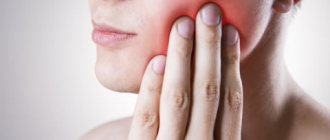
Dental diseases of the teeth are a group of pathologies that affect the enamel, gums, oral cavity, as well as the jaw and chewing muscles. Dentists call them harmless to humans, since the mortality rate is negligible. However, it is these disorders that determine the patient’s overall well-being: they affect self-esteem and an internal sense of comfort.
Experts recommend not to let the situation get worse and to closely monitor the condition of your teeth. Due attention allows you to avoid serious complications, as well as promptly identify defects that need correction.
In this article we examine in detail the most common problems, as well as methods of treating them.
Basic terms in dentistry: names and causes of inflammatory diseases of the teeth and oral cavity
It plays a vital function in the digestion process: it is responsible for grinding food and influencing its subsequent breakdown by enzymes. In addition, there is a huge number of microorganisms that create natural microflora, but are considered opportunistic. This nuance determines the fact that any dental disorders progress extremely quickly.
Among the key factors in the occurrence of dental diseases are:
- poor hygiene;
- mechanical injuries and damage;
- chronic illnesses;
- metabolic disorders and abnormalities;
- the presence of thermal irritants in the body;
- bad environment;
- hereditary predisposition.
The branch of medicine that studies the emergence and development of pathologies is called etiology. It is partially correlated with diagnostic procedures, as it helps in determining pathogenesis.
ODONTOGENIC AND PERIODONTAL INFECTION
PULPITIS
Main pathogens
Viridans streptococci ( S. milleri
), non-spore-forming anaerobes:
Peptococcus
spp.,
Peptostreptococcus
spp.,
Actinomyces
spp.
Choice of antimicrobials
Antimicrobial therapy is indicated only in case of insufficient effectiveness of dental procedures or spread of infection into surrounding tissues (periodontal, periosteal, etc.).
Drugs of choice
: phenoxymethylpenicillin or penicillin (depending on the severity of the disease).
Alternative drugs
: aminopenicillins (amoxicillin, ampicillin), inhibitor-protected penicillins, cefaclor, clindamycin, erythromycin + metronidazole.
Duration of use
: depending on the severity of the current (at least 5 days).
PERIODONTITIS
Main pathogens
Microflora is rarely detected in the periodontal structure and is usually S.sanguis, S.oralis, Actinomyces
spp.
In periodontitis in adults, gram-negative anaerobes and spirochetes predominate. P.gingivalis, B.forsythus, A.actinomycetemcomitans and T.denticola
are highlighted most often.
In juveniles, there is rapid involvement of bone tissue in the process, with A. actinomycetemcomitans
and
Capnocytophaga
spp.
P.gingivalis
is rarely isolated.
In patients with leukemia and neutropenia after chemotherapy along with A. actinomycetemcomitans
C.micros
is isolated , and in prepubertal age -
Fusobacterium
spp.
Choice of antimicrobials
Drugs of choice
: doxycycline; amoxicillin/clavulanate.
Alternative drugs
: spiramycin + metronidazole, cefuroxime axetil, cefaclor + metronidazole.
Duration of therapy
: 5-7 days.
For patients with leukemia or neutropenia after chemotherapy, cefoperazone/sulbactam + aminoglycosides are used; piperacillin/tazobactam or ticarcillin/clavulanate + aminoglycosides; imipenem, meropenem.
Duration of therapy
: depending on the severity of the course, but not less than 10-14 days.
PERIOSTITIS AND OSTEOMYELITIS OF THE JAWS
Main pathogens
With the development of odontogenic periostitis and osteomyelitis, S. aureus
, as well as
Streptococcus
spp., and, as a rule, anaerobic flora prevails:
P.niger, Peptostreptococcus
spp.,
Bacteroides
spp.
Specific pathogens are less commonly identified: A.israelii, T.pallidum
.
Traumatic osteomyelitis is often caused by the presence of S.aureus
, as well as
Enterobacteriaceae
spp.,
P.aeruginosa
.
Choice of antimicrobials
Drugs of choice
: oxacillin, cefazolin, inhibitor-protected penicillins.
Alternative drugs
: lincosamides, cefuroxime.
When P.aeruginosa
, antipseudomonas drugs (ceftazidime, fluoroquinolones) are used.
Duration of therapy
: at least 4 weeks.
ODONTOGENIC MAXILLARY SINUSITIS
Main pathogens
The causative agents of odontogenic maxillary sinusitis are: non-spore-forming anaerobes - Peptostreptococcus
spp.,
Bacteroides
spp., as well as
H.influenzae, S.pneumoniae
, less often
S.intermedius, M.catarrhalis, S.pyogenes
.
Isolation of S.aureus
from the sinus is characteristic of nosocomial sinusitis.
Choice of antimicrobials
Drugs of choice
: amoxicillin/clavulanate. For nosocomial infection - vancomycin.
Alternative drugs
: cefuroxime axetil, co-trimoxazole, ciprofloxacin, chloramphenicol.
Duration of therapy
: 10 days.
Classification: types of main dental diseases of the teeth
Conventionally, all violations can be divided into two broad categories - those associated with damage to the enamel, and those caused by processes occurring in periodontal tissues. There is another classification principle - based on “carious” affiliation:
- with existing caries lesions;
- non-carious deviations.
The second option is considered the simplest and most convenient to treat if the problem is detected at an early stage. In addition, it is easier for patients to comply with prescriptions, which simplifies the process of rehabilitation measures.
Note that congenital and hereditary pathologies stand apart. They are formed in the womb and are very difficult to correct.
Treatment
The primary diagnosis of the disease based on clinical manifestations is carried out by a dentist, who also carries out local symptomatic treatment based on the primary diagnosis. But in complex and unobvious cases, laboratory diagnostics are carried out, and doctors of other specialties (allergists, dermatologists, immunologists) are brought in for treatment.
Local symptomatic treatment usually involves the use of rinses with solutions of painkillers and antiseptic drugs (including herbal origin), the use of drugs for tissue regeneration (after the acute phase subsides), if necessary, the elimination of irritating and provoking factors (removal of dental plaque, hard deposits, treatment dental caries).
Local drug treatment is carried out only after an accurate diagnosis has been established (the cause of stomatitis) and usually includes treatment with antiviral, antibacterial, antifungal drugs in the form of ointments and applications.
General treatment is carried out, as a rule, in a hospital and is aimed at eliminating the internal causes of stomatitis (treatment of internal pathologies), taking therapeutic drugs according to special regimens, and physiotherapy.
Developmental and eruption disorders
What are the different dangerous dental diseases? Perhaps the most serious problems are caused by congenital or hereditary causes. In these cases, the exacerbation does not begin due to pathogenic bacteria, pathogens or other factors. Such pathologies, as a rule, play a symptomatic role. They signal about malfunctions occurring in the body and force you to take immediate action.
Edentia
A pathological process accompanied by the loss of individual units or complete rows. In extremely severe stages, complete tooth loss occurs. The disease begins to develop during intrauterine development. The main factor is poor heredity or serious illnesses suffered by the mother while carrying the fetus. Due to jaw overload, even healthy teeth can become loose.
Superset
A disease in which excess elements appear in the oral cavity, which have an extremely negative effect on the state of the bite, and provokes additional speech and chewing problems. Dentists recommend removing such units, and the sooner the better.
Fusion and Merger
A rather rare deviation, expressed in the connection of tooth buds with dental cement. The main processes unfold during the formation of elements. Only 1% of people are prone to this disease. As a rule, the disease is diagnosed as part of a temporary dentition. In adulthood, pain is approximately 5-6 times less, so patients often do not suspect the presence of a problem until a visit to the dentist.
Spear-shaped teeth
A disease that affects crowns. It is considered infrequent - it occurs in one case in a thousand. It causes both aesthetic, psychological and physiological inconvenience. Usually it is a consequence of chronic injury to the inner surface of the cheeks, lips, and tongue. In some patients it is accompanied by hypoplasia.
Impacted teeth
An ailment in which the teeth are unable to fully erupt. As medical practice shows, the problem begins to bother people due to the gums being blocked by bone layers. Another reason is premature loss of elements.
Dystopian teeth
A disease in which the incorrect location of units is recorded. The displacement occurs as a result of deviations of the alveolar ridges from the norm. It causes psychological discomfort, disrupts the beauty of a smile, and interferes with the correct functioning of the masticatory muscles.
Macrodentia and microdentia
Congenital hereditary anomalies that determine the specifics of development. Manifested in improper formation and scaling of teeth. They have a negative impact on the bite and the aesthetics of the smile. They are also extremely rare - in every 20 people (in the context of not a single state, but the whole world).
NON-ODONTOGENIC AND SPECIFIC INFECTION
NECROTIC STOMATITIS (VINCINS ULCER-NECROTIC GINGIVOSTOMATITIS)
Main pathogens
Fusobacterium is concentrated in the gingival sulcus
, pigmented
Bacteroides
, anaerobic spirochetes. With necrotizing stomatitis, there is a tendency for infection to quickly spread into surrounding tissues.
The causative agents are F. nucleatum, T. vinsentii, P. melaninogenica, P. gingivalis and P. intermedia
.
In patients with AIDS, C. rectus
.
Choice of antimicrobials
Drugs of choice:
phenoxymethylpenicillin, penicillin.
Alternative drugs:
macrolide + metronidazole.
Duration of therapy:
depending on the severity of the current.
ACTINOMYCOSIS
Main pathogens
The main causative agent of actinomycosis is A.israelii
, association with gram-negative bacteria
A. actinomycetemcomitans
and
H. aphrophilus
, which are resistant to penicillin but sensitive to tetracyclines, is also possible.
Choice of antimicrobials
Drugs of choice
: penicillin at a dose of 18-24 million units/day, with positive dynamics - transition to step-down therapy (phenoxymethylpenicillin 2 g/day or amoxicillin 3-4 g/day).
Alternative drugs
: doxycycline 0.2 g/day, oral drugs - tetracycline 3 g/day, erythromycin 2 g/day.
Duration of therapy
: penicillin 3-6 weeks IV, drugs for oral administration - 6-12 months.
Diseases associated with enamel
What are the possible dental problems due to enamel damage? In the vast majority of cases, they are associated with violations of the structure and integrity of the rows. Usually they are a consequence of internal failures in the body. They are eliminated only at the moment when the factor that provoked their exacerbation disappears.
Fluorosis
A chronic disease that develops during eruption. Accompanied by structural damage, darkening, and loss of dentin. The reason is excess fluoride, which is explained by long-term use of fluoridated water, medications, etc.
Cracks
Deviation, usually localized on the anterior chewing row. It looks like vertically or horizontally located defects of a non-carious nature. Appears due to sudden temperature changes, aggressive chemical exposure, or due to injury.
Turner teeth
An advanced form of local manifestations of hypoplasia. It is fixed long before the first eruptions and has nothing to do with carious lesions. Externally it looks like an enamel layer. Sometimes accompanied by excessive sensitivity.
Hypoplasia
Pathological phenomena of a congenital nature, which is characterized by a complete or partial absence of enamel. If we talk about the external picture, then it is worth noting depigmented or white spots, rather large grooves based on the surface, as well as small depressions. Caused by incorrect structure of hard dental tissues. If left untreated, it is complemented by acute forms of caries.
Wedge-shaped defect
Disadvantages expressed in wedge-shaped cavities in the cervical region of the units. This occurs due to the fact that certain areas of the enamel disappear, resulting in wedge holes. The problem can be registered on any element, but more often on fours and fives.
Infectious causes
Stomatitis can be caused by infection with viruses or bacteria. In these cases, stomatitis is usually considered as an independent disease.
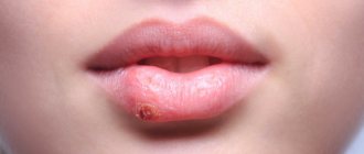
This is especially true for infection with herpes simplex viruses, which leads to very painful inflammation of the mucous membrane. There are two types of herpetic stomatitis - acute herpetic stomatitis (with primary infection) and recurrent herpetic stomatitis (which can pass through the acute stage or manifest itself as a general infection of the body in the presence of provoking factors). Herpetic stomatitis can lead to an aphthous form, in which erosions, aphthae, and even painful ulcers appear on the mucous membrane.
A specific type of stomatitis is chronic recurrent aphthous stomatitis, which begins almost immediately with the formation of aphthae, and the nature of which is not yet clear. But viral and bacterial infections and vitamin imbalances are also suggested as initiating factors.
The specific infection that causes stomatitis includes a fungal infection (fungi of the genus Candida) and the disease itself is called candidiasis (thrush). Thrush is characterized by the formation of whitish dots on the mucous membrane, and as the fungal infection multiplies, a whitish coating. Candidiasis develops, as a rule, as a result of violations of hygiene rules, physiological disorders in the body, and the use of certain medications. It is often observed in children and its danger cannot be underestimated. This infection, which initially appears on the oral mucosa, can be spread to all organs and systems of the body, and in this case already poses a significant threat to health.
Purely bacterial causes of stomatitis are less common. As a rule, bacterial infections affecting the oral mucosa accompany viral types of stomatitis (for example, streptostaphylococcal lesions).
Other dental diseases: list of dental diseases with photos
Next, we propose to analyze ailments that may appear as a result of teething or age-related changes.
Grinding
A pathology in which there is a rapid decrease in hard tissue. Provokes thinning of enamel and damage to dentin. It can affect both individual units and full-fledged rows.
Erosion
A process in which a violation of enamel integrity is recorded. Some areas are damaged symmetrically. Main signs: changes in pigment, various cosmetic defects, painful reaction to cold or spicy food.
Pathological resorption
Loss of root tissue caused by chronic diseases, advanced periodontal disease or progressive pulp disease. It can be one-sided or combined. In the absence of rehabilitation and therapeutic therapy, tooth loss occurs.
Deposits and build-ups
They are mineralized residues that form above or below the gums. The main material for formation is food particles, the results of the activity of pathogenic microorganisms, as well as components of saliva. They trigger processes that provoke periodontitis.
Medicines for the treatment of diseases of the mucous membrane of the mouth, tongue and lips
Diagnosis, treatment and prevention of diseases of the mucous membrane (OM) of the mouth, tongue and lips remain an important problem in clinical medicine. Changes in the CO of the mouth, tongue and lips in many systemic diseases occur long before the appearance of general clinical symptoms. Therefore, the correct interpretation of such changes and the choice of appropriate medications are of great importance not only for dentists, but also for other specialists.
When presenting the material, we used the classification of diseases of the oral cavity by E.V. Borovsky and A.L. Mashkilleyson [7], which provides for the identification of 9 groups of this pathology, taking into account etiological and pathogenetic factors, which, due to the impossibility of citing them, will be omitted in this publication. We limited ourselves only to issues of medical tactics, drug treatment of major diseases of the oral mucosa, tongue and lips.
The authors have made every effort to ensure the accuracy of the information presented, including drug dosages. Aware of our responsibility associated with the preparation of the publication, and taking into account the constant changes occurring in medical science, we recommend that the dosage of medications be updated according to the appropriate instructions.
Basic principles of treatment of diseases of the mucous membrane of the mouth, lips and tongue:
- Rational treatment requires contact between the dentist and other dental and non-dental specialists.
- Treatment must be carried out in compliance with the principles of bioethics, these diseases must be considered from the standpoint of the state of the whole organism, therefore in most cases one cannot limit oneself to local influences only.
- An axiom for the dentist should be the elimination of all unfavorable irritating factors in the oral cavity in the patient, which can support and provoke the development of the pathological process. It is unacceptable to use so-called cauterizing agents and long-term use of the same mouthwashes.
- Treatment should begin only after at least a preliminary diagnosis has been established and meet the following requirements: be comprehensive; provide a pathogenetic approach; do not disturb the anatomical and physiological characteristics of the oral mucosa; eliminate the pain factor; promote rapid epithelization of lesions; provide for the active involvement of the patient in performing medical procedures at home.
Traumatic lesions
Medical tactics for traumatic lesions of the mucous membrane due to the action of mechanical factors, high and low temperatures, radiation, unfavorable meteorological factors, and chemicals boils down, first of all, to eliminating the effect of the traumatic factor, which not only facilitates differential diagnosis, but is also a therapeutic intervention.
Decubital ulcer
Heals, as a rule, 10-12 days after eliminating the mechanical cause. The surface of the ulcer is treated with oxygen-containing antiseptics (hydrogen peroxide, potassium permanganate, sodium hypochlorite), and after cleaning its surface from necrotic plaque, keratoplasty agents are used. It should be remembered that with prolonged existence (more than 2-3 months), especially in older people, a decubital ulcer can become malignant.
Chemical injury CO
It occurs as a result of its immediate immediate contact with concentrated solutions of acids, alkalis, some medications and agents sometimes used by dentists (silver nitrate, arsenic paste, resorcinol-formalin mixture). In case of acute chemical injury, the CO drug that caused the burn is immediately neutralized (in the absence of a neutralizer, the mouth is washed with water). Subsequently, the affected areas are treated with anesthetic substances, weak antiseptic solutions, proteolytic enzymes, and then keratoplasty agents are applied.
Physical trauma CO
It can also be acute and chronic. Painkillers, antiseptics, and drugs that promote tissue regeneration are used. In some cases, it is necessary to resort to excision of the affected area.
Leukoplakia
A fairly common disease of the oral mucosa and red border of the lips, pathomorphologically characterized by chronic inflammation with pronounced hyperplasia and keratinization of the epithelium.
The scope of treatment measures is determined both by the form of the disease and the size of the lesion and the speed of development of the process. Among the medications for flat form of leukoplakia, applications are made to the lesion and vitamin A is prescribed orally (3.4% oil solution of retinol acetate or 5.5% retinol palmitate, 10 drops 2-3 times a day for 1. 5-2 months). Courses are repeated 2-3 times a year. For verrucous and erosive forms, the same is done within one month, and in the absence of positive dynamics, the lesion is excised, followed by mandatory histological examination, which determines further medical tactics.
The manifestations and severity of changes in the mucous membranes of the mouth, tongue and lips during infectious diseases depend both on the type of pathogen and on the individual characteristics of the patient’s body, which determines the dentist’s tactics.
Adenoviral diseases
In case of influenza and adenoviral diseases, the lesions of the mucosa are not specific; after a few days from the onset of the disease, hyperemia with “granular” rashes on the mucous membrane is replaced by petechial rashes, and by the 7-9th day the pathological changes disappear.
Chronic recurrent herpes
It appears at any age in persons previously infected with the herpes simplex virus.
Medical tactics for viral infections include the use of drugs from several groups that affect various parts of the etiology and pathogenesis of diseases: antivirals, immunocorrectors and immunomodulators, vaccines, interferons and their inducers. The most pronounced therapeutic effect is observed with the combined use of 2-3 drugs of general and local action with different mechanisms of antiviral effect.
Ointment and cream are started to be used when the first signs of infection activation appear and continue until the erosions are epithelialized. Early treatment can prevent the development of vesicles. A large group of antiviral drugs includes nucleoside analogues, similar in structure to intermediate products of DNA and RNA biosynthesis (iododeoxyuridine, chlordeoxyuridine, fluorodeoxyuridine), effective against generalized forms of herpes and herpes zoster. An effective antiviral agent is acyclovir (Zovirax, Virolex). In addition to acyclovir, the group of abnormal nucleosides includes: valacyclovir (Valtrex) in tablets of 500 mg; take 2 times a day for 5-7 days; famciclovir (famvir) tablets 250 mg; take 3 times a day for 7 days.
It should be noted that the therapeutic effect of antiviral drugs is most effective when prescribed in the first hours and days of development of the lesion elements and in the prodromal period of the viral disease.
Local antiviral drugs include: adimal, bonafton, florenal, riodoxol in the form of ointments and liniments (gossypol). Ointment and cream are started to be used when the first signs of infection activation appear and continue until the erosions are epithelialized. Moreover, early treatment can prevent the development of vesicles.
The leading role in the development of antiherpetic immunity belongs to interferon. The antiviral drug "Infagel" (it includes recombinant interferon alpha-2) has a wide spectrum of antiviral activity, bacteriostatic and anti-inflammatory effects, as well as antitumor and immunomodulatory activity. It is used in the form of applications to the lesions 2 times a day with an interval of 12 hours and dried for 10-15 minutes to form a protective film [10].
Interferon inducers (poludan, larifan) and antiviral vaccines (in the form of a cycle of intradermal injections to prevent relapses and during remission of the disease) have found widespread use in dental practice. The therapeutic effectiveness of the vaccine increases significantly when combined with poludanum or other interferon inducers.
In addition to these drugs, salicylates, analgesics, antihistamines and antiseptic solutions, multivitamins, EF radiation and helium-neon laser radiation are used in the treatment of herpes infection. To stimulate the processes of regeneration of the mucous membrane of the mouth and the red border of the lips, applications of oil solutions of vitamins A, E, ointment and jelly of solcoseryl and Actovegin, aerosols “Livian”, “Spedian”, “Gipozol” are used. After eliminating the elements of the lesion, sanitation of foci of chronic infection is mandatory.
In case of herpes zoster, along with the measures mentioned above, the doctor’s task includes eliminating the pain syndrome and preventing postherpetic neuralgia. For this purpose, analgesics, ganglerone, and B vitamins are prescribed.
Vincent's ulcerative-necrotizing gingivostomatitis (YANGS)
This is an inflammatory disease of the mouth, characterized by necrotic decay of the affected areas. Medical tactics for YANGS in some cases (especially in severe cases) include hospitalization of the patient, local and general measures, and medical examination.
Local treatment is important and begins with the patient’s first visit to the doctor, and then is carried out daily and in stages:
- Pain relief (applications, oral baths).
- Treating the mouth with antiseptic solutions (more effective - those that release atomic oxygen and chlorine-containing preparations: hydrogen peroxide, potassium permanganate, hypochlorite).
- Mechanical (and after application of proteolytic enzyme solutions to the lesion) removal of necrotic tissue, removal of mineralized dental deposits and elimination of other local traumatic factors.
- After cleansing the lesions from necrotic plaque, it is possible and advisable to use (in the form of applications) keratoplasty agents and preparations that promote tissue regeneration (oxycort aerosol, methyluracil ointment, oil solutions of vitamins A and E).
- It is better to postpone the removal of damaged teeth and roots, treatment of chronic forms of pulpitis and periodontitis until the ulcers are completely epithelialized.
- Subsequently, final sanitation of the oral cavity (planned treatment of caries, its complications, periodontal and mucosal diseases).
General treatment (especially for severe disease) involves the use of antimicrobial drugs. Orally prescribed: metronidazole (Flagyl, Klion) 0.25 g 2 times a day for 7-10 days, in some cases it is necessary to use broad-spectrum antibiotics. General strengthening and stimulating therapy is also carried out: ascorutin, vitamin C in large doses (up to 3.0 g per day), and antihistamines are prescribed. The nutrition of patients with YANGS should be complete and balanced.
Fungal infections of CO
Fungal infections of the mouth (oral mycoses) are most often caused by fungi of the genus Candida albicans.
General treatment includes:
- Ingestion of antifungal drugs for 10 days: diflucan (capsules) 50-100 mg 1 time per day; the use of polyene antifungal drugs (nystatin, levorin, amphotericin B) is traditionally included in general treatment, although it is known [11] that polyenes are practically not absorbed in the gastrointestinal tract, thus exerting a local effect on oral mucosa, and therefore It is recommended to dissolve the tablets in the mouth after meals (nystatin 1 tablet 500,000 units every 6-8 hours, levorin 1 tablet 500,000 units every 8-12 hours); Amphotericin B preparations are used mainly for severe systemic mycoses due to their high toxicity [11].
- Vitamins C, PP, group B taken inside.
- Use of antihistamines.
- Treatment together with other specialists of concomitant and background diseases.
Local treatment includes:
- Antifungal drugs for application and lubrication of lesions: 0.5% decamine ointment, 1% ointment and clotrimazole solution (canesten); 1% pimafucin ointment; for yeast infections and cheilitis, it is advisable to use 5% levorin ointment.
- Aqueous solutions of aniline dyes: 1-2% solution of gentian violet, 2% solution of methylene blue.
- Agents that alkalize the oral cavity: rinse with a 2% sodium bicarbonate solution; applications and lubrication of lesions with a 20% solution of borax in glycerin.
- A decoction of wild rosemary for mouth rinsing and oral baths.
Lesions of the oral mucosa are observed in all periods of syphilis, and dentists with similar manifestations have become much more common. Treatment of patients with syphilis is carried out in specialized venereological institutions. Dental interventions are usually associated with the sanitation of the oral cavity and are carried out in strict compliance with the disinfection and sterilization regime in health care facilities.
Treatment of fixed and widespread allergic and toxic-allergic diseases includes the “exclusion” of the allergen, for example, a drug, the use of painkillers, antiseptics, desensitizing and keratoplasty agents, if necessary. Sanitation of odontogenic foci of chronic infection is mandatory.
Erythema multiforme exudative
A fairly common dermatosis affecting the oral mucosa, observed mainly in young and middle-aged people. Stevens-Johnson syndrome is a severe variant of exudative erythema multiforme (also called acute mucocutaneous-ocular syndrome).
Medical tactics depend on the severity of the disease. In some cases, patients need to be hospitalized.
General treatment is usually prescribed by other specialists (dermatologist, immunologist) and includes:
- Detoxification therapy: drinking plenty of fluids or infusion therapy if it is impossible to take liquids orally.
- Taking glucocorticoids: prednisolone in an initial dose of 20-60 mg per day orally for 5-7 days (the dose depends on the severity of the process and the form of the disease, with Stevens-Johnson syndrome higher dosages are required), then the dose of prednisolone is reduced by 5 mg every 2 —3 days before complete withdrawal of the drug.
- Immunomodulatory therapy: for example, Lavomax (Tilorone) is prescribed 125 mg (1 tablet) orally in the first two days, then 125 mg after 48 hours. The dose of the drug per course is 2.5 g [15].
- Prescription of antihistamines, B vitamins, ascorutin.
- Antibiotics are prescribed when there is a threat of purulent-inflammatory complications and the doctor is confident that there is no connection between the development of the disease and the use of these antibacterial agents.
Local treatment is aimed at relieving inflammation and accelerating epithelization of the affected areas of the mucous membrane of the mouth and lips. It includes:
- The use of painkillers (1-2% solutions of trimecaine, lidocaine, anesthetics in aerosols) before each manipulation in the oral cavity (baths, rinses, applications) and before meals.
- Antiseptic treatment of the mouth (solutions of hydrogen peroxide, potassium permanganate, chloramine).
- To improve the rejection of necrotic tissues, apply solutions of proteolytic enzymes (trypsin, chymotrypsin) to the lesions.
- After cleaning the eroded surfaces from purulent-fibrinous plaque, use keratoplasty preparations (sea buckthorn and rosehip oil, oil solutions of vitamins A and E, the drug “Gipozol”).
- EF irradiation of the oral mucosa.
After the elimination of acute phenomena, the foci of infection are sanitized. In the inter-relapse period - specific desensitizing therapy.
Chronic recurrent aphthous stomatitis
One of the most common diseases with an autoimmune component of pathogenesis, it is characterized by periods of remission and exacerbation with the formation of aphthae (a superficial painful defect of the mucous membrane of a round or oval shape, covered with fibrinous plaque and surrounded by a bright inflammatory rim).
General treatment is prescribed after consultation with an immunologist:
- Immunocorrective and immunomodulatory therapy with thymogen, levamisole.
- Metabolic drugs for 10 days (calcium pantothenate orally 0.1 g 4 times a day; potassium orotate 0.5 g 3 times a day an hour before meals, lipamide 0.025 g 3 times a day after meals).
- Sedatives and adaptogens.
- Exclusion from the diet of hot, spicy foods, prohibition of smoking and drinking alcohol.
- In the inter-relapse period - specific and nonspecific desensitizing therapy.
- Vitamins: C, group B.
Local treatment:
- If there are aphthae, apply painkillers to the aphthae.
- Injections under the base of the aphthae with a mixture of 0.1 ml of 0.1% atropine sulfate and 1 ml of 0.25-0.5% solution of novocaine or trimecaine.
- Applications to aphthae of biopolymer soluble films (for example, oblekol films).
- Applications to aphthae of oil solutions of vitamins A, E, Oblekol, “Aevita”.
Pemphigus
Among the pathological processes of the oral mucosa with the formation of blisters (vesical dermatoses), the central place is occupied by pemphigus - an autoimmune process in which intraepithelial blisters appear on the skin and mucous membranes.
Treatment is coordinated with a dermatologist and begins in the hospital. Glucocorticoid therapy (first in shock and then in minimal, maintenance doses) interrupts the progression of pemphigus and leads to the disappearance of clinical symptoms of the disease. It should be remembered that local use of glucocorticoid drugs is ineffective. Sometimes cytostatics (mainly methotrexate) are also used to treat pemphigus.
Local treatment is aimed at preventing secondary infection of erosions and ulcers. Non-irritating CO solutions of antiseptics and keratoplastics are used.
Lichen planus
Mucocutaneous reaction - lichen planus occurs in 1-2% of the population. When treating lichen planus, an integrated approach is required, the basis of which is psychotherapy, especially at the initial stage, supplemented by the prescription of tranquilizers, sedatives, adaptogens, desensitizing agents, vitamins (A, PP). An effect was detected when prescribing the anxiolytic “Tenoten” (from 1 to 4 tablets per day for 3-6 weeks) and the antioxidant “Kudesan” [9].
Smoking and taking allergenic products is prohibited. It is necessary to exclude dissimilar metals in orthopedic structures and replace metal fillings. With exudative-hyperemic and erosive-ulcerative forms, it is sometimes necessary to resort to steroids (prednisolone) and drugs such as delagil.
Local events include:
- Local anesthetics.
- Applications of 1% emulsion of dibunol, vitamins A, E, Solcoseryl ointment.
- Irradiation of lesions with a helium-neon laser in combination with oral administration of tigazone capsules (10 mg 3 times a day for a course of up to 6-8 weeks).
- Electrophoresis of vitamin PP from dimexide medium.
In his medical practice, the dentist must proceed from the fact that lichen planus can become malignant.
Changes in the mucosa of the mouth and the red border of the lips often occur in pathologies of various organs and systems of the body and metabolic disorders.
It should be noted that in most cases, the manifestation of systemic diseases in the oral cavity is not specific, however, some changes in the oral cavity clearly indicate one or another type of organ or systemic disorder and are of great clinical significance.
Vitamin A deficiency
Causes disturbances in the epithelial structures of the skin and oral mucosa. Retinol is prescribed (in the form of a concentrate or pill) up to 50,000-100,000 IU per day.
Vitamin B1 deficiency
Accompanied by hyperplasia of the fungiform papillae of the tongue, paresthesia and allergic reactions of the oral mucosa. For treatment, preparations of thiamine chloride and thiamine bromide are used in courses of up to 30 days. It is unacceptable to mix a solution of vitamin B1 with other B vitamins in one syringe or to administer them sequentially through one needle.
Vitamin B2 deficiency
It manifests itself as a peculiar change in the skin, red border of the lips, ulceration in the corners of the mouth (angular stomatitis), weeping, maceration of the epithelium. For medicinal purposes, riboflavin is prescribed orally at a dose of 0.01 g 3 times a day for 4-6 weeks.
Vitamin B6 deficiency
With a deficiency of vitamin B6, symptoms of disorders of the nervous system (polyneuritis) and gastrointestinal tract, angular stomatitis, cheilitis, and glossitis are observed. The daily therapeutic dose of pyridoxine for adults, administered primarily parenterally, is 0.05-0.1 g.
Vitamin B12 deficiency
Accompanied by neurological disorders, changes in hematopoiesis (B12-deficiency anemia). Hunter-Meller glossitis is characteristic. For therapeutic purposes, 1000 mcg of vitamin B12 is administered intramuscularly daily until hematological disorders are completely eliminated, during the same time (4-6 weeks) manifestations in the oral cavity are stopped. Subsequently, lifelong maintenance therapy is prescribed (one injection of vitamin B12 per month).
Vitamin PP deficiency
Severe PP (nicotinic acid) deficiency is described as “pellagra,” which is characterized by a triad of symptoms: dementia, dermatitis, diarrhea. Treatment consists of taking nicotinic acid orally after meals (0.1 g 2-3 times a day for a course of 2-3 weeks). At the same time, thiamine, riboflavin and pyridoxine are usually prescribed, since in most cases polyhypovitaminosis occurs.
Vitamin C deficiency
It manifests itself as hemorrhagic syndrome (petechial hemorrhages in various parts of the oral mucosa, swelling and bleeding of the gums) and complications caused by the addition of a secondary infection (ulcerative necrotizing gingivitis). For therapeutic purposes, ascorbic acid is prescribed up to 1.5 g per day (after meals) with simultaneous intake of rutin (50-100 mg 2-3 times a day).
Most diseases of the blood and hematopoietic organs have typical manifestations in the oral cavity: changes in the color of the mucous membrane, the phenomenon of hemorrhagic diathesis, paresthesia, ulcerative-necrotic processes. The mucous membrane of the mouth in acute leukemia is almost constantly affected. In more than half of the patients, changes in the mucosa are ulcerative-necrotic and hemorrhagic in nature.
The dentist’s task when treating such patients (usually in a hematology hospital) remains pain relief, proper professional antiseptic treatment of the oral cavity, careful removal of necrotic plaque (mechanically and with the help of proteolytic enzymes), and stimulation of epithelization of lesions.
Salivary dysfunction
Disorders of this kind are of great clinical significance, especially associated with a decrease in the secretion of the salivary glands, up to hyposialia or even xerostomia.
Medical tactics:
- Distinction between “subjective” (i.e., developing against the background of somatic pathology without the death of the glandular epithelium) and “objective”, in which, due to the death of salivary gland tissue, there is a decrease in the secretory activity of the salivary glands.
- Identification and elimination of organ disorders; correction of disorders of “general” and “local” immunity.
- Elimination of foci of chronic odontogenic infection.
- Rational prosthetics.
- Medications include vitamins A, E, C, group B, and antidepressants.
- To stimulate salivation - orally (2-4 drops 15 minutes before meals) 1% solution of pilocarpine; galvanization of the salivary glands.
The doctor’s tactics for neurogenic diseases of the tongue, lips and mucus (glossalgia, glossodynia, stomalgia, burning mouth syndrome) include:
- Sanitation of the mouth to eliminate irritating factors (galvanism phenomena, defects in fillings and prosthetics) and eliminate chronic odontogenic infection; treatment of fungal infections of the mouth and tongue.
- Treatment of somatic diseases.
- Normalization of the function of the central and autonomic nervous systems; for somatized depression - antidepressants (for example, amitriptyline according to the scheme starting with one-fourth tablet at night, increasing weekly by one-fourth tablet, leading to a whole tablet per day by the end of the month).
- B vitamins.
- Vasoactive drugs and vegetotropic drugs.
- Applications of local anesthetics.
- Galvanization of the upper cervical sympathetic nodes, in some cases - ultratonotherapy, ozone therapy, etc.
If conservative treatment of erosive and ulcerative lesions (for example, in chronic trauma, leukoplakia, lichen planus) within 10-14 days is ineffective and there is no tendency to their healing, surgical or cryosurgical excision of the lesion with mandatory histological examination should be used.
It must be remembered that all precancerous conditions must be treated surgically. There is no need for wait-and-see tactics and long-term drug therapy. This is permissible, as mentioned above, only in cases of damage to the mucosa of the mouth, tongue, and lips, where reverse development of the pathological process is possible under the influence of only therapeutic treatment.
This article is intended both for practicing dentists and for students, interns, residents, and young teachers of medical universities. However, it will be a shame if the reader only absorbs some of the information and concepts presented in it. To expand your own knowledge and for the benefit of our patients, it is advisable to read other publications on this issue [1-8, 12-14, 16-18].
LITERATURE
- 1. Banchenko G.V. Combined diseases of the oral mucosa and internal organs. - M.: Medicine, 1979. - 190 p.
- Banchenko G.V., Maksimovsky Yu.M., Grinin V.M. Language is the “mirror” of the body: Clinical guide for doctors. - M., 2000. - 408 p.
- Borovsky E. V., Danilevsky N. F. Atlas of diseases of the oral mucosa. — 2nd ed., revised. and additional - M.: Medicine, 1991. - 320 p.
- Danilevsky N. F., Leontyev V. K., Nesin A. F., Rakhniy Zh. I. Diseases of the oral mucosa. - M., 2001. - 271 p.
- Diagnosis, treatment and prevention of dental diseases / V. I. Yakovleva, E. K. Trofimova, T. P. Davidovich, G. P. Prosveryak. — 2nd ed., revised and supplemented. - Mn.: Higher. school, 1994. - 494 p.
- Dmitrieva L. A., Glybina N. A., Glybina T. A. et al. Experimental substantiation of the use of a new antioxidant drug in the treatment of erosive and ulcerative lesions // Periodontology. — 2012, No. 3 (64). - pp. 52-58.
- Diseases of the mucous membrane of the mouth and lips / Ed. prof. E. V. Borovsky, prof. A. L. Mashkilleyson. - M.: Medicine, 1984. - 400 p.
- Diseases of the oral mucosa: Textbook / Ed. L. M. Lukinykh. - N. Novgorod: Publishing House of NGMI, 1983. - 212 p.
- Karakov K. G., Vlasova T. N., Oganyan A. V., Pisarev G. Yu. Optimization of complex therapy of lichen planus of the oral mucosa // Dentist-practitioner. - 2012, No. 1. - P. 35-37.
- Karakov K. G., Vlasova T. N., Oganyan A. V., Polyakova O. V. The role of complex therapy in the treatment of herpetic infection of the dental system // Practitioner dentist. - 2012, No. 2. - P. 48-49.
- Practical guide to anti-infective chemotherapy / Ed. L. S. Strachunsky, Yu. B. Belousov, S. N. Kozlov. - M.: Borges, 2002. - 384 p.
- Rybakov A. I., Banchenko G. V. Diseases of the oral mucosa. - M.: Medicine, 1978. - 232 p.
- Therapeutic dentistry: textbook: 3 hours / ed. G. M. Barera. - M.: GEOTAR-Media, 2055. - Part 3. - 288 p.
- Tretyakovich A. G., Borisenko L. G., Pishchinsky I. A. Differential diagnosis and principles of treatment of diseases of the oral mucosa: educational method. allowance. — 2nd ed., revised. and additional - Mn.: BSMU, 2005. - 66 p.
- Udzhukhu V. Yu., Minatulaeva M. A., Kubylinsky A. A. Pathogenetic rationale and clinical effectiveness of the use of Lavomax in patients with exudative erythema multiforme // Farmateka. - 2008, No. 9. - P. 60-62.
- Tsepov L. M., Nikolaev A. I. Medical tactics for erosive and ulcerative lesions of the mucous membrane of the mouth, tongue and lips (Training manual). - Smolensk: SGMA, 2005. - 16 p.
A complete list of references is in the editorial office.
List of neurotic dental diseases
Stressful periods have a particularly detrimental effect on the state of the body. In addition, as practice shows, the quality of oral hygiene deteriorates sharply. A person simply forgets about cleaning or does not do it thoroughly enough, which increases the risk of developing caries. The parallel use of sedative medications disrupts the production of saliva and causes a feeling of dryness.
Gradually, all this is reflected in psychosomatics - muscle spasms and pain are detected. The most common occurrence is excessive clenching of the jaw.
Bruxism
A disorder characterized by uncontrollable grinding of teeth caused by emotional stress. As a rule, it makes itself felt during sleep. The causes of its appearance include stress, prolonged depression, and malfunctions of the masticatory apparatus.
Grinding
This is not so much a full-fledged disorder as a symptom. Occurs when muscle spasms occur when the patient clenches his jaw tightly. In the vast majority of cases, the person does not even know there is a problem. It is detected during routine dental examinations.
Periodontitis
Periodontitis is an inflammation of periodontal tissues, which includes the teeth, ligaments, cementum and gums. Periodontitis as a disease is a consequence of gingivitis - a minor inflammation of the gums, the main cause of which is neglect of oral hygiene. If with gingivitis the inflammation spreads exclusively to the soft mucous membranes, then with periodontitis the ligaments that hold the teeth in the sockets are affected. This is why in 90% of cases when this disease is diagnosed, tooth mobility is observed, which eventually leads to their loss.
The most common causes of the disease are the following:
1. Improper or irregular oral care
. Dental plaque, which is present on the surface of the teeth and in the spaces between teeth, is not as safe a substance as it might seem at first glance. Soft and easily removed at the beginning, it goes through certain cycles of “development”. The result is the mineralization of plaque and its transformation into hard tartar. This process in most cases is observed in those who do not pay due attention to daily oral care or use an incorrectly selected toothbrush, toothpaste and mouthwash.
2. Poor blood supply to the gums
. Periodontitis is one of the most common problems among smokers. Substances contained in tobacco smoke lead to a narrowing of the blood vessels in the oral mucosa and their fragility, which impairs the blood supply to the gum tissue and supporting apparatus of the teeth. A slowdown in blood circulation and, as a consequence, the development of periodontitis is also facilitated by a lack of chewing load caused by eating habits (for example, the predominance of soft foods in the diet).
3. Nutrient Deficiency
. The lack of fresh vegetables, fruits, herbs, a sufficient amount of fish, meat and dairy products in the diet quickly leads to a lack of essential substances in the gum tissue. If poor nutrition is a permanent habit, then over time the metabolic processes in the gums are disrupted, which creates the ground for inflammation and periodontitis. A deficiency of vitamins A, C and group B can lead to negative consequences.
Treatment of periodontitis
Professional teeth cleaning is an integral step in the treatment of periodontitis. This procedure removes physical obstacles (plaque and tartar) that prevent the gums from returning to their previous position and tightly covering the teeth.
Drug treatment - the use of antiseptics for topical use. This need is due to the high risk of spread of inflammation and infection to other tissues.
Surgery
At an advanced stage of periodontitis, when the inflammation has spread deep into the bone tissue, surgical intervention becomes necessary. Such manipulations include partial excision of the gums (gingivectomy), washing of periodontal pockets with medicinal solutions, removal of stones, and flap operations. In some cases, surgical treatment of periodontitis involves the implantation of bone tissue substitutes or the application of collagen or artificial membranes to restore the supporting apparatus of the tooth.
Compliance with oral care rules
Without regularly removing plaque and protecting the oral cavity from bacteria, it is impossible to achieve sustainable results in the treatment of periodontitis. Hygiene procedures twice a day with properly selected products, the use of dental floss and rinses will help make recovery faster.
The main dental diseases in humans: what they are and how to treat them
Modern medical practice, implemented in the Dentika dental clinic, actively uses innovative methods of diagnosis and treatment. Almost any disease can be eliminated provided that you seek help in a timely manner, as well as follow all doctor’s prescriptions and prescriptions. Among the measures used by dentists are:
- filling;
- surgical intervention;
- prosthetics;
- implantation;
- aesthetic correction.
In addition, professional hygiene methods are used: bleaching, restoration, etc.
Gum diseases in adults
Gingivitis
Inflammation of the mucous membrane is localized around the diseased tooth; the pathological process can be acute or chronic.
Types of Gingivitis:
- catarrhal,
- atrophic,
- hypertrophic,
- ulcerative-necrotic.
If you start treatment on time, you can defeat the disease very quickly and without consequences in the form of tooth loss. If the situation is neglected, the disease can develop into a more dangerous form - periodontitis.
Periodontitis: what is it?
Periodontitis is a gum disease that destroys the supporting structure of the affected tooth, as well as the ligament between the bone and the root of the tooth. As a result of the inflammatory process, the gum partially peels off from the tooth, forming a gum pocket in which food debris accumulates, which is a breeding ground for pathogenic microorganisms. It is not possible to remove these accumulations with a toothbrush.
Without proper treatment, bleeding and swelling of the gums quickly develops, and pus can form in periodontal pockets. Teeth become loose and eventually fall out. The acute phase is characterized by general malaise, weakness and severe pain in the affected area.
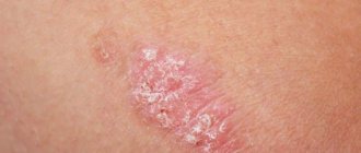
Periodontal disease: photo
Periodontal disease, like periodontitis, affects the supporting part of the tooth, but inflammation does not occur. There is a disruption in the blood supply to soft tissues, their dystrophy develops, the necks of the teeth are exposed, and the gums are slowly destroyed. Periodontal disease does not remain local - over time it affects both jaws, so it is very difficult to fight this disease. For periodontal disease, physiotherapy, local treatment and splinting of loose teeth are effective.
Prevention
Above, we examined in detail what dental diseases are called and what are the main reasons for their occurrence. Now let's talk about preventive measures to avoid serious problems and pathologies.
If you experience any strange sensations in your mouth, it is recommended that you make an appointment with your dentist immediately. He will conduct an initial examination and draw up an individual treatment program. However, in some cases it is possible not to resort to aggressive measures of influence and limit oneself to supportive manipulations.
So, what needs to be done to prevent the development of diseases?
- Do not ignore the symptoms that appear - go to the doctor immediately.
- Do not let the disease worsen, follow all dental recommendations and prescriptions.
- Get professional examinations every six months.
- Maintain hygiene at home, use only high-quality products.
- Don’t forget about professional hygiene - ultrasonic cleaning should be done once every six months to a year.
In addition, it is important to adhere to the principles of proper nutrition and give up bad habits.
Periodontal disease
Dental periodontal disease is a serious disease in which the last stage of gum inflammation occurs. This is often the cause of the development of infectious diseases, gastritis, stomach ulcers or cirrhosis of the liver. Even more often, the patient’s teeth simply fall out, and he cannot lead his usual lifestyle or eat his favorite food.
How to recognize periodontal disease
The signs of this dental disease are unclear and blurred. The patient is most often concerned about:
- exposure of the necks of the teeth;
- presence of tartar;
- burning gums;
- discomfort when eating.
There are 3 stages of periodontal disease:
- Easy. The patient has no complaints; very rarely there is a reaction to cold or hot food. The presence of periodontal disease can be determined during a dental examination. The mild stage of the disease is best treated.
- Average. The roots of the teeth are exposed by an average of 4-6 mm. The patient begins to experience a burning sensation in the mouth, and there is an acute reaction to eating hot, cold or sour foods.
- Heavy. The roots of the teeth are exposed by 8-10 mm. Chewing food causes severe pain.
Treatment methods
Diagnostics
Before starting treatment for periodontal disease, the dentist conducts an initial examination, during which he determines the extent of damage to the teeth and gums: which teeth can be restored and which will have to be removed. This is necessary in order to draw up an algorithm for further actions. The patient is then sent to the diagnostic room to take targeted and panoramic X-rays. Using them, the periodontist determines the depth of the pockets and the condition of the bone tissue.
Removing plaque and tartar
Inflammation of the gums, which is always observed with periodontal disease, mainly occurs due to soft plaque, subgingival and supragingival calculus. The main reason for their appearance is poor oral hygiene. Therefore, the specialist’s task is not only to treat the disease, but also to teach the patient proper hygiene.
General and local therapy
To increase immunity, the patient is prescribed a complex of vitamins and anti-inflammatory drugs. If the inflammation is minor, the dentist will prescribe a course of local therapy, which can be done independently at home.
Splinting teeth
An increase in tooth mobility indicates that the jawbone and soft tissue around them have begun to rapidly deteriorate. To prevent the teeth from changing position and falling out (for example, they may fan out), they are held together with fiberglass tape and filling material. This is also necessary before surgical treatment.
Surgical operations
If periodontal pockets reach 5-10 mm, it is impossible to prevent the progression of the disease without surgical intervention. First, the pockets are cleaned of granulations and food deposits. This procedure is called curettage. It comes in two types - open and closed.
Closed is carried out with special instruments, curettes. It is carried out only for periodontal disease at the initial stage (pockets reach 3 mm), when there is slight inflammation of the gums.
Open curettage is necessary in advanced stages of periodontal disease. With its help, all granulations and food deposits are completely removed. This operation is more difficult to perform. To completely clean out the pockets, incisions are made in the gum. Flaps of the mucous membrane are peeled away from the bone and the root surface is cleaned with curettes and an ultrasonic scaler. To restore bone tissue, the periodontist implants synthetic bone.
Next, the patient undergoes flap surgery to prevent receding gums. The doctor removes a 1.5 mm marginal strip of gum, since after prolonged inflammation the gum changes in such a way that it can no longer adhere normally to the tooth. After this, the mucous membrane flaps are pulled to the neck of the tooth.
Timely diagnosis and choosing the right treatment will help stop periodontal disease and maintain healthy teeth!
Let's sum it up
Dentistry studies the etiology, symptoms, and treatment features of the most common dental diseases. There are a huge number of reasons that provoke oral problems. Some of them are easily eliminated, others are subject to only partial correction. However, for both cases the rule is true: the sooner you start working on dental treatment, the higher the likelihood of a favorable outcome. That is why it is important not to let things take their course and undergo regular medical examinations.
Brief conclusions
- The most common oral diseases are tooth decay, gum disease, infectious diseases and oral cancer.
- Caries and gum disease are highly treatable in the early stages.
- The causes of infectious diseases of the oral cavity vary - from viruses to poor diet and uncontrolled use of medications.
- For prevention, it is important to review “life hygiene” and regularly visit the dentist.
Anti-Aging Medicine Seminars Gain knowledge based on evidence-based medicine first-hand from the world's leading experts. As part of the Anti-Age Expert Modular School, in-person two-day seminars are held every month, where the intricacies of anti-age medicine are revealed to doctors of more than 25 specialties
Find out more
If inflammation is not stopped at the very beginning, full recovery may not occur?
Absolutely right. At the stage of inflammatory changes in the deep periodontal tissues, complete recovery cannot be achieved; the disease can only be brought into stable remission - a state when patients do not complain. It is characterized by the absence of bleeding gums, bad breath and decreased tooth mobility. Further therapeutic and preventive measures are aimed at maintaining the protective forces of the periodontal tissues and preventing the formation of dental plaque. To do this, control courses of maintenance therapy are carried out at intervals of two to three months. Then the interval between them increases to five to six months.