from February 4, 2021
Keratosis or hyperkeratosis is a non-inflammatory form of dermatosis, which is accompanied by excessive keratinization of the skin.
For healthy skin of any person, the process of keratinization is a normal physiological state. The process of keratinization provides one of the most basic functions of the skin - protective.
MECHANISM OF KERAINIZATION IN NORMAL AND IN HYPERKERATOSIS
Normally, as a result of their vital activity, skin cells gradually move from the lower layer to the upper, while gradually accumulating keratin - a protein that makes them more durable and resistant to external influences. The top layer of skin is nothing more than completely keratinized cells or horny scales, which are peeled off and replaced with new ones during human life. Such cells lack viability, but at the same time, thanks to keratin, they provide protection to living cells located underneath them from negative environmental factors.
With the development of hyperkeratosis as a pathological condition of the skin
- on the one hand, there is an active accelerated division of epidermal cells that produce keratin;
- on the other hand, there is a delay in the process of normal physiological exfoliation of horny scales from the surface layer of the skin.
What are destructive (ablative) methods?
All skin tumors must be examined by a dermatologist before removal due to the risk of skin cancer. Treatment performed in a timely manner will help prevent the development of basal cell carcinoma (BCC) or squamous cell carcinoma of the skin (SCC).
- Cryosurgery using liquid nitrogen is the most common method for treating keratoses, but is not suitable for hyperkeratotic lesions. Liquid nitrogen is sprayed directly onto the affected areas of the skin using a cryodestructor or applied using the “reed” method (application with a cotton swab on a wooden stick). Cryosurgery is easily performed on an outpatient basis, shows excellent cosmetic results and is well tolerated.
- Radio wave, electro- and diathermo-laser destruction.
- Photodynamic therapy involves the application of a photosensitizing agent, methyl aminolevulinate, and then exposure to light of a specific wavelength, which leads to tissue necrosis. Photodynamic therapy is well tolerated and has excellent cosmetic results.
- Surgical removal . The surface of the skin is cleaned with a special instrument (curette).
- Chemical peeling:
- Jessner's solution (resorcinol, lactic and salicylic acids in ethanol);
- trichloroacetic acid solution 35%.
- Dermabrasion . The affected areas of skin are removed using a fast-moving abrasive brush.
- Phototherapy (IPL) and fractional photothermolysis - coagulation of keratosis elements using light energy. Suitable for the initial manifestations of actinic keratosis.
TYPES OF KERATOSIS
Most often, the phenomena of hyperkeratosis are localized on the legs (heels, feet, knees) and arms (palms, elbows), less often - on the scalp, face in the area of the nose, cheeks, forehead. Basically, this pathology is formed in areas of irritation by any external mechanical or environmental factors. However, hormonal imbalances, heredity and improper skin care can also provoke hyperkeratosis.
The most common types of manifestations of hyperkeratosis are:
- Actinic (solar) keratosis on the face and body (keratomas). It develops due to excessive exposure of the skin to ultraviolet rays and photodamage. Appears as flat, rough yellow or brown spots;
- Palmar and plantar keratoderma - calluses, corns on the soles of the feet and palms, heel cracks. Formed in areas exposed to prolonged friction or pressure, during infectious and fungal diseases;
- Follicular or pilaris keratosis (“goose bumps”). Dermatosis occurs as a result of both accelerated keratinization and disruption of the physiological desquamation of horny scales. Against this background, the normal secretion of sebum is disrupted and local inflammation of the hair follicles on the body occurs. It looks like a rash of small multiple nodules of bright pink and gray color on the face, shoulders, legs, buttocks;
- Seborrheic keratosis is the most common form of hyperkeratosis. It is a hyperpigmented “plaque” with clear boundaries from light to dark brown, covered with keratinized skin. It can be either single or multiple.
- Actinic keratosis - develops in old age in the form of beige-brown spots and is localized on the face, shoulders, back, and back of the hands.
Follicular keratosis (parafollicular keratosis, Kirle disease - CD) is a rare skin disease from the group of perforating dermatoses, which is based on a violation of keratinization with intradermal invagination of the epidermis [1] (synonyms: follicular and parafollicular hyperkeratosis, penetrating the skin, follicular and parafollicular hyperkeratosis with penetration into the skin (hyperkeratosis follicularis et parafollicularis in cutem penetrans), penetrating hyperkeratosis [2, 3]).
The disease was first described in 1916 by the Austrian dermatologist J. Kyrle, who observed this dermatosis in a patient with diabetes mellitus [3-5].
Epidemiology.
The disease is rare. The age of patients is 20-70 years. Women get sick more often, children - very rarely. Concomitant diseases: often - diabetes mellitus and chronic renal failure, less often - heart failure, hypothyroidism, type II hyperlipoproteinemia (increased blood levels of cholesterol and triglycerides) [1, 3-6].
Etiology and pathogenesis
have not been fully studied. The previously assumed role of a viral or bacterial infection, a violation of vitamin A metabolism in the development of this dermatosis is only of historical interest [4]. A certain significance in the development of the disease, in addition to impaired carbohydrate metabolism, is attributed to liver damage (chronic hepatitis) with the development of secondary vitamin A deficiency [5].
The genetic predisposition to the development of CD has not been conclusively proven, although cases of the disease among relatives in the same family with consanguinity (first-degree relatives) have been described [1, 3-5].
According to the original definition by J. Kirle [7], this is a disease in which an atypical clone of keratinocytes penetrates through the epidermis into the dermis. It is believed that the basis of the pathological process is a violation of keratinization, differentiation and keratinization of keratinocytes (formation of dyskeratotic foci and acceleration of the keratinization process). This leads to the formation of keratotic plugs with areas of parakeratosis. Keratification begins already at the border of the epidermis and dermis. The rate of differentiation and keratinization exceeds the rate of cell proliferation, therefore the parakeratotic cone partially penetrates deeper into the damaged epidermis and causes its perforation into the dermis [1, 3-6].
Unified classification
There are no follicular keratoses. Among the independent nosological forms, papular, atrophying and vegetative follicular keratoses are distinguished [8]. A number of authors [4, 6] draw attention to the similarity of CD with lenticular persistent hyperkeratosis of Flegel and consider the latter as a variant of CD, although the clinical manifestations differ. Other authors [5, 8] classify CD as vegetative follicular keratoses, and persistent keratosis follicularis (Flegel's disease) as papular.
The concept of reactive perforative keratinization disorder against the background of kidney, liver, and diabetes mellitus is considered. A concept has emerged - perforating skin diseases; it includes a group of diseases in which the elimination of altered skin components occurs through the epidermis (a process called transepidermal elimination). Some authors dispute CD as an independent nosological entity [7].
In the International Classification of Diseases, 10th revision, in class XII (diseases of the skin and subcutaneous tissue), heading L87 is allocated - transepidermal perforated changes, in which CD is assigned a separate subheading L87.0.
Pathomorphology
very characteristic and has diagnostic significance. The recesses of the epidermis and the dilated openings of the hair follicles are filled with hyperkeratotic plugs. Under the plugs in the epidermis, the growth of the granular layer is pronounced, and in places where there is no hypergranulosis, there is parakeratosis, penetrating the entire epidermis to the dermis. Subsequently, thinning of the epidermis occurs and penetration of horny masses into the dermis with the formation of inflammatory infiltrates such as granulomas from lymphocytes, leukocytes, histiocytes and giant cells of foreign bodies in the dermis. Death of the sebaceous glands in the area of the affected follicles, degeneration of collagen fibers, and hyperelastosis are observed [1, 2, 5, 6].
Clinical picture.
The onset of the disease is gradual, new rashes appear as old elements disappear. Follicular or parafollicular papules are characteristic, first the color of healthy skin, and subsequently grayish or brownish-red in color, with a diameter from a pinhead to 1 cm. In the center of the elements there is a horny plug, when removed, a crater-shaped depression is formed. Papules tend to grow peripherally and merge with the formation of dry polycyclic plaques covered with layers of scales and crusts. The consistency is dense, the surface is uneven, warty. Fresh rashes are accompanied by mild itching (more often in patients with diabetes) or do not bother the patient. Older, larger lesions are painful, especially when pressed. In the places of calculations there is a linear arrangement, which indicates the possibility of the occurrence of the Koebner phenomenon. A secondary infection may occur. Localization of rashes - extensor surfaces of the limbs, torso, buttocks. The mucous membranes are not affected [1-6]. Rashes in the mouth, palms, soles, and genitals are rare [4].
Flow
CD is chronic, relapsing. The disease is difficult to treat. The prognosis largely depends on the underlying disease.
Diagnostics
based on anamnesis, clinical and histological picture of the disease. Differential diagnosis of CD should be carried out with Devergy's lichen pilaris, Darier's follicular dyskeratosis, Mibelli's porokeratosis, transepidermal perforated skin changes (perforating creeping/serpiginous elastosis, reactive perforating collagenosis), Flegel's disease [8].
Flegel's disease
(hyperkeratosis lenticularis perstans), like CD, is a rare form of keratosis pilaris. It is believed that the basis of this disease is a hereditarily determined (autosomal dominant type) pathology of keratinization, expressed in a violation of the synthesis of keratin 55K, the content of which is sharply reduced in the lesions of Flegel's disease, while in normal skin this type of keratin dominates. Histologically, thinning of the epidermis, follicular orthokeratosis with foci of parakeratosis, and spongiosis in places are revealed; a band-like lymphocytic (mainly T-helper) infiltrate is expressed in the dermis. The previously attached great importance in the pathogenesis of dermatosis to the absence or disruption of the structure of Odland bodies [4] is now being questioned. Odland's granules appear in the upper rows of the spinous layer in keratinocytes, occupy a peripheral position and release their contents by exocytosis into the intercellular space, where it takes on a lamellar structure (intercellular cement) - a component of normal keratinization. Unlike CD, this dermatosis appears more often in adolescence and middle age, and is characterized by the formation of disseminated, smaller (1–5 mm) and more superficially occurring horny papules (without a tendency to form plaques). In CD there is no impairment of keratin 55K [6].
Treatment:
vitamin A, long-term aevit, retinoids (neotigazone, etretinate, acitretin at the rate of 0.5 mg/kg per day). Externally - keratolytic ointments, 2% sulfur salicylic ointment, retinoid cream, 0.1% tigazone cream, 5% fluorouracil cream (for an area of no more than 500 cm2 at a time), electro-, cryo- and laser destruction (carbon dioxide laser) of individual lesions, ultraviolet irradiation . The effect of treatment is temporary. After removal of the horny plug, an atrophic, unevenly pigmented scar remains; relapse may occur nearby. Correction of visceral pathology is necessary [3-6].
We present our own clinical observation.
Patient B., 57 years old, retired (military orchestra musician), was admitted to the purulent surgery department on September 13, 2010 for surgical treatment for osteomyelitis of the left foot. The attending physician prescribed a consultation with a dermatologist due to skin rashes.
Anamnesis.
The first rash on the skin of the front chest appeared about 20 years ago. The rashes practically did not bother me; minor itching was rarely noted; I was more “worried” about the cosmetic aspect. Diagnoses were made: dermatitis, xerosis, keratosis. There was no effect from the recommended salicylic ointment, creams with chamomile, string, or taking vitamin A orally, which was explained by the “peculiar structure of the patient’s skin.” According to the patient, the best effect was noted from the use of cindol (“the chatter dries well and peels off the plugs” on the skin). The rashes did not disappear completely, no seasonality was noted.
Since 1995, he has been suffering from type 2 diabetes mellitus and has been taking oral hypoglycemic drugs (diabetes). In 2009, he received inpatient treatment at his place of residence for diabetic foot syndrome, a purulent-necrotic wound on the left foot; healing by secondary intention. Since the summer of 2010, the rashes have increased significantly, which the patient associated with extreme heat. In July, I received a puncture wound in my left foot (I stepped on a nail at the dacha), which did not heal for a long time, and my blood sugar level increased. Due to worsening severe pain, the patient was hospitalized.
Skin status.
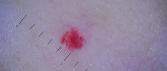
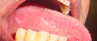
On the skin of the body there are multiple follicular and parafollicular papules of a red-brown color, in places grouped into small plaques covered with dense scales. In the center of the papules there are horny plugs. Where the plugs have come off, there are crater-shaped depressions filled with the previously applied zindol. In place of the resolved elements there are small scars. On palpation, the rashes are dense, the skin resembles a “grater”, and is moist. Several intradermal nevi are round, dome-shaped, soft in consistency, and brown in color (Figure a-c).
Figure 1. Patient B., 57 years old: manifestations of CD.
The patient suffers from diabetes mellitus and diabetic nephropathy. a—c—many pinkish-red and grayish-colored papules and nodules with horny plugs. In those places where the patient scratched the skin, the elements of the rash are located linearly (Koebner's symptom). Several intradermal nevi. Skin of the head, limbs, visible mucous membranes without rashes, physiological coloring. Results of special examinations.
Clinical blood test: hemoglobin - 124 g/l, erythrocytes - 4.0 1012/l, leukocytes - 6.7 109/l (upon admission 12.8 109/l), neutrophils - 67%, segmented - 65 %, band - 2%, eosinophils - 3%, lymphocytes - 23%, monocytes - 9%, erythrocyte sedimentation rate - 35 mm/h (on admission 53 mm/h). ASTL-0 titer and rheumatoid factor were not detected. PSA -10 mg/l. Urinalysis upon admission: specific gravity - 1020, protein - 0.132‰, sugar - 0.3 mg/l, acetone - no, leukocytes - 3-5 in the field of view, erythrocytes - 4-5 in the field of view, cylinders - 4 in drug. Upon discharge, urine analysis was normal.
Radiography
chest organs - without pathology. X-rays of the left foot show complete destruction of the head of the third metatarsal bone, partial destruction of the second metatarsal joint. Electrocardiogram - sinus rhythm, heart rate 76 per minute; changes in the myocardium; phenomenon of early ventricular repolarization.
Endocrinologist.
Diabetes mellitus type 2, severe, insulin-requiring, decompensated. Diabetic angiopathy of the vessels of the lower extremities. Diabetic nephropathy. CRF-0.
Ophthalmologist.
Proliferative diabetic retinopathy. Initial cataract of the right eye.
Surgeon.
Flaccid granulating wounds of the plantar surface of the left foot. Osteomyelitis of the II-III metatarsal bones of the left foot.
Dermatologist.
Keratosis follicular and parafollicular, penetrating (KK). Intradermal nevi. There are no contraindications to surgical treatment.
On September 20, 2010, the patient underwent transmetatarsal resection of the head of the third metatarsal bone. Amputation of the third toe of the left foot. Received antibacterial therapy, humulin, lantus; Dressings with dimexide, water-soluble ointments, and physiotherapy were carried out. The skin of the body was treated with cindol once a day.
Recommendations for discharge.
Observation by an endocrinologist or surgeon at the place of residence; humulin 18 units 3 times a day before the main meal, lantus at 22:00 16 units. Dressing of residual wounds with betadine ointment. Laser coagulation of the retina after normalization of sugars in a separate hospitalization, then see a dermatologist.
Discussion
Every dermatologist encounters strange patients. This is especially true for rare dermatoses, some of which a doctor may encounter only once during his practice. “It’s better to see once than to hear a hundred times,” says Russian folk wisdom. “The main tool of a dermatologist is his eyes” [3].
The fact that our patient had severe symptoms of hyperkeratosis of the trunk was obvious, but we had not encountered either CD or Flegel’s disease before. Only on the basis of anamnestic data, a characteristic clinical picture and information obtained from literature sources (the patient refused the proposed skin biopsy) was the final diagnosis made. Our patient suffers from diabetes mellitus and diabetic nephropathy. If the recommended screening of all patients with hyperkeratoses to detect latent diabetes [4] had been done earlier, the diagnoses might have been made earlier. This is precisely the purpose of this publication.
REASONS FOR THE DEVELOPMENT OF KERATOSIS
Factors causing the development of hyperkeratosis are divided into external and internal.
External factors that provoke hyperkeratosis include chronic inflammatory processes on the skin, infections, ultraviolet radiation, exposure to aggressive chemicals, as well as strong pressure, for example, tight shoes, a tight hat, hairpins, headbands, improper skin care and incorrectly selected cosmetics, which causes dry skin, as well as a lack of vitamins A, C, D, etc.
If the pathology has formed without prior irritation or injury, then skin hyperkeratosis is probably caused by internal factors. In this case, it may be hereditary diseases, dry skin due to metabolic disorders, liver disease, gall bladder, thyroid disease, diabetes mellitus, congenital pathologies. In addition, manifestations of hyperkeratosis occur and accompany some chronic skin diseases - eczema, psoriasis, lichen planus, seborrhea, erythroderma, keratoderma.
What links in pathogenesis are affected by Clindovit®?
Clindovit® is a topical antibiotic that is active against most strains of propionibacteria6. When applied to the skin, it reduces free fatty acid levels from approximately 14 to 2%6. The main active ingredient of the drug is clindamycin6. The base also contains auxiliary components: emollient and allantoin6.
Since Clindovit® is an antibiotic, it is recommended to combine it with other drugs, for example, benzoyl peroxide or azelaic acid (Azelik®)12. In particular, azelaic acid normalizes keratinization processes and helps eliminate follicular hyperkeratosis40.
HOW TO TREAT HYPERKERATOSIS
Symptoms of hyperkeratosis usually do not cause pain and are considered a cosmetic defect. The exception is calluses and corns on the feet, which cause discomfort when walking. In this case, you should immediately seek qualified help, especially if the manifestations of hyperkeratosis cause pain, discomfort, there are symptoms of infection (redness, swelling, accumulation of pus) or if you have diabetes.
Hyperkeratosis will not go away on its own; in any case, it is necessary to start treatment.
The method of treating hyperkeratosis is chosen by the doctor, taking into account the cause, location, prevalence and shape of the thickenings, as well as compensation for the underlying chronic disease as a result of which the pathological process was formed.
To treat hyperkeratosis, the following are most often used:
- ointments, creams with a softening effect; creams rich in fat;
- taking acitretin;
- Daivonex ointment (calcipotriol);
- medical hardware manicure (if the process is localized on the palms, feet, arms and legs);
- creams with urea;
- peeling, including chemical and laser peeling (face and body);
- radiosurgical removal (keratomas);
- surgical cutting (calluses, corns).
You cannot remove calluses, corns or warts on your own, as there is a risk of infection and other complications.
Diagnosis and treatment of the disease
If characteristic skin problems appear, you should consult a dermatologist as soon as possible. The specialist examines the skin and collects the patient’s complaints. It is important to differentiate hyperkeratosis from other skin diseases, for example, psoriasis or lichen, which also cause peeling of the skin. If the diagnosis is in doubt, a biopsy of suspicious skin areas may be performed.
To treat the disease, ointments with topical corticosteroids are used to treat the damaged areas. This may include prednisolone or hydrocortisone ointment, fluacinolone-based formulations, or clobetasol. In addition, glucocorticoid drugs are prescribed, which have an exfoliating and anti-inflammatory effect, thoroughly cleansing and disinfecting the skin.
Patients with hyperkeratosis of any form are advised to take warm baths with the addition of salt, baking soda or starch and further moisturizing with cosmetic creams.
If hyperkeratosis is detected, mechanical peeling is contraindicated, as it can aggravate the patient’s condition. Only acid (chemical) peeling based on creams with a mild effect is allowed. These are products consisting of salicylic, lactic, citric and other acids. Vitamins A and C are also prescribed orally.
Plantar hyperkeratosis is treated with antifungal ointments, and also requires the elimination of all pathological factors of the disease. It is necessary to replace shoes with more comfortable ones, as well as receive orthopedic help in case of flat feet or club feet. In addition, salt baths, mechanical grinding using a hard sponge and pumice at home are useful. After this, the feet are lubricated with a moisturizing and nourishing cream.
As for possible complications of the disease, they can be observed with plantar hyperkeratosis, when cracks, corns appear, and a secondary fungal infection begins to develop. Follicular hyperkeratosis in some cases causes pyoderma. Formations with a warty form sometimes develop into a malignant tumor. The prognosis for timely and high-quality treatment of hyperkeratosis is favorable, although in some cases the fight against the disease can last a lifetime with alternating stages of exacerbation and remission.