A tumor is a new growth whose cells have lost the ability to grow under controlled conditions. This group of diseases is very extensive, so it is divided into a separate branch of medicine - oncology. Oncology studies the mechanisms of development, diagnosis, treatment, and prevention of not only malignant, but also benign neoplasms.
Tumors are presented in a very wide variety and can develop in any organ or tissue. For each type of neoplasm, a number of signs are identified, according to which classification is carried out and a diagnosis is established.
- Tumor atypia
- Features of tumor growth
- Types of tumors
- Why do tumors occur?
- Classification of tumors
- Diagnostic methods
- Treatment of tumors
Tumor atypia
Any tumor consists of stroma and parenchyma. The stroma consists of the extracellular matrix, blood vessels, and nerve endings. Parenchyma is the tumor cells themselves. Their structure, as a rule, differs from normal cells - this is atypia. The difference may lie not only in the structure of the cells themselves, but also in their functioning, metabolism, etc. Therefore, there are several types of atypia:
- Morphological atypia. Can be cellular or tissue. In the first case, tumor cells lose the ability to mature and differentiate, as a result of which they acquire a large nucleus, irregular shape, and other characteristics that distinguish them from normal cells. With tissue atypia, the ratio of various tissue elements changes, for example, the ratio of the thickness of the epidermis and dermis.
- Biochemical atypia. Characterized by changes in tumor metabolism. It is this feature that plays an important role in the uncontrolled growth of newly formed tissue. In tumor cells, almost all types of metabolism change, but the most significant is the change in carbohydrate metabolism, which increases several tens of times. Other features include the predominance of protein synthesis over its breakdown, increased absorption of amino acids and water, accumulation of potassium ions and loss of calcium ions.
- Immunological atypia. Tumors are characterized by changes in antigenic structure. For this reason, the immune system cannot effectively “attack” the changed cells, as a result of which the latter can multiply and grow uncontrollably.
- Functional atypia. This is a consequence of the changes described above. Changes in the structure and metabolism of tumor cells inevitably lead to changes in their functions. This may be increased secretion of hormones, loss of the ability to phagocytose, production of substances that are not normally formed, etc.
Some types of atypia have made it possible to develop specific antitumor drugs. In particular, some cytotoxic drugs used during chemotherapy block the uptake of glutamine and glucose. These substances are used by certain tumors in large quantities and are necessary for the growth and division of pathological cells. Immunological atypia formed the basis for cancer immunotherapy. With the help of special drugs, it is possible to eliminate the “defense mechanisms” of the tumor, which make it “invisible” to the immune system.
"Lump" under the skin
The prerequisites for the occurrence of lipomas, or as they are popularly called - wen, in the vast majority of cases are created during the period of embryonic development. When the adipose tissue of the embryo is formed, islands of cells are formed in which metabolic processes are absent or sharply slowed down. Lipomas grow from such cells - most often single, less often multiple. Often this disease is hereditary. Wen can also occur as a result of a bruise or constant mechanical irritation of some part of the body. Lipomas develop mainly in the subcutaneous tissue of the head, neck, back, and armpits. Wen are usually painless. However, sometimes when they grow, the nerve endings are compressed and pain occurs. Lipomas grow very slowly, taking decades. Although these tumor formations are only a cosmetic defect, doctors usually recommend removing them. The fact is that sometimes lipomas begin to grow rapidly, reaching enormous sizes, put pressure on surrounding tissues and can fester.
Features of tumor growth
The second important feature that is characteristic of all neoplasms is uncontrolled and autonomous growth. That is, the tumor can grow as long as the body exists. At the same time, the regulatory mechanisms that apply to normal cells do not work. The following types of growth are distinguished:
- Expansive growth. Characteristic of benign tumors. The primary focus has clear boundaries due to the capsule. As the tumor increases in size, pushing and compression of surrounding tissues is noted.
- Infiltrating growth. It is characterized by tumor growth into surrounding tissues, which is why it is also called invasive growth. In this case, the boundaries of the primary focus are erased, due to which difficulties arise in determining the exact dimensions. The tumor invades not only healthy tissues of the organ, but also lymphatic ducts and blood vessels, which is a prerequisite for metastasis. This type of growth is observed in malignant neoplasms.
- Exophytic and endophytic growth. It is noted in hollow organs. In the first case, the tumor grows into the lumen of the organ, and in the second - into its wall.
- Unitetric growth - a neoplasm develops from a single focus located in one organ.
- Multicentric growth - the neoplasm originates from several foci located in one organ.
Each type of tumor is characterized by certain growth characteristics. Benign neoplasms in most cases grow slowly, may not increase in size for a long time and even regress. With malignant neoplasms, on the contrary, rapid growth is often observed with infiltration and destruction of healthy tissue.
Book a consultation 24 hours a day
+7+7+78
Hidden in the “thicket” of hair... are atheromas
Atheroma is a benign tumor that occurs as a result of blockage of the sebaceous gland duct. The mushy contents of the tumor (hence the name) consist of fatty substances and epithelial cells. The size of atheroma can be from a pea to a chicken egg and larger. Atheroma usually occurs on any part of the body where hair grows, but most often occurs on the scalp, face, and back. Externally, atheroma looks like a painless, round, dense formation with clear contours, while the skin above the atheroma does not fold. The contents of the tumor can become infected, resulting in pain, redness, swelling, tenderness, and fever. When suppuration occurs, the contents of the atheroma soften. Treatment is opening the abscess with subsequent drainage of the cyst cavity. Complete excision of the atheroma in this case is possible only after the inflammatory changes have subsided.
Types of tumors
Tumors are classified according to various parameters. One of the most important is the degree of differentiation. Based on this feature, benign and malignant neoplasms are distinguished. Benign tumors are composed of differentiated cells. Their cells are as similar as possible to the cells of normal tissues. For this reason, such diseases do not pose a serious health threat in most cases. The following signs are characteristic of benign tumors:
- Slow growth.
- No metastases.
- No infiltration of surrounding tissues.
- No tendency to relapse.
Benign tumors respond very well to treatment, which is carried out surgically. Examples of such neoplasms are fibroids, lipoma, adenoma, papilloma, and atheroma.
Malignant tumors consist of poorly differentiated cells that have lost their function and acquired the ability to divide uncontrollably. They have a negative effect on the body, grow into surrounding tissues, quickly spread throughout the body and lead to severe disruption of the functioning of internal organs.
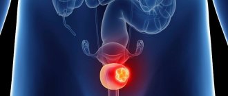
The key difference between malignant and benign tumors is their tendency to metastasize. Metastasis is a secondary tumor site that occurs in a distant organ or tissue as a result of the movement of cancer cells from the primary site. Each type of cancer is characterized by certain localizations of metastases. The liver, lungs, bones, and brain are most often affected. The spread of tumor cells can occur in the following ways:
- Through the blood (hematogenous metastasis).
- Through lymphatic ducts (lymphogenous metastasis).
- Along nerve fibers (perineural metastasis).
- Upon contact with other organs and tissues (contact metastasis).
In some cases, a combination of several options may occur.
Among other features of malignant tumors, there is a tendency to relapse, difficulty in treatment and a significant impact on body functions due to metabolic disorders. In particular, cancer cells actively absorb glucose, vitamins and other nutrients, disrupt natural biochemical reactions and release toxins that are harmful to healthy cells. As a result, such general symptoms of cancer as cachexia (exhaustion), hypoxia, anemia, and intoxication syndrome develop.
Removal of benign skin tumors
Dermatologists are convinced: it is necessary to get rid of benign formations, with the exception of small scatterings of moles or other minor defects throughout the body. This is especially true for the face, because the growth attracts attention, spoils the overall impression and gives a lot of unpleasant emotions to its owner.
There are several methods for removing benign skin tumors:
- Electrocoagulation
Under local anesthesia, the growth is cut off with a special surgical coagulator, which creates a high-frequency current. Simultaneously with removal, the tissues are soldered together, which avoids bleeding and infection. The crust at the treatment site disappears after 7-10 days, sometimes leaving a slightly noticeable scar. The method is effective if the defect is small.
- Cryodestruction
Liquid nitrogen can only be applied to formations in the upper layers of the epidermis. If it is flat, apply applications with liquid nitrogen. In case of deeper occurrence, a cryodestructor is used. After the procedure, the body begins a reaction of rejection of the treated defective tissue. The resulting crust disappears and heals within a month and a half.
- Laser removal
A powerful beam of light destroys neoplasm cells and evaporates them from the skin. The laser does not affect healthy neighboring tissues. The manipulation is carried out under local anesthesia, it is low-traumatic and bloodless. Laser treatment parameters are selected individually. Damaged tissues are evaporated layer by layer until the beam reaches healthy skin. The formed crust disappears on its own after 1-2 weeks.
- Radio wave method
High frequency radio waves cut and coagulate tissue. The crust disappears after a week. The method is contraindicated in patients with a pacemaker, herpes, or elevated body temperature.
- Surgical removal of benign skin tumors
This method is used when the growths are too large and other technologies cannot cope. The formation is excised with a scalpel and removed, capturing a small area of healthy skin. The scar after surgery takes several weeks to heal, but the wound requires long-term careful care. The description of this traumatic technology is not encouraging, so it is better to choose another method to eliminate defects on the face.
On the face, neoplasms are removed using laser or cryodestruction
Why do tumors occur?
Until now, scientists have not been able to establish the exact causes and mechanisms of tumor formation, however, thanks to modern molecular diagnostic methods, important details of the process of oncogenesis are known. Disruption of the process of cell division and differentiation develops as a result of changes in DNA molecules. Such a failure can be provoked by various factors called carcinogens. They are divided into three main groups:
- Chemical carcinogens. They are represented by harmful substances that are formed during the combustion of tobacco, are used in production, are present in food, and are released into the external environment. This group includes a very large number of substances, the number of which is approaching 2000. From this list, there are several dozen chemical carcinogens that have the most significant effect on the body and most likely lead to the development of tumors.
- Physical carcinogens. These include radioactive, x-ray and ultraviolet radiation in doses that exceed permissible values. Physical carcinogens, like chemical ones, do not directly affect the formation of tumor cells. They provoke various changes, including disruption of the DNA structure, which cause the development of various neoplasms.
- Biological carcinogens. This group is represented by specific viruses that promote oncogenesis. This group includes some types of human papillomavirus, retroviruses, adenoviruses, hepatitis B and C viruses, etc. Biological carcinogens, unlike the two previous groups, directly affect the genetic apparatus of the cell and change it.
Understanding the mechanisms of tumor development and the discovery of various carcinogens allowed doctors to develop preventive measures. In particular, in order to reduce the risk of developing benign and malignant neoplasms, it is necessary to stop smoking, monitor your diet, avoid working in hazardous working conditions, strengthen your immune system, etc.
Book a consultation 24 hours a day
+7+7+78
Classification of tumors
The simplest and most understandable classification of benign neoplasms. In this case, it is only necessary to know the tissue from which the tumor developed. Taking this feature into account, a diagnosis will be formed. If a benign neoplasm developed from cartilaginous tissue, then the diagnosis will be “chondroma”, from glandular tissue - adenoma, etc.
The division of malignant tumors is much more complex. New growths from epithelial tissue are called cancer or carcinoma (follicular cancer, adenocarcinoma). If the tumor developed from connective tissue, then it is classified as sarcoma (chondrosarcoma, myosarcoma), from nervous tissue - glioma, etc.
The next indicator that is taken into account when making a diagnosis is the degree of cell differentiation. It is graded on a Grade scale and includes the following options:
- GX—cell differentiation cannot be determined.
- G1 - cells are highly differentiated.
- G2 - cells are moderately differentiated.
- G3 - cells are poorly differentiated.
- G4 - cells are undifferentiated.
As the score increases, the aggressiveness of the tumor increases, as does the degree of its malignancy.
Depending on the characteristics of the primary lesion, the presence of metastases in regional lymph nodes and distant organs, malignant tumors are classified according to the international TNM system. The letter T describes the size and growth characteristics of the primary tumor, N indicates the presence or absence of metastases in nearby lymph nodes, and the letter M indicates metastases in distant organs. For each letter a certain index is assigned, as a result the diagnosis may look like this - T3N1M0 or T1N0M0, etc.
Based on the TNM classification, malignant tumors are divided into stages. A localized primary lesion, which does not extend beyond the organ and is small in size, corresponds to the first stage. If the tumor grows into adjacent organs or has distant metastases, then the fourth stage is set.
Diagnosis via injection
The diagnosis of a benign neoplasm is usually made on the basis of a clinical examination. In difficult cases, cytological puncture with a thin needle can be used to distinguish them from other tumors. Such a puncture is practically painless; on the other hand, it can provide very valuable information for the diagnosis. Due to the fact that our Center has its own clinical and biochemical laboratory, puncture results can be obtained in the shortest possible time. Timely ultrasound examination, which is used in our Center along with other diagnostic methods, can also be of great importance for making an accurate diagnosis.
Diagnostic methods
For modern oncology, the problem of timely detection of tumors, primarily malignant ones, continues to be relevant. In the initial stages, neoplasms do not manifest themselves in any way. The first signs are noted as the tumor grows and the functions of certain organs are impaired. In such cases, patients complain of pain, discomfort, weakness and other nonspecific symptoms, which depend on the exact location of the tumor.
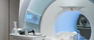
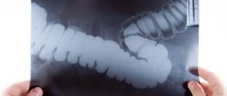
If the tumor is located superficially, for example, in the breast tissue, then it can be palpated. This feature makes diagnosis a little easier, but in order to understand the true nature of the tumor, it is necessary to undergo a comprehensive examination. It includes various imaging methods that help detect the primary lesion: CT, MRI, ultrasound, endoscopy, x-ray.
At the next stage, the doctor is faced with the task of determining the type of tumor, the degree of cell differentiation and other important features. A biopsy, the gold standard for diagnosing tumors, can answer these questions. As a rule, without a biopsy in oncology it is impossible to make an accurate diagnosis and select treatment.
Additionally, to assess the general condition of the patient or to determine sensitivity to a particular drug, laboratory and molecular genetic research methods are prescribed. The exact diagnostic program is always selected individually.
Treatment of tumors
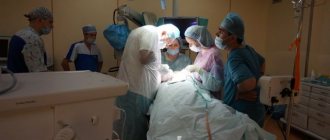
The second pressing problem in oncology is the effective treatment of tumors. If a benign tumor can be removed surgically relatively easily, then with a malignant process this is not always possible. Due to the absence of pronounced symptoms, tumor invasion into neighboring organs and vital vessels, and a tendency to metastasize, many patients remain inoperable. In such situations, conservative treatment is used, which includes methods such as chemotherapy, radiation therapy, targeted therapy, immunotherapy, etc. However, in this case, the patient’s chances of a full recovery are reduced. If the tumor has spread beyond the organ, then conservative therapy in most cases will help delay relapse and increase the patient’s life expectancy.
The most effective treatment is possible when the tumor is detected at the earliest stage, after surgical removal of the primary lesion, after which, for some diseases, conservative treatment methods can be used.
Currently, all the efforts of doctors and scientists are focused on solving two pressing problems. On the one hand, methods for early detection of cancer are being developed, and on the other, new and effective treatment methods are being developed.
Book a consultation 24 hours a day
+7+7+78
Ball on a leg
Fibroma consists of fibrous connective tissue. If smooth muscle fibers are mixed with this component, the tumor is called fibromyoma. This benign formation grows slowly, over the years, has a dense consistency, in most cases it is spherical in shape; sometimes “sits” on a stalk (polyp). Treatment for atheroma is surgical only. In our Center, surgery to remove such a formation is performed on an outpatient basis, and it is completely painless. The wound heals very quickly (if the atheroma is really small and shallow). The formation of scars primarily depends on the individual properties of human tissue; usually, after such an intervention there are practically no noticeable marks.