Surgeon
Bohyan
Tigran Surenovich
Experience 36 years
Surgeon of the highest category, Doctor of Medical Sciences, member of the International Association of Surgeons, Gastroenterologists and Oncologists
Make an appointment
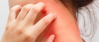
Hemangioma is a benign vascular formation that appears due to an embryonic disorder in the development of blood vessels. The tumor appears on any part of the skin and looks like a red, purple or bluish spot rising above the surface of the skin. Most often detected at birth or formed during the first weeks of life. Treatment is surgical and conservative.
Types of acquired red moles on the body
- Simple (capillary). Proliferation of newly formed capillaries, small venous and arterial vessels. Looks like a red spot.
- Cavernous. A spongy cavity with blood - a red or bluish nodule. Often forms under the skin.
- Branched (racellose). A plexus of tortuous dilated capillary trunks. They pulsate, noise and trembling are detected. It is rare and occurs on the extremities or face. If injured, life-threatening bleeding may occur.
How to identify hemangioma? Press on top of it and it should fade or disappear.
What is infantile hemangioma?
This is a tumor-like formation of a qualitative nature, which is formed by fragments of blood vessels of varying thickness. The nature of the origin is as follows: during the process of intrauterine development, the formation of blood vessels is disrupted. As a result, almost immediately after the birth of the child, or some time (about a month) later, a hemangioma appears on his body.
Cavernous hemangioma
By the way. Most often, this formation appears on the skin, at any point. But in 20% of cases, the tumor can localize itself to internal organs, muscle and even bone tissue.
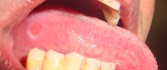
The neoplasm looks like this:
- a thickened tumor-like spot of various sizes and degrees of elevation above the surface of the skin or depression;
- the color is either pale burgundy, closer to pink, or bright burgundy;
- the edges are uneven;
- the structure is dense;
- composition – atypical and normal cells.
New growth on a child's cheek
Important! The key point is that although this is a full-fledged tumor, it is always benign. Therefore, there is no need to do anything. Over time, in most cases, the hemangioma subsequently resolves on its own, without a trace.
In children, quite often hemangioma simply resolves over the years.
Of course, exceptions, deviations and anomalies occur, since each person’s body is individual, including a newborn baby. The most important thing for parents when a neoplasm is detected is not to panic and carefully study the issue of how the local pediatrician and this material can help them.
Are red moles dangerous?
In themselves, these formations are harmless and are not precancers.
If you have a lot of red moles on your body, the cause may be a serious liver or pancreas disease. Pay attention to this - this is a reason for examination.
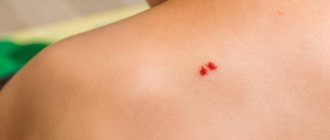
Problems may arise in case of traumatization of hemangiomas. Even fairly small formations threaten heavy bleeding, which is not easy to stop.
Clinical case
Patient L, 26 years old, presented with a traumatized mass in the axillary region. According to her, she tore off a convex hemnagioma with the edge of a rigid corset of a wedding dress almost a few minutes before the start of the wedding ceremony. The hemangioma bled very heavily and a large blood stain appeared on her white dress. She had to wear the witness's jacket over her wedding dress. It was in such a strange outfit that the wedding took place.
Hemangioma in children - symptoms and treatment
Currently, there are various ways to treat infantile hemangiomas. Many of them have already lost their significance, others are successfully used at the present time. The main vector of treatment is active observation or conducting one or another type of therapy from the moment of diagnosis.
When choosing treatment tactics, you should be guided by several factors:
- age of the child (the younger the patient, the higher the risk of further growth of the hemangioma);
- type of hemangioma and its location;
- presence of complications;
- parents’ desire to conduct this or that therapy;
- doctor's experience [24].
To help doctors decide on treatment tactics for infantile hemangioma, Russian surgeons D. A. Safin and D. V. Romanov proposed a special rating scale. With its help, you can assess the need for drug treatment with beta-blockers. This scale takes into account both the age of the child and the location of the hemangioma, as well as its size, quantity and thickness (presence of pathological volume) [28].
Rating scale for determining indications for systemic drug treatment of infantile hemangiomas with beta-blockers
| Criteria | Description | Points |
| Age | 0-4 months | 4 |
| 5-8 months | 3 | |
| 9-12 months | 2 | |
| 1 year and older | 1 | |
| Localization | Orbit, nose, lips, ears | 4 |
| Perineum, buttocks, genitals | 4 | |
| Hemangiomatosis | 4 | |
| Parenchymal organs and glands | 4 | |
| Scalp and other parts of the face | 3 | |
| Neck | 3 | |
| Joint area | 2 | |
| Hands and feet | 2 | |
| Torso | 1 | |
| Limbs | 1 | |
| Size (diameter) | More than 5 cm | 2 |
| 1-5 cm | 1 | |
| Up to 1 cm | 0 | |
| Pathological volume | Eat | 1 |
| No | 0 | |
| Quantity | 5 or more elements | 1 |
| 1-4 elements | 0 | |
| Complications | Eat | 1 |
| No | 0 |
The doctor examines the patient, evaluates the specified criteria and sums up the points. When the score is from 9 to 14, systemic drug treatment is indicated, and when the score is from 4 to 8, drug therapy is not required.
When assessing the location of a hemangioma, it is necessary to take into account not only the cosmetic defect, but also the risk of various complications. These “critical zones” include the ears, nose, lips and eyelids (orbits of the eye). For example, if the cartilage of the ear is destroyed, a permanent deformation of the auricle will occur, the correction of which will require plastic surgery. When infant hemangiomas are located in the perineum and buttocks, ulcerations often appear that are difficult to heal, so this localization is also critical.
The large size of the tumor indicates an active process of proliferation, which is accompanied by the risk of further growth of the hemangioma and a high probability of various complications.
In the treatment tactics of infantile hemangioma, it is also necessary to take into account the presence of a deep (subcutaneous) component of the tumor. It is not amenable to local treatment and laser treatment, which leads to tumor progression in the deep layers of the subcutaneous fat layer. Therefore, in this case, it is recommended to resort to drug therapy.
The greater the number of hemangiomas on the skin, the more difficult it is to carry out local therapy. The presence of more than five vascular tumors significantly increases the risk of hemangiomas in the internal organs (diffuse neonatal hemangiomatosis).
All methods of treating infantile hemangiomas can be divided into two categories:
- Conservative (non-invasive) treatment : oral medication, lotions, watchful waiting (time treatment) and laser treatment.
- Surgical (invasive) treatment : injections (sclerotherapy) or surgery (removal of hemangioma).
Conservative (non-invasive) treatment
Waiting tactics
This treatment was widely practiced before the advent of beta blockers. Now this method can also be used, but it is necessary to take into account: the younger the child, the higher the risk of tumor growth. Watchful waiting requires frequent and systematic follow-up examinations to determine the possible growth of hemangioma.
Drug treatment
Beta blockers. In 2008, French dermatologist C. Leaute-Labreze discovered the effect of propranolol on infant hemangiomas. From this period began the “golden age” of beta-blockers in the treatment of infantile hemangiomas. Given the low risk of side effects and the high effectiveness of treatment, they have become the “first line” of treatment for this disease.
The exact mechanism of action of beta-blockers is still unknown. Presumably, it includes vasoconstriction, suppression of VEGF-A (vascular endothelial growth factor), stimulation of apoptosis (the natural “disassembly” of pathological tissues). Studies have revealed the presence of β2-adrenergic receptors and VEGF-A in the capillaries of expanding infantile hemangioma, which decrease when β2-adrenergic receptors are suppressed.
Beta blockers can be used topically as a lotion or systemically by administering the medication orally. For local treatment, timolol-based eye drops or eye gel are used. For systemic therapy, the drugs Propranolol or Atenolol are used. Each of them has its own characteristics in dosage, frequency of administration and the risk of side effects.
Comparison of the pharmacological activity of the drugs Propranolol and Atenolol.
| Indicators of pharmacological activity | Propranolol | Atenolol |
| Bioavailability | 20-30 % | 40-60 % |
| Half-life of the drug | 3-5 hours | 6-9 hours |
| Lipophilicity (permeability through the cell membrane) | +++ | – |
| Removing the drug from the body | Liver 100% | Kidneys 90%, liver 10% |
Steroid hormones are one of the oldest methods of drug treatment for infantile hemangiomas. This method has been used from 1960 to the present and is a “second line” of therapy. Until 2008, it was the main method of treating hemangiomas, especially with active tumor growth.
The mechanism of the effect of glucocorticosteroids on infantile hemangioma is still not completely clear. However, it is known that steroid hormones influence adipogenesis, inhibit the formation of new vessels and reduce the production of proangiogenic proteins (VEGF-A, etc.).
Steroid hormones in tablets can be used as systemic drug treatment. Given the high risk of side effects of glucocorticosteroids and the emergence of beta-blockers, this method has become much less common. The main indications for its use are complicated hemangiomas and unresponsiveness of the formation to treatment with beta-blockers. The average course of treatment is from 4 to 12 weeks (maximum dose). Sometimes steroid hormone therapy can last up to 9-12 months of the child’s life.
It is also possible to inject glucocorticoids into the tumor. Typically, this treatment uses Triamcinolone injections or a mixture of Triamcinolone and Betamethasone. As a rule, such treatment is prescribed for small volumetric formations.
Cytostatics (Cytoxan, Vinblastine and Avastin), according to foreign authors, help stop the division of tumor cells in metaphase. However, this method has not found application in Russia, since Cytoxan acts on the growth of sensitive, rapidly proliferating cells, thereby inhibiting the erythrocyte lineage of the blood and the development of the egg. There is also an opinion that after using Cytoxan there is a risk of developing a secondary malignant tumor - angiosarcoma.
Vincristine is a cytostatic drug, an alkaloid from the pink periwinkle plant (Vincarosea). It causes apoptosis (the regulated process of cell death) of endothelial cells and reduces the production of their growth factors. As a rule, treatment with Vincristine is carried out for vascular pathology, which is not a true infantile hemangioma, but is associated with kaposiform hemendothelioma or tufted angioma with Kasabach-Merritt syndrome. The medication is given weekly through a central catheter.
This treatment method may be useful if steroid hormone therapy does not respond. Its appointment is carried out with the direct participation of oncologists. Side effects of the drug include skin irritation and rash, neurotoxicity, constipation, cranial nerve palsies, bone pain, alopecia, and muscle weakness. Typically, side effects are short-lived.
Interferons (Interferon-alpha-2a, Interferon-alpha-2b and Imiquimod - 5% Aldara ointment) stimulate the secretion of interferons, which suppress endothelial and fibroblast growth factors, and also promote tumor necrosis. They are used when steroid therapy is ineffective. The effect of these drugs is noticeable only after four months of use.
Since interferons cause a lot of complications (anemia, neutropenia, hypothyroidism, fever, neuroplegia), their use in the treatment of young children is not justified.
Laser correction
To treat vascular pathology, lasers with wavelengths of 532 and 585 nm, less often 1064 nm, are used. These lasers do not damage the skin, therefore they are considered conservative treatment.
The operation of lasers is based on the theory of selective photothermolysis, which was described in 1980. Laser radiation has a constant wavelength, but is absorbed differently by tissues. This is explained by the presence of chromophores (water, melanin and hemoglobin) in the skin. The result of this energy absorption is heating of the tissue. For example, wavelengths of 532 and 585 nm are best absorbed by oxyhemoglobin, due to which isolated heating of the vessel occurs, leading to damage to its endothelium without harm to surrounding tissues. As a result, the vessel closes.
After laser treatment, there are no scars or scars.
Surgical (invasive) treatment
Sclerosis of hemangioma
This method can be used for small surface formations. To glue tumor vessels together, a special drug is injected into them, which damages the endothelium of pathological capillaries and stimulates the formation of a blood clot. As a result, a fibrous subcutaneous scar is formed [4][5][7][16].
This method requires stages - carrying out a course of treatment at certain time intervals, for example, performing sclerotherapy once every 1-1.5 months. This is due to the rapid blood flow in the hemangioma, which reduces the time of exposure of the sclerosant to the endothelium, thereby reducing its effect.
The proposed sclerosants, for example, Aethoxysclerol and Fibro-vein, are intended for sclerosis of the veins of the lower extremities. A prerequisite for their use is the introduction of the drug into the bloodstream. They can cause disruption of microcirculation around the hemangioma up to tumor necrosis, which will lead to gross cicatricial changes. Alcohol should also not be used for sclerosis of vascular formations. Although it has a pronounced sclerosing effect, its use is often accompanied by side effects.
Cryodestruction (removal with liquid nitrogen)
The tumor focus can be destroyed using liquid nitrogen at a temperature of −195.6 °C. It is a colorless and odorless liquid that is sterile, non-toxic, inert to biological tissues and non-flammable. During cauterization of the hemangioma with liquid nitrogen, the tumor focus is clearly demarcated, which is replaced by an organotypic regenerate by 21-30 days after cryotherapy.
It is impossible to stop the growth and completely eliminate extensive and deep hemangiomas that have abundant blood circulation using cryodestruction. Usually it does not bring the desired result, as it leaves cosmetic defects, and in many cases leads to increased growth of hemangioma. Therefore, this method is best used in the treatment of local, non-extensive hemangiomas [5][8][21].
Surgical method of treatment
The radicality of surgical treatment of hemangiomas lies in the complete removal of all tissue affected by the tumor. Previously, it was believed that surgical removal of a tumor was dangerous due to complications: heavy bleeding during surgery and damage to the nerves, lymph nodes, and large arterial and venous trunks involved in the tumor. Now, with the advent of modern research methods, it has become possible to simultaneously remove a vascular tumor within healthy tissues, without affecting important anatomical structures [5][7][8].
Not all hemangiomas are subject to surgical removal: it should be used only in the presence of life-threatening conditions, for example, bleeding or when the airways are blocked, but most often it is resorted to at the final stage of involution, when there is almost no blood flow in the hemangioma - most often in 4- 5 years.
The scope of the operation is determined by the ability to perform it without irreversible cosmetic defects, the formation of a rough scar and the risk of dysfunction of nearby organs. In this regard, surgical treatment has its contraindications [5][7][8][21].
Outdated treatment methods
Radiation therapy (RBRT)
The essence of this method is to irradiate an area of skin with X-rays from a short distance. Such radiation is absorbed predominantly in superficial tissues and is effective only for infantile hemangiomas located exclusively on the surface of the skin.
As a result of X-ray therapy, “scars” form in the hemangioma, and telangiectasias (spider veins) form on the surface of the skin. As a result, to obtain a good cosmetic result in the future, either plastic surgery or laser treatment of the area of close-focus therapy is required [7][10].
How can the disease be diagnosed?
The capillary form in the vast majority of cases is diagnosed based on history and examination. The pediatric surgeon finds out when the tumor was first noticed, whether it has changed in shape, color and size, and whether there are signs of regression.
If this data is not enough and there are doubts about making a diagnosis, the specialist prescribes additional studies:
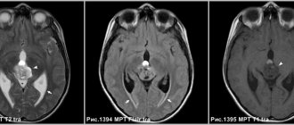
- ultrasonography;
- Dopplerography;
- computed tomography with contrast;
- magnetic resonance imaging;
- biopsy of the tumor site
- And so on.
Zolotov Sergey Alexandrovich
- Candidate of Medical Sciences, surgeon, specialist in laser surgery.
- From 2003 to 2009 completed training at the State Budgetary Educational Institution of Higher Professional Education "Russian National Research Medical University" named after. N.I.Pirogov Ministry of Health of Russia.
- From 2009 to 2011, he completed his residency at the Moscow State Budgetary Healthcare Institution, Research Institute of Emergency Pediatric Surgery and Traumatology, specializing in Pediatric Surgery.
- In 2011, he completed training under the program of additional professional education of doctors in laser medicine at the State Scientific Center for Laser Medicine of the Federal Medical and Biological Agency of Russia. From 2011 to 2014, he studied full-time graduate school at the Moscow State Budgetary Healthcare Institution Research Institute of Emergency Pediatric Surgery and Traumatology.
He has extensive experience in removing various benign formations of the skin and subcutaneous tissue using laser surgical methods.
More than 5 years of experience in surgery.
Forecast
Possible consequences of the disease
Hemangiomas can cause necrosis (death) of tissue, so the lesion becomes an entry point for infection.
The addition of a purulent process can cause sepsis. With hemangioma of bones, their destruction is possible. In addition, the process can contribute to blood clotting disorders and the formation of blood clots. Malignancy is possible - degeneration of hemangioma into cancer. With timely consultation with a doctor, the neoplasm is successfully treated in 100% of cases Source: What a pediatrician needs to know about infant hemangiomas. Zakharova I.N., Kotlukova N.P., Roginsky V.V., Sokolov Yu.Yu., Zaitseva O.V., Maykova I.D., Idrisova G.R., Pshenichnikov I.I.: Medical Council , 2021.
Price list for medical services of the laser surgery department
Consultation
| Name | Cost in rub. |
| Initial consultation | 2 000 |
Research
| Name | Cost in rub. |
| Cytological examination | 500 |
| Histological examination | 2 800 |
Anesthesia
| Name | Cost in rub. |
| Application anesthesia | 500 |
| Infiltration anesthesia | 500 |
| Conduction anesthesia | 1 500 |
Bandages
| Name | Cost in rub. |
| Adhesive bandage | 300 |
| Aseptic dressing | 500 |
| Bandage with medicines | 1 000 |
Removal of formations using laser radiation
| Anatomical zone | Cost in rub. for 1mm in maximum diameter |
| Scalp | 500 |
| Hands, forearms, feet, legs | 500 |
| Neck, chest, abdomen, back, armpits, shoulders, hips, crotch | 600 |
| Forehead, temporal region, ears, postauricular region | 600 |
| Eyebrows, nose, nasolabial triangle, chin, cheeks, cheekbones, red border of lips | 700 |
| Lower upper eyelids | 850 |
| Ciliary edge of the eyelids | 1 000 |
| External genitalia - single elements* *per 1 mm. 600 rub. | 1 000 |
Removal of papillomas with a diameter of up to 2.0 mm
| Name | Cost in rub. for a unit |
| Neck, chest, abdomen, back, armpits, shoulders, hips, crotch | 300 |
| Lower upper eyelids | 500 |
| Ciliary edge of the eyelids | 1 000 |
Scar treatment
| Name | Cost in rub. |
| Drug injection into the scar | 1 200 |
| Laser dermabrasion (resurfacing) per cm² | 1 200 |
Removal of vascular formations using laser radiation*
| Name | Cost in rub. |
| Photodestruction of the vessels of the wings of the nose | from 5 000 |
| Photodestruction of cheek vessels | from 10 000 |
*exact cost depends on capillary density, repeated stage of vessel destruction minus 30%
Treatment of ingrown toenails using laser radiation
| Name | Cost in rub. |
| Marginal resection of the nail plate | 6 000 |
| Removal of hypergranulations | 1 900 |
| Nail plate removal | 3 500 |
| Complex plastic surgery for ingrown toenails | 8 500 |
Removal of lipoma, atheroma
| Name | Cost in rub. |
| Lipoma removal | 18 000 |
| Removal of atheroma up to 5 mm | 6 000 |
| Removal of atheroma from 5 to 10 mm | 12 000 |
| Removal of atheroma more than 10 mm | 18 000 |
Mesenchymoma
Mesenchymoma
– a malignant formation related to sarcoma. Angiosarcoma and liposarcoma can be found in its composition.
The exact causes of the occurrence have not been clarified. Perhaps this is the effect of carcinogens on the fetus. Risk factors are:
- Vibration, hypothermia and ionizing radiation;
- Chemical exposure – toxic substances, drugs;
- Bacterial and viral infections.
The location is predominantly the chest and abdominal cavity, mediastinum and retroperitoneal space. The formation is manifested by pain, cough, heartburn, shortness of breath and a feeling of fullness.
Diagnosis is carried out using radiography, ultrasound, MRI, biopsy and endoscopic examination. Treatment is carried out surgically. Auxiliary methods are radiation therapy and chemotherapy.
Possible complications
With combined hemangiomas, deep ulcerations occur in 16% of cases. Ulcers are localized in the skin folds, groin, and lips, since the skin moisture is increased in these areas. After healing it is possible:
- scar formation;
- change in the shape of the lip, ear;
- partial destruction of cartilage.
Due to trauma to the tumor, bleeding occurs. In some cases it is difficult to stop them. Another common complication is dysfunction of the organ next to which the formation is located. When localized in such areas, hemangioma must be treated (most often with its removal) to avoid the risk of complications.
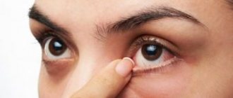
The most dangerous tumor is located:
- in the eye area - threatens the development of strabismus or partial loss of vision;
- neck - can compress the larynx, trachea and interfere with breathing;
- spine - causes acute pain;
- nose and ears - possible destruction of the nasal and ear cartilages;
- in the perineum, armpits - due to constant friction of the skin, the tumor may bleed;
- in the sacral area - a complication may be damage to the spinal cord with atrophy of the muscles in the legs, disturbances in the functioning of the intestines, and the genitourinary system.
The eyes are a dangerous location for hemangioma. Photo: JOVR / ResearchGate (Creative Commons Attribution-NonCommercial 3.0 Unported)