Cysts of the pineal gland (epiphysis) are common, asymptomatic, and in most cases are an incidental finding. Cysts, especially large and atypical-looking ones, are difficult to distinguish from cystic tumors, so patients with suspicious changes should undergo long-term observation. On MRI or MRI, a pineal gland cyst appears as a single-chamber fluid formation with the density of cerebrospinal fluid or with a signal intensity similar to that of cerebrospinal fluid. Peripheral contrast enhancement is typical for most cysts, and rim-shaped calcifications are found in 25% of cases.
Get an MRI of the pineal gland in St. Petersburg
Morphology
Germinoma (extragonadal seminoma) - arises from ectopic pluripotent stem cells that have not migrated from the yolk sac of the ectoderm to the gonads. It is a non-encapsulated, expansively growing formation, sometimes with infiltration (while there is edema in the adjacent areas of the brain) [2].
The structure of all germinomas is relatively homogeneous; there may be small cysts. In the center of the formation there is solid calcification, which can be located eccentrically, but is always surrounded by the tumor stroma. During the growth process, the tumor can infiltrate the thalamus and quadrigeminal plate, metastasize through the subarachnoid space, ventricular ependyma and spinal cord [2]. Spinal spread has been demonstrated at the time of diagnosis in 1/3 of patients [48]. On CT, this tumor appears as a → or ↑ lesion.
Fig. 1393-1395
A mass in the pineal region with small cysts in the stroma (arrow heads in Fig. 1393, 1395), accompanied by occlusive hydrocephalus with dilatation of the ventricular system (arrows in Fig. 1393) and transependymal liquor soaking (arrows in Fig. 1394).
When the disease manifests as bifocal masses (i.e., with simultaneous involvement of the pineal region and hypothalamus), germinoma is the most likely diagnosis [2]. Biochemical studies show a mild increase in serum levels of human β-gonadotropin (β-hCG) and placental alkaline phosphatase (ALP) [137].
2. Pineal tumors and their symptoms
Among the tumors of the pineal gland, the most common types of tumors are:
- pineocytoma is a neoplasm characterized by slow growth, consisting of pinealocytes and accounting for approximately half, or more precisely 45% of pineal tumors;
- pineoblastoma is a high-grade tumor consisting of poorly differentiated cells. It has a high ability to metastasize. This is, in fact, the second largest proportion (45%) of all pineal tumors;
- tumor of the pineal parenchyma (10% of cases), characterized by a poorly predictable course.
Among the symptoms of tumors of the pineal gland
the predominant ones are those associated with the factor of tumor pressure on the posterior parts of the third ventricle and the cerebral aqueduct:
- visual impairment (weakened reaction to light, motor paralysis of the eyeball);
- decreased hearing acuity;
- impaired coordination of movement and gait.
Visit our Neurosurgery page
Age-related features of visualization of the pineal gland on CT
Petrification in the corpus pineale is not detected until 6.5 years of age, and only in 10% of children from 10 to 11 years of age [6]. It is located in the midline, which should be taken into account in the differential diagnosis of lesions of the pineal region.
Fig. 1396-1398
Calcification of the pineal gland with age is of clinical significance. Absence of calcification up to 7 years (Fig. 1396). The appearance of petrification from 7 to 10 years in the form of an area of increased density (arrow in Fig. 1397). Persistent presence of calcific inclusions in the pineal region after 14 years (Fig. 1398).
Treatment
As in the case of oncological diseases of other organs, the greatest effectiveness in the treatment of tumors of the pineal gland is shown by an integrated approach, which involves the combination of several methods of therapy at the same time.
Radiosurgical treatment with the CyberKnife system
Treatment of brain tumors using the CyberKnife system
If the tumor of the pineal gland is small, the patient may be recommended radiosurgery using CyberKnife. This innovative cancer treatment method, which allows the treatment of brain tumors without surgery, has proven its effectiveness all over the world. Unlike traditional neurosurgery, CyberKnife radiosurgery does not require craniotomy, which, combined with the precision of delivering thin beams of radiation, significantly reduces the risk of damage to healthy tissues of this vital organ.
The radiosurgical treatment method involves delivering a high dose of ionizing radiation precisely to the boundaries of the tumor. As a result of receiving high doses of radiation, tumor cells die, and healthy tissues around the tumor focus remain maximally protected - a high dose is formed in the areas of intersection of thin radiation beams, which are specified according to a three-dimensional digital model of the location of the tumor and surrounding structures.
As a rule, radiosurgery of pineal tumors includes one or two sessions lasting up to half an hour, during which the patient remains conscious and can independently leave the clinic at the end of treatment.
Pineal gland tumor: treatment plan using the CyberKnife system
Bloodless, painless, no need for anesthesia, reduced risk of irreparable neurological damage - in comparison with surgical intervention in the deep structures of the brain, CyberKnife treatment allows the patient to carry out treatment on an outpatient basis, without interruption from the daily schedule of his life.
Surgery
The use of CyberKnife in the treatment of brain tumors is not always possible, since there is a limitation on the size of the tumor for radiosurgery.
Therefore, in the case of a large volume of brain tumor, the main treatment method will be surgery. Surgery for brain tumors involves minimally invasive surgery. During the operation, a sample of tumor tissue is taken for histological examination, and the maximum possible volume of the tumor is removed, so that no damage is caused to healthy brain tissue.
Radiation therapy
The modern Elekta linear accelerator with IMRT function allows for gentle radiation therapy with simulated ionizing radiation dose intensity
However, if the tumor is localized in a place of the brain that is difficult to access for surgical intervention, only part of the tumor is removed during surgery, and then the treatment is supplemented with a course of radiation therapy and the use of modern chemotherapy drugs, including to counteract possible metastases of brain cancer.
Radiation therapy for brain tumors is carried out on a high-precision linear accelerator Electa Synergy, equipped with an IMRT system (modeled by the intensity of ionizing radiation dose delivery). This makes it possible to significantly increase the accuracy and safety of treatment, as well as reduce the duration of each radiation therapy session, in contrast to treatment using linear accelerators of outdated models and cobalt devices.
IMRT allows high doses of ionizing radiation to be delivered precisely to the tumor boundaries with minimal impact on nearby healthy brain tissue.
Radiation therapy is also widely used for palliative treatment in cases where the tumor is incurable (multiple brain metastases, large tumor volume affecting critical brain structures, etc.). In this case, radiation therapy can reduce the severity of symptoms and improve the patient’s quality of life.
Contrast enhancement
The contrast enhancement is relatively homogeneous, except for the cyst contents. Solid areas of the tumor and the walls of cysts in tumors are enhanced.
Fig. 1399-1401
Intense contrast enhancement with adsorption of the contrast agent in the tumor stroma and in the walls of tumor cysts (Fig. 1399-1401)
Also, with contrast enhancement, leptomeningeal metastases are determined.
Fig. 1402-1404
Leptomeningeal contact spread of the tumor along the pia mater is determined by areas of contrast enhancement along the grooves (arrows in the figure). A scan of the spinal cord with contrast enhancement is also required to exclude the presence of carcinomatosis of its membranes (Fig. 1404).
Prevention
Preventive measures to prevent the development of a parasitic neoplasm include the prevention of a disease called echinococcosis.
- If there is a dog in the house, it must be dewormed in a timely manner. For this purpose, it is necessary to visit veterinary clinics with the animal or independently give the pet the necessary medications.
- It is necessary to separate the toys of the child and the dog.
- The dog must have its own plate and should not be allowed to share the dishes from which the rest of the family eats.
- The child must be taught to wash his hands after each interaction with a pet. Even if the baby pets his beloved dog just once, he should wash his hands thoroughly with soap.
Differential diagnosis
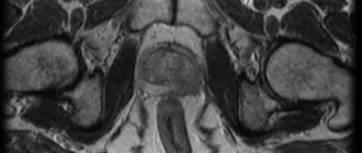
Pineal cyst
A pineal cyst is a benign variant of the normal pineal gland. At the same time, it differs from a tumor by a uniform thin wall (up to 0.2 cm) in which there may be petrification. The contents of the cyst can be → cerebrospinal fluid or gray matter, and can also be ↑ according to Flair (which is due to the lack of movement of cerebrospinal fluid in the cyst, as well as an increased amount of protein or hemoglobin derivatives).
In cases of large cysts or the presence of septations, short-term follow-up is recommended to assess changes in size. Large cysts can also lead to hydrocephalus, and the walls of the cysts accumulate the contrast agent due to the lack of a blood-brain barrier and the presence of pineal gland cells in the walls of the cysts. Pineal gland cysts are not accompanied by clinical manifestations characteristic of tumors: hypersexuality, diabetes insipidus, and rarely lead to hydrocephalus.
Fig. 1405-1407
A benign cyst of the pineal gland in the form of a homogeneous structure (arrow head in Fig. 1405) or with heterogeneous contents in the cavity (white arrow in Fig. 1405), accumulating contrast in the wall (black arrow in Fig. 1405). The pineal gland cyst has a uniformly thin wall, without thickening and without internal septa (arrows in Fig. 1406). On CT, the pineal gland cyst also has a thin wall, with petrification and contents somewhat denser than the cerebrospinal fluid (arrow in Fig. 1407).
Teratoma
Teratomas are characterized by the presence of fat and calcifications. Teratomas may be associated with corpus callosum and Chiari abnormalities.
Fig.1408-1410
Calcification (arrowheads in Fig. 1408, 1409) and fat (asterisk in Fig. 1408) against the background of abnormal development of the corpus callosum are signs of a mature teratoma. On MRI, a teratoma is detected as a formation of ↑ MR signal intensity on T1, in the presented case - in the pineal region (arrow head in Fig. 1410).
Gliomas
Pilocytic astrocytoma (often associated with NF type 1) is more common in this area, but glioblastoma and ependymoma may also be present. Astrocytoma arises from the brain stem, may contain cysts, and spreads to the midbrain peduncles and brain stem. Glioblastoma has central necrosis, a characteristic pattern of contrast enhancement (“corona effect”), and ependymoma arises from the wall of the aqueduct, concentrically stenotic it and elevating the quadrigeminal plate.
Fig.1411-1413
Pilocytic astrocytoma against the background of neurofibromatosis type I has unclear contours, diffusely spreads in the brain (arrowheads in Fig. 1411) and cysts are often found (asterisk in Fig. 1411). Glioblastoma is characterized by a ring-shaped pattern of contrast enhancement with central necrosis (arrows in Fig. 1412). Ependymoma, arising from the ependyma of the aqueduct, is accompanied by its blocking in the form of a mass in the lumen of the Sylvian aqueduct (arrows in Fig. 1413), complicated by occlusive hydrocephalus (arrow heads in Fig. 1413).
Pineocytoma
Pineocytomas are characterized by the deposition of petrification along the periphery (in the wall), and germinoma is always overgrown with petrification. In addition, pineocytoma grows slowly and never metastasizes.
Fig. 1414-1416
Pineocytoma is characterized by the presence of cysts in the stroma (arrow head in Fig. 1414) and multiple small petrificates along the edge of the formation (arrows in Fig. 1415, which is typical for a parenchymal tumor of the gland (arising from the parenchyma of the gland itself), thereby causing the “explosive" type germinoma overgrows the pineal gland, penetrating into the brain tissue, which leads to the preservation of central calcification in the form of a focus↑density on CT (arrow in Fig. 1416) and loss of the MR signal on MRI, which is especially demonstrably revealed on pulse sequences with gradient echo.
Metastasis
Fig. 1417-1419
Metastasis is accompanied by pronounced perifocal edema (arrows in Fig. 1417, 1419) and intensely homogeneous or ring-shaped accumulation of the contrast agent (arrow head in Fig. 1418).
Varicose veins of Galen
Fig. 1420-1422
Varicose dilatation of the great cerebral vein (vein of Galen) is a congenital vascular malformation, accompanied by pronounced dilatation of the indicated vessel (arrows in Fig. 1420, 1421) with a characteristic loss of the MR signal (loss of the MR signal from moving intravascular blood on spin-echo pulse sequences) and visualization of blood flow (Fig. 1422) on time-of-flight pulse sequences.
Meningioma and cavernous angioma (rarely occurring formations in this area).
3. Pineal cyst and its symptoms
>The cyst is a round-shaped formation filled with liquid contents produced by the pineal gland and is completely benign. It should be noted that the cyst never develops into a malignant tumor. In very rare cases, this neoplasm can have a negative impact on the functions of adjacent brain structures, and therefore is generally asymptomatic. Cysts are usually diagnosed accidentally during a medical examination.
In cases where the cystic formation reaches a large size and begins to put pressure on the surrounding brain structures, the following symptoms may occur:
- causeless headaches;
- nausea, vomiting;
- double vision, unfocused picture;
- impaired motor coordination, unsteady gait;
- hydrocephalus (if the circulation of cerebrospinal fluid is impaired due to compression of the duct);
- mental disorders.
About our clinic Chistye Prudy metro station Medintercom page!