A hematoma is an accumulation of blood in the tissues of the human body, and its occurrence is caused by damage due to falls and blows. In some cases, the appearance of a hematoma is not associated with physical effects, but is explained by the presence of a specific disease in a person.
In most situations (this applies to small hematomas), serious consequences do not occur, and the inflammation itself disappears after some time. However, there are often cases that require medical consultation and qualified treatment. Ignoring a bruise can lead to the formation of a cyst or tissue necrosis.
General information
Hematoma is a post-traumatic hemorrhage in a limited volume into soft tissue with the formation of a cavity filled with blood.
Habitual hemorrhages in the upper layers of soft tissues are often called “bruises” in everyday vocabulary, which is due to the bluish-purple color of the skin at the sites of hemorrhage. Most often, a bruise is a consequence of the rupture of several small capillaries resulting from a bruise. By bruise we mean injury to various soft tissues (skin, subcutaneous fat, muscle tissue and blood vessels) without violating the integrity of the skin. Most often, a hematoma is one of the clinical signs of a bruise resulting from domestic trauma. In most cases, hematomas go away on their own within 10-15 days and no medical help is required. With more extensive and deep hematomas (subfascial, intermuscular, subperiosteal), the function of the limb may suffer and their appearance may be accompanied by pain and swelling, which requires consultation with a traumatologist, and in cases of infection and suppuration of the hematoma, surgical treatment.
Hematomas appear especially often in children. In most cases, bruises on a child’s legs appear due to increased physical activity and frequent trauma to soft tissues. Much less often, bruises in a child are a manifestation of pathology. Also, frequent hematomas are typical for athletes due to high physical activity and for elderly people, the walls of blood vessels in which lose their elasticity and the connective tissue becomes thinner. In addition, even insufficient intake of vitamins C , D , and K .
Why it is not recommended to treat a hematoma on the leg at home
If there is large accumulation of blood under the skin, it must be removed. There are enough videos and articles on the Internet describing this procedure at home, but you absolutely cannot follow such advice. Firstly, a person risks damaging his leg even more and disrupting its normal functioning, even to the point of disability. Secondly, and more likely, you can introduce an infection into the body and provoke even greater inflammation. Thirdly, only an experienced doctor using special equipment will do this carefully, but independent attempts can permanently disfigure the skin and leave unaesthetic scars on it.
Pathogenesis
The provoking factor may be damage to the vascular wall as a result of a bruise, incomplete cessation of bleeding, disruption of the thrombus formation process, which leads to the outflow of blood from the vessels into the surrounding soft tissue and the formation of a cavity (hematoma). Bleeding from a hematoma in most cases stops on its own, which is caused by an increase in blood pressure in the cavity of the resulting hematoma, which exceeds the pressure in the damaged vessel and mechanical compression of the lumen of the bleeding vessel by loose fiber infiltrated with blood, and the resulting clots of coagulating blood quickly close the wall of the damaged vessel. At the same time, under the influence of tissue thrombokinase, some of the blood in the hematoma cavity also coagulates. Bleeding stops especially quickly in cases of venous hematomas.
Resorption of the hematoma occurs in stages. The resulting blood clots gradually become dense and decrease in volume due to the reabsorption of blood serum. Dead leukocytes, disintegrating, release various proteolytic enzymes, which soften and dilute the fibrin of the blood clot and destroy the red blood cells contained in it. Then the red blood cells become discolored and they disintegrate into lumps and grains, which, together with other products of cellular decay, undergo a process of phagocytosis by the structures of the reticuloendothelial system, which proliferate in the circumference of the hematoma.
Hemoglobin from erythrocytes gradually dissolves in tissue fluid and gradually turns into methemoglobin , which gradually transforms into verdohemoglobin (green) and, when disintegrating, turns into hemosiderin (yellow pigment), which is visually manifested by a change in tissue color, which is especially pronounced on non-pigmented skin with a superficial hematoma ( the so-called “bruise bloom”).
Color palette of superficial hematoma at different stages of the healing cycle
Initially, the superficial hematoma becomes red in color as oxygen-rich blood accumulates under the skin. After 1-2 days, the blood, gradually losing hemoglobin, changes color to blue-violet. After 5-10 days, the color of the skin changes to yellow or green, this is due to biliverdin / bilirubin , which are formed during the breakdown of hemoglobin . After 10-14 days, the color of the skin over the hematoma will acquire a light brown tint and begin to disappear. The whole process takes about 15 days.
The vast majority of hemoglobin is secreted by the liver and kidneys. Normally, the bulk of the blood in the hematoma is absorbed back during the coagulation process, enhanced regeneration of connective tissue cells occurs and the hematoma cavity is gradually filled with newly formed connective tissue.
In cases of insufficient absorption of blood accumulated in the hematoma, the process of fibrin formation intensifies, which leads to fibrous degeneration, hyalinization of the hematoma wall, compaction of the newly formed tissue and its adhesion to surrounding tissues, ultimately forming an encysted hematoma. an abscess / cellulitis may develop .
Possible consequences of improper treatment
If a person ignores the formed area of injured vessels for a long period of time or if incorrect measures are taken to eliminate it, irreversible consequences may arise that require serious treatment or even surgical intervention by qualified surgeons.
The most common ones include:
- traumatic cysts;
- tissue necrosis;
- purulent processes in the body;
- subcutaneous accumulations of pus in the affected area.
The above serious consequences can be avoided only if you consult a doctor in a timely manner and prescribe competent treatment. A hematoma on the leg that occurs as a result of a bruise or spontaneously without previous injuries clearly requires treatment.
It is acceptable to independently monitor the condition of the skin and the functionality of the limb in the first 2-3 days after the bruise appears. If there is no positive dynamics after the specified period, the “victim” must immediately consult a doctor to avoid irreversible consequences.
Article design: Oleg Lozinsky
Classification
The classification of hematomas is based on several principles.
- By localization in the soft tissue structure - subcutaneous, subfascial/aponeurotic, intermuscular, subperiosteal.
- According to the nature of the vessel - arterial, venous and mixed.
- Clinical signs are simple, pulsating.
- According to the degree of distribution - limited, diffuse, encysted hematomas.
Soft tissue hematomas are divided into:
- Lungs - appear within 24 hours after the injury. Painless or accompanied by mild pain. No treatment is required, they disappear in 10-15 days.
- Moderate - appear within 5-6 hours after injury, accompanied by severe pain and slight swelling. The motor function of the lower limb suffers slightly. Consultation with a traumatologist is necessary.
- Severe - appear within 2 hours after injury. There is widespread swelling and acute pain, and function suffers. Treatment is needed.
First aid for injury
If you bruise your leg with subsequent formation of a hematoma, you must do the following:
- during the first 15-20 minutes. after injuring an area of a limb, it is advisable to cool the damaged area, ideally with an ice compress or a special medical cold pack intended for use for bruises (if there is no damage to the integrity of the skin);
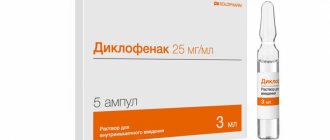
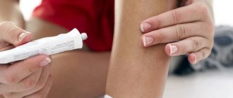
- if there is no tendency to allergic reactions, apply a thin layer to the bruised area with medical products containing heparin;
- limit the load placed on the injured leg by fixing it in a comfortable position for a while.
Qualified specialists do not recommend using anti-inflammatory absorbable ointments and gels earlier than 3 days from the moment of injury, due to the warming effect they have.
If there is an open wound, it is advisable to immediately show the leg to a doctor who can not only identify the presence of a real danger to the victim, but also provide assistance directly in stopping the bleeding.
Causes
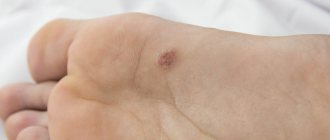
The most common cause of hematomas is soft tissue bruises resulting from domestic/industrial injuries (falls, after being hit with a blunt object, pinched limbs, compression of the foot - a bruise under the nail, often found on the big toe nail). However, a subcutaneous hematoma that occurs for no apparent reason can also be a sign of a number of diseases.
Bruises on legs without impacts
In some cases, bruises do not “fad” on their own, and sometimes patients complain that the bruise appeared on its own without prior injury (for no reason). Bruises on the legs appear especially often for no reason in women. This phenomenon, although commonplace, can be a sign of a number of diseases, among which may be:
- Congenital/acquired coagulopathies resulting from decreased levels of blood clotting factors.
- Vasopathies are disorders of the permeability of the walls of blood vessels caused by exposure to immunoallergic/infectious-toxic agents ( hemorrhagic vasculitis ).
- Thrombocytopenia is a decrease in the number of platelets due to inhibition of their production or increased consumption in DIC syndrome, hemangiomas , thrombocytopenic purpura .
- Thrombocytopathies are changes in the qualitative composition (structure) of platelets, characteristic of myeloma vitamin B12 deficiency , erythremia , liver/kidney disease, systemic lupus erythematosus , with a lack of protein component in the diet.
Also, the reasons why bruises appear on the legs without contusions can be:
- Old age - due to a physiological decrease in the elasticity of the vascular wall caused by age-related changes in the body.
- Vitamin and mineral deficiency, especially vitamins A, C, P and K. Another factor causing bruises may be insufficient intake of cobalt, calcium, and selenium into the body, which is caused by an unbalanced diet, including the practice of strict mono-diet.
- Viral infections (ARVI/flu). Under the influence of an infectious agent and its toxins, the permeability of blood vessels increases significantly.
- Hormonal imbalances/changes. Bruises on the legs can appear during pregnancy , which is associated with a lack of vitamins and increased stress on the lower extremities. Also, bruises on the legs often appear for no reason in women during menopause , which is associated with changes in hormonal levels and negatively affects the permeability of the vascular walls.
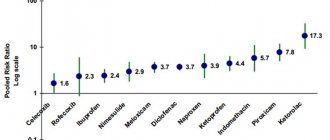
- Taking medications. The appearance of hematomas without blows can occur when taking blood thinning drugs ( Heparin , Aspirin , Thrombo ACC , etc.), headache medications ( Ibuprofen ). A provoking factor may be uncontrolled and long-term use of corticosteroids, NSAIDs ( Diclofenac , Ketanov , Naprasen ), some anti-asthmatic drugs and antidepressants.
- Varicose veins. A common cause of blue spots on the legs. Many patients note that a bruise appeared on the leg without a blow with increased/prolonged load on the lower extremities (long walking/standing/sitting).
What to do if there is a hematoma that occurs without a reason?
The vast majority of hematomas occur as a result of severe bruising or serious damage to bones and joints (fracture, rupture, tear, crack, etc.). However, there are cases of detection of an injured area of blood vessels without an objective reason for its formation.
In such a case, it is advisable for a person who regularly notices such phenomena on his body to examine the body for the presence of pathological processes. First of all, you should make sure that there are no diseases of the venous system, in particular thrombosis and thrombophlebitis.
It is also important to exclude qualitative changes in the connective tissue, usually caused by a general weakening of the body.
Separately, it is worth noting the natural occurrence of hematomas without previous bruises or injuries in people who have been taking steroids or other hormonal boosters for a long time. Such additives can not only negatively affect the functioning of the gastrointestinal tract, but can also result in thinning of blood vessels and vein walls, as well as disturbances in the circulatory system.
Based on the information above, if you are diagnosed with a bruise that has arisen for no reason, you should consult a doctor as soon as possible to identify abnormalities in the body’s functioning and their timely correction.
Symptoms
Clinical signs of hematoma are few. As a rule, immediately after an injury, limited/spread swelling appears, which can quickly increase in the absence of inflammatory phenomena. After 24-36 hours, due to rupture/crushing of small blood vessels of the skin and underlying subcutaneous tissue and soft tissue, bruises appear on the skin, which are clearly visible. The severity of pain can vary from its absence in case of minor household injuries to severe in case of severe injury. When pressing on the skin, pain is limited to the site of the bruise. Movement in the limb is preserved, and the victim can step on the leg. The swelling fluctuates.
After 4-5 days, fibrinous crepitus appears, a dense border ridge is determined along the periphery of the hematoma, and palpation reveals a clear fluctuation in the central part of the hematoma. This symptomatology is due to the presence of liquid blood in the hematoma cavity, the formation of blood clots and the deposition of fibrin on the walls of the hematoma. Local temperatures are elevated. Less commonly, enlargement of the nearest regional lymph nodes occurs. On the lower extremities, below the site of the bruise, a doughy, cold-to-the-touch swelling may appear, formed due to the transudation of blood serum into the tissue. Pain from pressure on the skin is usually limited to the site of the injury. Gradually, the color of the bruises becomes yellowish-green, then slightly brown and disappears.
If the internal hematoma is located deep in the large muscles, then infiltration and swelling forms in the deeper layers; accordingly, the clinical signs are somewhat different: local edema is usually absent, and an increase in the volume of the limb is more typical. When a hematoma becomes infected and a phlegmon/abscess forms, the pain syndrome sharply increases, pronounced local hyperthermia and hyperemia , and symptoms of general intoxication are added.
Diagnosis: when is medical help needed?
In order to avoid situations where traditional methods of treatment are useless and we are talking exclusively about surgical intervention, it is important to promptly determine the severity of a hematoma found on the leg after a bruise.
The most common signs of the need to immediately consult a doctor to prescribe an additional examination in order to establish a diagnosis and determine the correct treatment should be some factors:
They are as follows:
- impossibility or difficulty changing the position of the leg;
- severe pain when trying to touch the affected area;
- rapid spread of swelling over the area of the limb;
- the occurrence of weakness and elevated body temperature;
- loss of consciousness due to pain;
Treatment of a hematoma on the leg after a bruise should begin immediately if the pain leads to fainting. - lack of positive dynamics in the external state of the hematoma after 3-5 days.
The circumstances listed above should be the reason for prescribing an ultrasound, x-ray, MRI or CT scan, a general blood and urine test in order to identify the development of a purulent process in the victim’s body.
List of sources
- Urakov A.L. DIAGNOSTICS OF SOFT TISSUE DAMAGE DURING BLEEDING//Advances of modern natural science. – 2015. – No. 1-6. – pp. 951-957.
- Gumanenko E.K. Military field surgery. 2nd ed., rev. and additional 2012. - 768 p.: ill.
- Kornilov N.V. Traumatology and orthopedics: textbook. Ed. N.V. Kornilova. — 3rd ed., add. and p First Aid: Textbook/Under the general editorship of Doctor of Medical Sciences. Professor Vartanyan F.E. – M.: Russian Society of the Red Cross, 1997, – 215 p. reworked - 2011. - 592 p.: ill.