The visiting day had just begun when they knocked on the office door and asked in a quiet but anxious voice: “Doctor, can I come in?” I invited her: it was a girl who, limping on her left leg, said that her little toe hurt. As it turned out, on the morning of the previous day, Marina (that was the patient’s name) was actively getting ready and, leaving the room, hit her foot on the corner of the door. But, since there was no time, she got dressed and went to work, experiencing pain and terrible discomfort the entire time. In the evening, returning home, taking off my shoes, I saw a bruise and an enlarged little finger. “Tell me, is this a fracture of the little finger?” asked Marina. “Now let’s figure it out,” I replied.
Causes
According to statistics, foot fractures occur in 20% of all bone fractures. And all because we are actively moving throughout our lives, moving from place to place, doing some kind of work. And simply a banal rush, due to loss of attention, turns off our compensatory mechanisms, as a result of which our body loses control, which is why we get injured. As for the little toe, it has a number of anatomical features: firstly, it is the edge of the foot and, when moving the foot, our internal and external surfaces are exposed to the danger of impact. Secondly, the little finger is smaller than all other fingers, it is the thinnest and shortest, which translated from Latin digitus minimus means “smallest”.
Considering all these features, we should not forget about diseases associated with a decrease in bone mass density, which can lead to bone fragility (for example, osteoporosis). And also human age, because the older we get, the more bone strength decreases, this is due to a lack of calcium in the body, because it is the main component of bone tissue.
The adult body contains about 1.2 kg of calcium. Bone contains 99% of the body's total calcium, 87% phosphorus, 60% magnesium and about 25% sodium.
So what are the signs of a broken toe and what to do if it is broken?
Let's return to our patient. I asked Marina to describe what sensations she had after being hit by her little finger and what worried her most, in order to fully understand whether it was a bruise or a fracture. “At first there was a sharp pain, but I could move my little finger. Over time, the pain began to increase, and this made it impossible to move the finger. My little finger began to swell more and more, after which I completely stopped feeling it. It was very difficult to stand on my feet. By evening, my little toe turned blue, and the next morning half my foot turned blue. I got scared and went to the doctor.” How can we understand in our case that the patient has broken his little finger, because fractures are very similar to bruises on the leg.
In order to determine whether it is a fracture or a bruise of the little finger, let's start with their definitions:
A fracture is a partial or complete disruption of the integrity of bone tissue as a result of exposure to a traumatic force.
Symptoms of a bruised little toe
Identifying some key symptoms that occur in more than 90% of people in a given situation will help determine what type of injury has occurred.
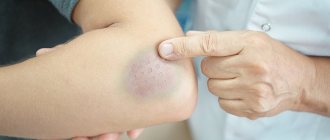
First of all, when a bruise occurs, an immediate strong swelling occurs, which subsides a little after a couple of hours. Also with pain. If your little finger is bruised, after a certain time it will hurt less and less. You will be able to walk comfortably with only slight discomfort.
Even with the most severe bruise, full mobility of the limbs is maintained. This is especially easy to check on both legs at the same time. If the little toe bends on one leg, but not on the injured one, then the injury is more serious.
It is also worth noting that bruises, in turn, are divided into several categories:
- Mild injury - slight pain; you can safely step on the foot and walk in closed shoes. A slight swelling may appear, which will subside within a couple of hours. A slight bruise may appear;
- The average degree of injury is a sharp, acute pain and the immediate appearance of severe swelling. Damage or complete removal of the nail plate is possible. It becomes more difficult to move the little finger, but it works. A severe hematoma also appears;
- Severe injury - in this case, the bruise and other consequences appear after several days. The hematoma also spreads to uninjured areas of the leg. When trying to move a finger, acute pain is felt and painful shock is possible;
- Serious damage - it is impossible to move a finger, a hematoma appears on the entire foot, as well as severe swelling that does not go away after a couple of hours. It hurts to step on. It feels very similar to a fracture or crack.
Classification
According to the classification, all fractures are divided into several types; we will describe the main ones:
- Traumatic - occur as a result of a strong mechanical shock to a bone that was previously unchanged and absolutely healthy.
- Pathological - when a fracture can occur as a result of a weak blow, but due to a pathological process in the bone itself, the bone becomes very fragile.
- Open - such fractures are accompanied by a violation of the integrity of the skin, rupture of soft tissue, which is often complicated by bleeding and infection.
- Closed – preserving the integrity of the skin.
- Complete - which in turn are divided into two types: with displacement and without displacement of fragments.
- Incomplete – this includes cracks and breaks.
A bruise is damage to superficial tissues, sometimes organs, but without significant damage to their internal structure, although there are serious changes when it comes to brain contusion.
After all the above, we have an idea of what each of the definitions means. But any diagnosis consists of the characteristics of the clinical course of the injury, so first we will touch on the symptoms.
Anatomy
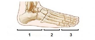
The structure of the foot is very similar to the hand. It refers to complex joints of small bones that form an arch and serve as a support for normal movement.
The little finger has a very complex structure. He is very fragile. A fracture can occur when a person twists his leg, drops a heavy object, or during active games. Such problems are very common in older people due to the fact that the amount of calcium decreases and osteoporosis develops.
The little finger consists of three phalanges, ligament muscles, capillaries and nerve fibers.
Diagnostics
It is impossible to diagnose a fracture of the little finger with 100% certainty based on symptoms, so in traumatology, X-ray examination is used to clarify the diagnosis.
The picture is taken in two projections, frontal and lateral, after which the doctor can determine that the little finger is broken and what the fracture looks like. But before determining whether it is a fracture or a bruise, it is necessary to provide first aid correctly.
First aid
As a rule, injuries are an unforeseen situation that can happen to us anywhere, often at the most inopportune moment. This happened to Marina at home, and since it is not at all easy to make a diagnosis without x-rays, it is necessary to take urgent action, because our future treatment and prevention of complications depend on it.
What help can be provided at home?
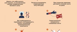
- In the first stages, it is necessary to completely eliminate the load on the injured leg.
- Inspect the damaged area. When it is an open wound, it must be treated with antiseptic agents. For this you can use betadine, hydrogen peroxide, chlorhexidine. If we are talking about alcohol solutions (iodine, brilliant green solution), they can only treat the skin around it, since they have a damaging effect, which in turn affects healing. If it is an open fracture, then after the wound has been treated, it must be covered with a sterile plaster or bandage.
- Apply cold to the area of impact for 10–15 minutes. This will relieve pain and reduce swelling for a short time. And to avoid frostbite of surface tissues, ice can be wrapped in any fabric, such as a towel.
- Pain relief with general analgesics. If you experience severe pain, you can take a drug with an analgesic effect - analgin, ibuprofen, ketorol. Only after this can a splint or bandage be applied.
- Immobilize with a bandage. It is impossible to independently determine the presence of a fracture and its type without special equipment and medical practice. Therefore, we must keep the leg in the same condition after which the impact occurred until we get to the emergency room. In order to fix the fracture site, we place the injured limb on a hard, flat surface. Then, together with the fixator, we bandage the leg, from the tips of the toes from the side of the sole to the middle of the shin. As a retainer, we use a tire or improvised means (thick cardboard, a block, a wooden strip). We bandage tightly, over clothing. The splint should run the entire length of the foot to the back of the shin. Another way is to bandage the broken little finger to the adjacent finger.
- Go to a trauma hospital.
Please note: as soon as you suspect a fracture, dislocation or rupture, do not waste time, but immediately seek medical help at the emergency room.
Treatment
To answer the question of how long it takes for a fracture of the little toe to heal, you need to understand that all injuries are completely different and treatment may differ in each case. Of course, there are general principles and approaches that should be followed, but do not forget that each case is individual.
Any treatment tactic depends on the severity of the patient’s condition; if the injury is accompanied by bleeding, then you need to start with its final stop.
- In case of a closed fracture of the little finger without displacement, a circular (circular) bandage is applied, which ensures complete immobilization of the limb. In order for the bone to heal correctly, you can walk during a fracture, but try as little as possible. Shoes should be true to size and not too tight. During treatment, all physical exercises on the foot are excluded and are possible only after healing. Working capacity is restored in 12-15 days. There is no need for plaster for this fracture.
- If, after an X-ray examination, the doctor diagnoses a displaced fracture of the little finger, then closed reduction of the fragments is necessary. This manipulation is performed immediately, it is carried out under conduction or intraosseous anesthesia. The traumatologist stretches the finger along its length to its correct position and eliminates the displacement of the fragments at an angle and width. After this, plaster casting takes place with a short plantar plaster splint, modeling the arch of the foot. Here it is necessary to achieve accurate reposition of the fragments and restore the function of the joint if it has been impaired. The duration of treatment and immobilization ranges from 4 to 6 weeks. The diagnosis is removed after a control image.
- With an open fracture of the little finger, surgical treatment methods are necessary. First, primary surgical treatment of the wound is performed with open reposition of fragments. Next, with the help of osteosynthesis (various fixing structures), the fragments are fixed in the correct position. After which immobilization bandages made of plaster or polymer bandages are applied. If the course of treatment is favorable, the wound heals on the 10th–12th day, after which the open fracture of the little finger can be considered closed. After fusion, the fixators are removed (in modern practice, there are special absorbable fixators that do not require removal). The treatment and recovery period is from two months to six months.
Is it possible to walk
If the little finger is fractured, the patient is advised to lie in bed more. Stepping on or moving on a broken limb is strictly prohibited. Do not forget that the patient's physical activity will be minimal. Therefore, he may become constipated. Carrots, beets, cabbage, and coarse fiber products help to avoid it. They improve the functioning of the gastrointestinal tract.
Types of orthopedic devices used for a broken finger
A patient with a broken toe needs to wear a plaster cast. It is worn for 2-3 weeks. There are also special orthopedic products that help fix the injured limb. This:
- A semi-rigid device that secures the foot on both sides. It is made from very dense material. It is attached to the leg with special tight fasteners.
- Hard bandage. It is worn in case of a displaced leg fracture. This device has special fasteners. They securely attach the toe when the little toe is fractured and provide proper tension to the foot.
Complications
As a rule, complications appear as a result of our wrong actions and mistakes. This is often associated with first aid, since, without experience, we do not know what to do in this situation. Another reason is that treatment was not started in a timely manner. While a person endures and is afraid to seek help from a medical institution, the pathological process can lead to irreversible consequences. An important point that is also worth paying attention to is violation of the doctor’s regimen and recommendations. As soon as we feel good, we immediately have reasons not to comply with the regimen and take medications, thereby increasing our recovery time.
But we should not forget that complications directly depend on the complexity of the injuries received. The most serious of them include:
Ankylosis
This is a violation of joint mobility that occurs as a result of a fracture. The destruction of the articular surfaces occurs, after which the inflammatory process begins. Inflammation causes connective or fibrous tissue to grow in the joint, making it completely immobile. Therefore, when this condition develops, it is very important to begin treatment as early as possible to stop the process and restore movement in the joint. In severe and advanced cases, they resort to prosthetics.
Osteomyelitis
This is a purulent-necrotic process of bone with the involvement of surrounding soft tissues. There are several types, but we are only interested in “post-traumatic”. Traumatic osteomyelitis most often occurs with open comminuted fractures, resulting in infection and a violent clinical picture, accompanied by high temperature (up to 39–40*), severe pain, weakness, decreased hemoglobin, and increased ESR. The peculiarity of this type is that two processes occur in parallel: the formation of callus and the destruction of bone tissue. Treatment is mainly surgical with antibiotic therapy.
The easiest way, as recommended by doctors
According to doctors, the most common and at the same time frequently used method is tapping. If such a test is carried out correctly, in most cases it is possible to distinguish a severe bruise of the little finger from a closed fracture. To do this, you need to tap the top of your finger in the direction of its base.
If the finger is broken, then the pain will be clearly defined in the place where the bone is deformed. If the bone is not damaged, then with axial tapping, pain at the site of the bruise will not be felt.
Such a test cannot be performed if there is a suspicion of a displaced fracture or damage to the joint, since this can lead to migration of bone fragments, which can lead to the development of serious complications.
Rehabilitation and recovery
After the wounds heal, rehabilitation measures begin, which speed up the patient’s recovery time. Our future ability to work depends on how quickly a fracture of the little toe heals. Physiotherapeutic treatment methods will help us with this.
These methods include:
- UHF (ultra high frequency) therapy. One of the most common electrotherapeutic methods, it reduces swelling and has an analgesic effect.
- Low frequency magnetic therapy. When exposed to magnetic fields, bones grow together much faster. Its main advantage is that the procedure can be performed through a cast. This is especially important for patients with complex fractures who have undergone osteosynthesis.
- Interference currents. Their main difference from other types of currents is that they are formed deep in the tissues, and are not produced by the apparatus. The effect of their influence is to improve trophic processes, relieve pain, and accelerate the resorption of edema (hematomas). For older patients whose injuries take a long time to heal, this method is especially necessary.
- Electrophoresis, just like currents, penetrates deeply into tissues, while a medicinal substance is applied to the electrode pads, which enhances the effect of the electric field. This substance accumulates at the site of inflammation, thereby reducing pronounced pain. Electrodes can only be applied to intact skin.
- Massage is necessary to improve blood circulation in the tissues and to eliminate congestion after a fracture; if your plaster has been removed, you can immediately go for the procedure.
How to determine a fracture
Reliable signs of a fracture are:
- pathological mobility of the finger. This means that the movements of the finger exceed the physiological norm, for example, it can hyperextend at the interphalangeal joint;
- the finger becomes visually shorter than before;
- bone deformation is visible to the naked eye or detected by palpation.
There are also probable (inaccurate) symptoms of a fracture:
- pain syndrome of varying intensity;
- skin redness and swelling;
- the little finger takes an unnatural position, hardly or at all;
- when you tap on the pad, a sharp pain occurs;
- the finger is hot to the touch.
