- Initial appointment with a dermatologist
1200 ₽ - Calling a dermatologist to your home
4000 ₽
Pityriasis versicolor –
a recurrent disease that affects only the horny surface of the epidermis. The pathology is called “lichen versicolor” because the spots have colors: yellow, whitish, brown. The skin begins to peel off and spots appear in the summer in children and adults. Residents of countries with high humidity and warm climates are susceptible to pathology.
In Russia, skin formations appear in 15% of the population. These are mainly people from 15 to 40 years old, more often observed in men. Children under 7 years of age rarely get shingles. For treatment, contact a dermatologist at our clinic, he will conduct an examination and prescribe medications that will improve the patient’s condition.
Types of pityriasis versicolor:
- Spotty-flaky. During the period of spread, it affects the body in different places: behind the ears, in the groin areas.
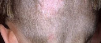
- Erythematosquamous. Often observed in adults and children. The rash is beige or coffee-colored, without inflammation. At first the formations are small, but soon they increase in size. This type appears on the head. It worsens in autumn and spring.
- Follicular. Lesions appear on the legs, back, chest, and arms. The spots spread in the hair follicles, their size is up to 3 mm, and itching is observed. This form can form during antibacterial therapy or the use of steroids in people with diabetes.
- Inverted. Characterized by peeling, itching, redness. This species is not common. When the disease occurs, symptoms are not expressed; spots appear on the head.
If you experience formations on the skin, it is better to seek help from qualified specialists at our clinic. Quickly identifying the disease in its early stages will help prevent its spread.
annotation
The genus consists of 14 Malassezia
of yeast-like fungi, 13 of which are lipophilic and 1 is non-lipophilic.
They are commensals and in predisposed individuals they usually cause a range of chronic, recurrent infections. In rare cases, they cause such serious diseases as infections that occur during vascular catheterization, during hemodialysis, and can cause peritonitis, etc. Although these mushrooms have been known to mankind for more than 150 years, due to the whimsical nature of mushrooms, their bulky cultures, and the ability to speciate, they have not been sufficiently studied. Since the last taxonomic revision, 7 new species have been added to this genus. Their ability to evade host immunity and increase in virulence, the range of diseases they cause has increased. Recently, Malassezia
have been implicated as a causative agent in the common disease atopic dermatitis. Although research on fungal cultures is complex, new molecular analysis techniques and advances in instrumentation have prompted an increase in research in this area. Therefore, we can pay more attention to this type of fungi to study in detail their characteristics and their growing impact on the disease clinic.
What was known?
Malassezia
causes superficial mycoses, but systemic infections are rare. They do not have descriptions of drug resistance.
Introduction
Malassezia
(formerly known as Pityrosporum) forms a cutaneous synanthropic flora that is associated with a variety of clinical manifestations, ranging from benign skin diseases such as pityriasis versicolor to hematogenous fungal infection in the immunocompromised individual.[1]
Currently, 14 species have been described, namely Malassezia furfur
,
Malassezia pachydermatis
,
Malassezia sympodialis
,
Malassezia globosa
,
Malassezia
o
btusa
,
Malassezia restricta
,
Malassezia slooffiae
,
Malassezia equina
Malassezia dermatis
, Malassezia japonica
, Malassezia nana
,
Malassezia capre
,
Malassezia yamatoensis
, recently
Malassezia cuniculi
was discovered .[2] Due to their lipophilic nature, these fungi are able to colonize seborrheic areas of the skin and survive on the fatty acids normally present in sebum.
They cause the following skin diseases: hyperkeratosis, invasion of hair follicles and inflammation. Although, by definition, superficial mycoses do not extend beyond the horny epithelium, these fungi are detected in the mouth, central and deep layer of the hair follicle. Malassezia
infections can be superficial and localized, and can also be systemic in immunocompromised people.[3]
Spread on skin
Malassezia skin colonization density
depends on age, body area and comorbid skin conditions, as well as on the person’s place of residence.
Being lipophilic, the highest prevalence of Malassezia
is found in sebaceous areas of the skin such as the scalp, face and upper torso.
Malassezia
most often found in younger people, who tend to have oilier skin.[3]
Factors affecting the reproduction of Malassezia
Geographical factor: depending on the geographical area, the density of different species of the genus Malassezia
on the skin.
The most favorable zones for the growth of Malassezia
are subtropical and tropical climates.
Research shows that the growth of Malassezia globosa
increases during the summer when temperatures are high and sweating is increased.[4]
Age factor: pityriasis versicolor affects people from 20 to 40 years old. However, in India, it is seen in people in the age group of 10 to 30 years. Pityriasis versicolor is rare in children and the elderly. [5,6,7,8]
Hormonal factor: corticosteroid therapy, malnutrition, increased serum cortisol concentrations contribute to the occurrence of pityriasis versicolor in patients.[9]
Pathogenesis: Human sebum, which is a complex mixture of lipids, is the source of nutrition for this type of yeast. Sebum consists of triglycerides of fatty acids, waxes, esters, sterol esters, cholesterol, cholesteryl esters and squalene. Sebum is utilized by breaking down triglycerides and esters into diglycerides, monoglycerides and free fatty acids. Thus, changes in the secretion of sebum and its breakdown causes the development of dandruff. Malassezia infection
, is also associated with increased sweating.[10
a
,
b
,11,12,13]
Clinical scenarios
Yeast of the genus Malassezia
cause the development of chronic recurrent dermatoses such as pityriasis versicolor, seborrheic dermatitis and folliculitis.
Malassezia
has recently been shown to play a role in the pathogenesis of atopic dermatitis and psoriasis, especially in cases involving the scalp.
In addition, studies mention Malassezia
, which is the causative agent of confluent reticular papillomatosis and onychomycosis.
This yeast is also associated with catheter-associated infections among neonates receiving total parenteral nutrition and in frail older adults receiving parenteral lipid supplements. Also, such patients may experience hematogenous fungal infection and sepsis caused by Malassezia furfur
and
Malassezia pachydermatis
.[1,14]
Pityriasis versicolor
Malassezia infection
.
Pityriasis versicolor, also known as pityriasis versicolor, is a common superficial mycosis that is a chronically recurring infection of the stratum corneum of the skin. It is characterized by small white scales, hypo- or hyperpigmented macules, which are unevenly distributed throughout the body and are most often found on oily areas of the skin on both the trunk and extremities. Some patients may experience itching, but most patients are asymptomatic.[1] The Latin name for pityriasis versicolor (or multicolored) is pityriasis versicolor
.
It comes from the Greek. “ P ityron
” - “flakes, scales.”[3,15]
Back in 1871 Neumann
et al.[16]
revealed that it is the mycelial forms of Malassezia
that are responsible for the appearance of pityriasis versicolor.
Successful cultivation of Malassezia
is described in the works
of Neumann
et al.
and Gordon
et al.[17]. It was also found that yeast forms can be seen both on areas of skin affected by pityriasis versicolor and on healthy areas of skin. It was found that in order for the clinical picture of pityriasis versicolor to manifest itself, the necessary conditions are a large proliferation of fungal filaments, and they must secrete several serovar A antigens.[1]
Differential diagnosis
Skin lesions caused by pityriasis alba, pityriasis rosea, vitiligo, hypopigmented mycosis fungoides, erythrasma and seborrheic dermatitis look similar.
Diagnosis
Pityriasis versicolor is diagnosed clinically; only in rare cases is a biopsy or Malassezia
. Under a Wood's lamp, pityriasis versicolor fluoresces yellow. Examination of mushrooms with 10% potassium hydroxide solution shows single-celled, short, curved, straight mycelium filaments and spore clusters. All of the above should resemble “meatballs and spaghetti.”
Histopathological examination
Although pityriasis versicolor is rarely biopsied, it has spores (yeast form) and short hyphae (mycelium) in the stratum corneum of the skin, which are detected using diazonium blue, periodic acid-Schiff, or silver nitrate-methenamine complex. Moderate hyperkeratosis and acanthosis are rare.[3]
Malassezia species that do not cause pityriasis versicolor
Pityriasis versicolor is caused by several species of Malassezia
[18,19,20,21],
M. globosa
is the most common causative agent of lichen with a probability of 58.2%, 55%, 55% and 53.3%, respectively.
A study conducted in north central India found 54% M. globosa
, and 30%
M. furfur
.
Several studies have shown that M. furfur
predominantly causes pityriasis versicolor.
Makimura
et al.
identified M. furfur
and
M. sympodialis
in temperate climates;
however, in tropical countries, they isolated M. globosa
.[22,23,24]
Pathogenicity of M. globosa
due to its esterase and lipolytic activity. Unlike other mycoses, here, in the center of the lesions there are more viable cultures.[25]
Seborrheic dermatitis
Seborrheic dermatitis is the second most common infection associated with Malassezia
, which was originally described by
Unna PG
in 1887. It is a superficial eczematous dermatitis, can be subacute or chronic, and is characterized by the presence of an erythematous plaque with dry or greasy scales. It is found in areas of the skin rich in sebaceous glands, such as the scalp, face, ears, chest and armpits. Weak corticosteroid drugs have proven effective in treatment.
However, the relapsing nature of this disease makes it difficult to treat. Antifungal therapy should be the mainstay in the treatment of this disease, since the use of corticosteroids does not reduce the number of Malassezia
.[3,26]
Seborrheic dermatitis is common during periods of a person's life when their glands are most active:[15]
- First 3 months of life (infantile seborrheic dermatitis)
- During puberty
- When sebum production declines after age 50.[15]
This disease affects 3-5% of the population, with more men than women. In immunocompetent individuals, seborrheic dermatitis usually begins during periods of high sebum secretion, i.e. during puberty and become chronic and recurrent forms. Stress is known to aggravate this type of dermatitis. In patients with AIDS, resistance to antifungal therapy for seborrheic dermatitis is observed from 30 to 80%, in contrast to immunocompetent individuals. [27]
In HIV-infected patients, the appearance of seborrheic dermatitis is a surrogate marker of CD
4+ T lymphocytes.
M.
restricta
and
M. globosa
are observed primarily on the scalp, both during health and illness.[28]
Pathogenesis
The immunopathogenesis of seborrheic dermatitis is complex and multifactorial.
It has been proven that the increased density of colonies is higher in damaged skin than in undamaged skin. However, techniques must be standardized to prove this. Recently, “quorum sensing”—the ability of some organisms to communicate and coordinate their behavior through the secretion of molecular signals—has been actively discussed in science. Malassezia
also operates according to the “quorum” principle.
Also Malassezia
has a complex antigenic structure, represented by both protein and carbohydrate antigens, which are associated with the stages of their life cycle;
protein antigens appear in the early stages of growth and are associated with the development of cell walls and cytoplasmic organelles, while carbohydrate antigens are derived from mannan and mannoproteins. Research by B ergbrant
et al.
showed an increase in NK
cells, impaired T cell function in addition to an increase in total serum
IgA
and IgG among
patients
with seborrheic dermatitis.
However, the titer of Malassezia
-specific antibodies was not elevated, so it was suggested that hypergammaglobulinemia may occur as a reaction to toxins contained in yeast and to lipases.
People suffering from dandruff have loosely connected corneocytes or a small number of dysmosomes. The level of secreted lipids is reduced, which in turn affects the epidermal water barrier. Itching associated with histamine also increases, causing damage and reducing the effectiveness of the skin's defense mechanism. All this makes it easier for Malassezia
into the skin.
Malassezia
produces lipases that break down sebum into arachidonic and oleic acids, which are irritants and have a desquamative effect on keratinocytes. Thus, this contributes to the onset of the inflammatory process and creates vicious circles.
In some cases Malasseziae
have the ability to exist as pathogenic microorganisms that cause seborrheic dermatitis, and in other cases exist as normal flora. They are capable of causing an immune response. They activate the classical and alternative pathways of the complement cascade. Complement component C3 has been found in damaged skin and has been postulated as one of the causes of inflammatory reactions. Although yeast should ideally be phagocytosed and eliminated by our immune system, it is capable of suppressing the secretion of cytokines and interleukins.
Malassezia
alters and evades the local immune response by producing secondary metabolites that contribute to seborrheic dermatitis.[28] Environmental factors, stress, UV radiation, climate change, hormonal changes and trauma also likely contribute to the maintenance of seborrheic dermatitis.[28]
Milky crusts: Infantile or neonatal seborrheic dermatitis is also called pityriasis capitis, which is a nonpruritic dermatosis ( Cradle
cap directory
,
WebMD
.
Retrieved
August
26 , 2012).
Infantile seborrheic dermatitis usually occurs from the 2nd week of life to 6 months. The rashes can be eczematous and psoriasis-like in nature, rare localizations of seborrheic dermatitis are the face, torso and sternum area. The lesions can be single or multiple, and can merge, especially on the face in the fold area.[15]
Neonatal cephalic pustulosis (neonatal acne)
A child has skin colonization with Malassezia
begins on the first day of life.
Malassezia
is transmitted to the baby from the mother and from health care workers, and colonization increases during the first weeks of life.
Environmental factors and maternal hormonal levels influence the colonization process. The high prevalence of M. sympodialis
in newborns is observed from birth and is the cause of neonatal acne.[29]
Folliculitis caused by Malassezia
Synonyms: pityrosporum folliculitis.
This intensely pruritic, follicular papulopustular rash is seen usually on the upper torso in the young age group and is rare in older people.
This disease was first described in 1969 by Weary
etal., based on the histological ultrastructure
of Potter
etal. in 1973 it was discovered that it was a separate type of disease. This is a chronic infection in which patients typically present with erythema, papules and pustules on the chest or back, and a follicular pattern. The distinctive symptom is itching. Rare localizations are the face, neck, shoulders and arms.[6,15,30,31]
Risk factors for folliculitis:[32,33,34,35,36,37]
- Immunosuppression
- Diabetes
- Broad-spectrum antibiotics
- Steroids
- Puberty,
- Pregnancy
- Cosmetics, lotions, sunscreens, emollients, olive oil, which are the cause of skin clogging.
Diagnosis: The diagnosis of folliculitis is based on the clinical picture and positive response to antifungal therapy.
Some differences in the distribution of Malassezia
according to epidemiological data:
Low number of Malassezia
, compared to normal skin, causes other diseases.
There are pathogenic strains that are not isolated in psoriasis.
Histopathological findings: According to microscopy, the discharge from the pustules represents budding yeast forms and spores, and not hyphae, as in the study of pityriasis versicolor. to diagnose pityrosporum
folliculitis
, but histology shows dilated hair follicle openings with large amounts of cellular material and keratin plugs.
The inflammatory response is caused by lymphocytes, histiocytes and neutrophils. The mouth of the sebaceous gland ducts is surrounded by monocytes and mucin deposits. Yeasts of the genus Malassezia
can be detected in hair follicles; however, they are more often found in the orifices of the sebaceous gland ducts. Periodic acid-Schiff and Grocott-Gomori staining can be used to identify unipolar budding yeast cells; mycelial forms were not detected by these methods. [38],[39],[40]
Treatment
Treatment for pityrosporum
follicilitis
uses oral antifungal medications, since topical medications do not penetrate the hair follicles. Since there is a high relapse rate, there is a need for maintenance therapy.
Atopic eczema/dermatitis syndrome
Atopic dermatitis is a chronic, inflammatory skin disease. Patients with atopic dermatitis often have bronchial asthma. The pathogenesis is not entirely clear. Patients suffer from itching, which may be the body's response to irritants or microorganisms, due to the loss of surface barrier mechanisms in dry skin [Fig. 1]. [3],[41],[42]
Rice. 1: Malassezia
in the pathogenesis of atopic eczema/dermatitis syndrome
After the successful therapeutic use of ketocanazole in the treatment of atopic dermatitis, a number of studies have proven the role of Malassezia
as one of the etiological factors in the occurrence of exacerbation of atopic dermatitis. [43]
Their multilayer membranes and cell wall mannans contain antigens. of Malassezia among patients
-specific
IgE
, and they have positive skin prick tests.
Malassezia
may act as a persistent allergic irritant.
The combined cellular and humoral immune response leads to increased sensitization. The cellular response leads to the activation of T h
2 lymphocytes, this leads to an increase in the levels of IL 4 and IL5 compared to the control group of healthy people.
Due to allergen-specific IgE
and cytokines release histamine from mast cells.
Symptoms of itching due to histamine release lead to further sensitization of Malassezia
.
Antifungal drugs lead to a decrease in the yeast population on the skin, thereby reducing the antigenic load, which prevents the inflammatory process.
IgE
specific are determined to
M. sympodialis
,
M. furfur
and, more recently, to
M. globosa
.
Isolation and speciation
Several studies have been conducted on M. globosa
,
M. restricta
,
M. sympodialis
and
M. furfur
.
M. globosa
the allergen is responsible for the production of heat shock protein 70.
In 2002 Sugita
et al.
described new species
of
M. dermatis
isolated from patients with atopic dermatitis in Japan.
However, the researchers suggest that more studies should be conducted in other countries as well to confirm the fact
that
M. dermatis
can be detected in patients with atopic dermatitis not only in Japan.
The pathogenesis of the disease is not very clear. Genus Malassezia
promotes the production of
IgE
-specific antibodies, and this plays an important role in pathogenesis.
More research is needed to understand the role of Malassezia
in the mechanisms of disease, chronicity and exacerbation. [3],[15],[41],[42],[43],[44],[45],[46],[47],[48],[49],[50],[51 ],[52]
Psoriasis
It has been documented that chronic plaque psoriasis and psoriasis vulgaris are caused and aggravated by Malassezia
.
Rivolta
et al.
in 1873 he first proved this fact. Later, in 2007, Narang
et al. [53] treatment with fluconozole is effective for psoriasis.
Among patients with psoriasis, especially those whose affected area involves the scalp, eyebrows, ears, and trunk Malassezia
plays a big role.
Elewski
et al.
in 1990 [54] reported a case of guttate psoriasis in areas affected by Malassezia folliculitis
.
Some differences have been noted in the epidemiological data and distribution of the genus Malassezia
.
The pathogenesis is not fully understood. Triggers of T cell activation and cytokine release are multifactorial - stress, infections, trauma, drug use, etc.
Autoantigens: 13 keratin has homology to K17, which is an identified candidate autoantigen that is not normally found in adults but appears in response to injury.
This explains the Koebner phenomenon, where new lesions appear at sites of injury. Neutrophils are found in psoriasis in the skin infiltrate and increase chemotoxic reactions.
This phenomenon does not occur when the skin is damaged by staphylococci.
Superantigens are those antigens that cause a cytokine storm. Superantigens are staphylococci and streptococci, which aggravate the course of atopic dermatitis, guttate psoriasis and chronic psoriatic plaques.
It can be said that the superantigen promotes inflammation by activating complement, producing inflammatory cytokines and recruiting neutrophils, but this fact is not fully proven.
Malassezia colonies
on skin affected by psoriasis may be due to the fact that keratinocytes produce antimicrobial peptides.
These peptides destroy the normal bacterial microbiota in the affected area and allow only selective overgrowth of Malassezia
.
This proliferation itself promotes an increase in the number of keratinocytes and an increase in the level of LL
37 (human cationic antimicrobial protein).
This can cause the development of psoriatic rashes. However, the number of colonies is always lower compared to the healthy control group. The reason for this is not very clear, but may be due to the inhibitory effect of LL
37 noted in patients with psoriasis.[3,15,55]
Indirubin, an indole produced by Malassezia
, have been successfully used in treatment, further strengthening the association
of Malassezia
with this disease.[3,15,55,56]
Onychomycosis
In Malassezia
there are no enzymes with keratolytic activity.
They are lipophilic and so nails are not a good source of lipids, there are no lipids under the nail plate, Malassezia
does not colonize them.
Malassezia
is not considered to be the primary causative agent of onychomycosis.[2,56]
Table 1. Diseases rarely caused by Malassezia
.
Superficial infections | Deep infections |
| Confluent reticular papillomatosis | Mastitis[61] |
| Dacrocystitis[57] | Sinusitis[62] |
| Vulgar acne[58, 59] | Septic arthritis[63] |
| Seborrheic blepharitis[59, 60] | Malignant otitis externa[64] |
| Nodular hair infection |
The role of Malassezia in the occurrence of systemic diseases
There have been reports of a role for Malassezia
as an infectious agent during peritoneal dialysis in a patient with chronic renal failure.
Several cases have been reported implicating Malassezia
in the development of peritonitis.
Hassal
et al.
notes pulmonary embolism and pulmonary infection due to Malassezia
in a patient who was on urokinase therapy.
Role of Malassezia in catheter-associated blood infections
Catheter-associated sepsis due to Malassezia
, was found in low birth weight infants and in intensive care unit patients who were on total parenteral nutrition and in those with predisposing conditions.
two
species, namely M. furfur
and
M. pachydermatis
are proven causative agents of systemic disease.[65,66,67,68,69,70,71,72,73]
Involvement of Malassezia pachydermatis in the development of human infections
This species was isolated in 1925 by Weidman
from a captured Indian rhinoceros.
He named it pityrosporum pachydermatis
. [74]
Dodge
et al.
in 1935 assigned it to the genus Malassezia
. This species exists as normal flora in domestic animals such as dogs, cats, and also in wild animals. Skin colonization is common among pet owners
For the first time M. pachydermatis
was described in 1983 as a causative agent of peritonitis during peritoneal dialysis in a patient with insulin-dependent diabetes.
M. pachydermatis
was the cause of a hematogenous fungal infection in the neonatal intensive care unit in 8 low birth weight preterm infants receiving total parenteral nutrition with lipid supplements.
M. pachydermatis
was isolated from the blood of 6 of 8 newborns, from the tracheal aspirate of 2 of 8, from the eyes of 1 of 8, from nasal secretions in 1 of 8, and from urine in 1 of 8 newborns.[75]
M. pachydermatis
may lead to outbreaks as it can persist on the surface of the incubator for up to 3 months, despite standard disinfection.
Thus, the following were implemented: careful personal hygiene of medical and neonatal care personnel, as well as modifications to cleaning procedures.[72] Colonization of pet owners by Malassezia
was demonstrated by high rates of amplified DNA compared to those who did not own pets.
Thus, M. pachydermatis
is a potential pathogen.[15]
Malassezia
pachydermatis
was isolated from a facial granuloma in a healthy woman and her dog from skin scrapings and earwax. The skin lesions healed after oral fluconazole and cryotherapy.
conclusions
Genus Malassezia
known to mankind for more than 150 years as a synanthropic group of opportunistic fungi.
Although they are generally not life-threatening, causing only chronically recurring superficial mycoses in most cases, this fact is taken into account when searching for the causes of a wide range of diseases, including systemic infections. It is a potential pathogen among predisposed patients and among patients receiving intensive care. The taxonomy of Malassezia
has been supplemented with 7 new species. More research is needed to understand their behavior in clinical settings. The minimum inhibitory concentration of antifungal drugs used against newly identified species should also be studied.
What's new?
New relationship between pathogens and diseases such as atopic dermatitis and psoriasis, etc.
Documentation of high minimum inhibitory concentrations obliges us to understand the spectrum of diseases caused by the genus Malassezia
, including knowledge of rare diseases caused by these pathogens.
Symptoms of pityriasis versicolor:
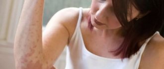
- Formation of pink spots. At first they are single, then they increase in size and merge. Rashes are observed on the stomach, neck, under the hair, on the chest.
- As the disease spreads, they become dark in color. May be red or brown. The colors vary on the affected areas, which is why lichen is called tinea versicolor. In some cases, the formations merge into one whole.
- After a certain time, peeling occurs. The scales resemble bran.
- Itching is observed, but not in all cases.
- After healing, white or light spots remain on the skin; tanning does not affect them; this is caused by cell damage as a result of fungi.
What is erythrasma
Erythrasma is a chronic bacterial disease affecting the epidermis layer in the deep folds of the skin. It is characterized by a long course - in some cases, symptoms develop for at least 10 years, without causing significant discomfort to the patient. The clinical picture of erythrasma is similar to a fungal infection of the skin, but modern dermatology classifies it as a group of pseudomycoses.
The following main stages are distinguished in the development of the disease:
- Progression. The first characteristic spots appear on the skin, their size slowly increases, and additional symptoms develop. In some cases, secondary infections occur. The spots gradually merge with each other, forming large areas of damage.
- Stabilization. New spots do not appear, and existing ones stop growing. Peeling of the skin begins. This stage is usually associated with a change in external conditions, for example, cold weather, during which the intensity of sweating decreases and the skin condition stabilizes.
- Exacerbation or relapse. Usually associated with the beginning of the warm season. But in the case of prolonged erythrasma, the disease constantly develops in waves, and after a slight decline its symptoms again actively appear.
- Remission. Occurs with a favorable microclimate, compliance with preventive measures and proper skin care. The color of the affected areas gradually returns to normal, itching and flaking disappear, and the skin is restored.
Without timely, well-chosen treatment, erythrasma can lead to the development of serious complications.
For example, it can provoke eczema and secondary infection in patients with diabetes or obesity. Also, the course of the disease is aggravated by increased humidity and contamination of the affected areas. As a result, its typical symptoms are complicated by burning, itching and pain.
Diagnosis of pityriasis versicolor
The disease is diagnosed by a dermatologist. He performs a visual examination of the patient, studies the presence of spots and their distribution, and makes a diagnosis at this stage. If the form of lichen is not pronounced, then the clinic specialist uses the following methods. Using a Wood's lamp, he makes several tests. After highlighting areas of the skin, they may change color to yellow-red or green.
To clarify the diagnosis, laboratory diagnostics will be required. Specialists using special equipment will be able to detect the presence of the disease after additional research.
Pathogens and carriers of lichen
The content of the article
Dermatologists distinguish several types of lichen, caused by different pathogens.
Most often, the appearance of lichen on human skin is initiated by fungi:
- zooanthropophilic - transmitted to humans from an infected animal, including from pets;
- anthropophilic - live only on human skin;
- geophilic - live in the soil and come into contact with human skin upon contact with the ground.
In addition to fungi, viruses are often the causative agent of lichen. Their cunning lies in the fact that they can live asymptomatically in the human body for a number of years and become more active when the immune system is weakened. This is exactly how, for example, those familiar to many with herpes or shingles behave.
Differential diagnosis of pityriasis versicolor with other diseases
To make a diagnosis, the doctor performs the following procedures:
- Conducting an examination under a microscope to determine the scraping. The specialist scrapes the scales from the skin area, processes it and examines it. Using a microscope, it is possible to detect spores and fungi that are round in shape. The mycelium threads are white in color and long in shape, like a thread.
- Balzer test. A 5% iodine solution is used, which is applied to or near the infected areas. This study will take time; the affected skin gradually acquires a different color.
Treatment of pityriasis versicolor
The dermatologist prescribes treatment depending on the nature and form of the disease, the characteristics of the lesion, the age of the patient, and the presence of other pathologies. Applies complex treatment, which includes ointments, sprays, creams, shampoos. They consist of components that effectively affect the disease. Depending on the complexity of the pathology, the patient is prescribed tablets. Therapy will help overcome lichen in a short time and eliminate complications.
For treatment, the doctor prescribes ointments that contain terbinafine. Treatment of the affected skin area is prescribed 2 times a day. This substance quickly destroys microbial cells and eliminates the appearance of new formations. Imidazole, clotrimazole and others are effective for treatment. The substances are included in creams and gels. Tolcyclate and ciclopirox are ointments prescribed to ensure that the pathogens quickly die.
When lichen appears in the hair, the doctor prescribes shampoos containing ketoconazole or nizoral. In the initial stages, patients are recommended salicylic and zinc ointments. The products quickly relieve itching. You should not use medications without consulting a specialist. Qualified doctors of our clinic, using modern equipment, will diagnose and prescribe a course of treatment.
A visit to a dermatologist is necessary 21 days after the prescribed treatment. The doctor will assess the patient’s condition, prescribe additional medications and preventive measures, these include physiotherapy and others.
Reasons for development
Versicolor versicolor is a type of fungal skin infection that affects the stratum corneum of the epidermis and hair follicles. Its causative agents are two types of fungi, and infection is possible only through prolonged and close contact with the patient. And in this case, provoking factors play a big role. These include:
- weakened immunity;
- hyperhidrosis;
- disruption of the sebaceous glands;
- diseases of the endocrine system (obesity, diabetes, Itsenko-Cushing syndrome, etc.);
- hormonal imbalance due to pregnancy, menopause or taking hormone-containing medications;
- vegetative-vascular dystonia;
- abuse of antibacterial personal hygiene products;
- excessive exposure to ultraviolet rays (intense tanning, frequent visits to the solarium) and regular overheating of the body.
It is noteworthy that patients with pityriasis versicolor over 60 years of age are extremely rare. This is due to natural age-related changes in the skin, which make it less susceptible to pathogens.
In children under 10 years of age, the main causes of pityriasis versicolor infection are neglect of personal hygiene rules or improper skin care. At this age, with the protective functions of the skin intact, the body independently copes with pathogenic microorganisms attacking it, so the development of the disease does not occur. But closer to adolescence, when hormonal changes begin, the body’s susceptibility to bacteria, viruses and fungi increases, so children over 10 years old become infected with pityriasis versicolor just like adults.
Prevention of pityriasis versicolor
To reduce the disease, preventive measures play an important role. They include regular washing of skin areas, removal of sweat, sebaceous fat, and regular change of bedding and clothing. In hot weather, antifungal shampoos are an excellent method. They will eliminate the occurrence of relapses. The specialist prescribes products for regular hair washing. As a preventive measure, it will take 5 to 7 days to use the drugs. The skin should be wiped with salicylic alcohol every day.
It is better to wash things not with powders, but with soap, adding soda to the water. You can avoid deprivation; for this you need to strengthen your immune system, eat right, and take vitamins once a year. Experts recommend playing sports and taking walks in the fresh air for treatment. In case of illness, it is recommended to include vegetables, herbs, fermented milk products in food, and exclude spicy, sweet and smoked foods.
Gastrointestinal disorders can cause pityriasis versicolor, so you need to undergo a course of treatment.
Are all types of lichen contagious?
Many people believe that absolutely all types of lichen are dangerous to others. Meanwhile, this is not entirely true. So, with pink, ringworm or shingles, the patient will really need to be isolated. But, for example, pityriasis or scaly ones are not dangerous for others.
And this is another reason why any change in the skin should be the reason for going to a specialist - only he will be able to correctly determine the type of lichen, assess the degree of its danger, give recommendations for treatment, and finally select a set of medications and therapeutic measures aimed at including general strengthening of the body.
Complication of pityriasis versicolor
The pathology affects the upper layer of the skin, but is often severe. It affects internal organs.
In case of severe itching, the patient scratches the severely damaged skin. All this causes discomfort and leads to inflammation, in addition to infectious pathology. Contact a specialist immediately. You can’t just start the disease and start solving the problem. A specialist will help and prescribe medications to improve the condition.
If the disease returns after all measures, we recommend that you undergo examination by our specialists.
Lichen ruber planus
Flat multi-colored (from pale pink to purple) nodules of lichen red often appear on the mucous membranes, nails (they crumble), and delicate skin of the chest and abdomen. Their appearance is accompanied by severe itching, which, as a result of scratching, leads to the rapid growth of foci of skin damage.
Dermatologists find it difficult to determine the cause of the appearance of lichen red, but note that it is quite common in patients with diabetes, as well as those suffering from diseases of the stomach, liver, and biliary tract. Moreover, women over 40 years of age are primarily at risk.
Pityriasis versicolor in children
The disease is not typical for children under 6 years of age. During this period, the skin has protection mechanisms that prevent the dermis from being exposed to moisture and high temperatures. However, if caring for the baby is not correct, parents lubricate the skin with oils: shea butter, cocoa, then this disrupts the functioning of the defense mechanisms. For this reason, the disease appears in preschool age.
In children during the prepubertal period, hormonal changes begin in the body. The skin is very sensitive to high humidity and exposure to the sun, like adults. Thanks to changes, fungi can provoke the appearance of disease.
Ringworm in children occurs with the same symptoms as in adults. The treatment is no different. If redness appears, it is advisable to immediately consult a doctor. The specialist will conduct an examination and prescribe a drug that is suitable for the child’s age. It is not recommended to use the products without the advice of a specialist, as this can lead to negative consequences.
Causes of tinea versicolor
The content of the article
Ringworm affects middle-aged people. Medicine identifies the causes that lead to the appearance of this disease in humans. These include:
- Predisposition to fungal skin diseases.
- Disruptions in the course of physiological processes in the epidermis.
- Endocrine diseases.
- Lack of a good night's sleep.
- Lack of rest.
- Time zone change.
- Excessive hygiene, using antiseptics.
- Improper functioning of the human immune system.
- Frequent bathing in salt water.
- Lack or excess of vitamins.
- Wearing clothes made of synthetic materials.
- Improper functioning of the lymphatic drainage system.
- Long-term use of steroid hormones.
- Nervous system problems.
- Hormone imbalance.
- Lack of hygiene measures in public places.
Pityriasis versicolor during pregnancy
As a result of hormonal changes, the appearance of pathology is possible, but it does not harm the mother and fetus. The disease causes discomfort, but self-medication and folk remedies are unacceptable. If pathology appears, contact a dermatologist, he will choose treatment and, if necessary, prescribe diagnostics. Pregnant women are prescribed therapy for ringworm, taking into account the danger of the situation.
The doctor prescribes drugs that do not enter the bloodstream, does not prescribe anti-fungal tablets, they are toxic and harmful to the unborn child. Nizoral is prescribed; it is not recommended to use the medicine without a prescription.
Many dermatologists advise delaying the full course of treatment until after the baby is born. The fungus is not harmful to the fetus and will not affect its development. Oral medications are prohibited during breastfeeding, as they can pass into the milk and harm the baby's health.
Why should you contact our clinic?
Dermatologists of the highest category
Experienced dermatologists in Moscow. Experience of specialists from 15 years.
Modern equipment
Modern equipment from the world's best manufacturers.
Expert class equipment
Ultrasound is performed using expert-class equipment manufactured by General Electric, SONY, Mindray.
Tests and ultrasound on the day of treatment
Tests, x-rays, ultrasound with interpretation, dressings on the day of treatment in Moscow.
Consultations for adults and children
A network of dermatological clinics in Moscow. Clinics near the metro.
Experienced doctors
Our experienced doctors with over 15 years of experience. Candidates of Medical Sciences.
Correct treatment of pink dandruff in Gibert's disease
Treatment of pityriasis rosea is primarily based on the diagnostic results and recommendations of the dermatologist based on his knowledge and experience. Independent attempts to cure pathology usually lead to the appearance of accompanying symptoms, sometimes very dangerous.
Considering the symptoms, the dermatologist prescribes:
- ointments with corticosteroids acting in several directions - relieve skin tightness and accelerate the healing of the rash;
- antiallergic - antihistamines - relieve itching;
- immunomodulators – stimulate natural immunity;
- antiseptic solutions – prevent secondary infection;
- dandruff softening ointments.
During treatment, the dermatologist recommends:
- Avoid hot water procedures and swimming. Showering with a soft sponge is allowed;
- Wear loose, natural underwear. Synthetics do not allow the skin to breathe, exacerbating symptoms;
- Eliminate allergens: eggs, chocolate, red fruits.
- Reduce fried, flour, smoked foods;
- Do not use cosmetics.