Signs and symptoms
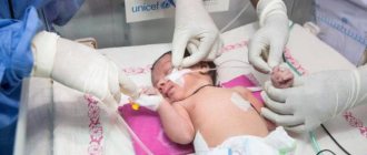
The skin of babies with Harlequin ichthyosis is covered with thick, scaly, diamond-shaped plates (see photo above). The tightness of the skin stretches around the eyes and mouth, causing the eyelids and lips to turn inside out, exposing the red inner lining. The baby's chest and abdomen may be severely restricted due to tight skin, making breathing and feeding difficult. The arms and legs may be small, swollen, and partially bent. The ears may appear misshapen or missing, but are actually fused to the head due to thick skin. Children born with Harlequin ichthyosis may also have:
- flat nose (depressed nasal bridge);
- hearing impairment;
- frequent respiratory infections;
- decreased joint mobility.
Premature birth is common, leaving babies at risk for complications during early labor. These infants are also at high risk for low body temperature, dehydration, and hypernatremia (high levels of sodium in the blood). Narrowing and swelling of the mouth may interfere with absorption, and infants may require tube feeding. The infant's corneas need to be lubricated and protected if the eyelids open under the pressure of tight skin.
Frequently asked questions about ichthyosis
How is ichthyosis inherited from sick parents?
The disease ichthyosis is hereditary. Pathology is caused by mutation of genes on the X chromosome, so girls are carriers, and boys are most often affected.
Is ichthyosis transmitted during contact with a sick person?
No. Ichthyosis is not an infectious disease. They cannot be contracted through contact with sick people. The pathology is caused by gene mutation.
How do people with ichthyosis live?
The disease requires constant skin care. The main tasks are to reduce moisture loss, intensively moisturize the skin, and control the thickness of the stratum corneum. Due to impaired thermoregulation in such patients, it is important to avoid overheating and prolonged stay in rooms with dry air.
Causes
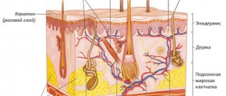
Harlequin ichthyosis is caused by changes (mutations) in the ABCA12 gene, which provides instructions for making a protein necessary for normal skin cell development. The ABCA12 gene plays a key role in transporting fats (lipids) to the most superficial layer of the skin (epidermis), creating an effective skin barrier. When this gene mutates, the skin barrier is destroyed.
Harlequin ichthyosis is inherited in an autosomal recessive manner. Recessive genetic disorders occur when a person inherits an abnormal gene from each parent. If a person receives one normal gene and one abnormal gene for a disease, the person will be a carrier of the disease, but usually asymptomatic. The risk that two carrier parents will both pass on the abnormal gene and therefore conceive an affected child is 25% in each pregnancy. The risk of having a child who is a carrier like the parents is 50% with each pregnancy. The chance for a child to receive normal genes from both parents is 25%. The risk is the same for men and women.
Can it be cured?
At the current stage of development, medicine is not yet able to cope with genetic diseases, so modern treatment of ichthyosis is aimed at reducing clinical manifestations and alleviating the patient’s condition. For this purpose they prescribe:
- vitamin complexes, iron-containing compounds, immunoglobulin;
- blood plasma transfusions;
- hormone therapy (in difficult cases);
- baths with the addition of various drugs, emollient ointments and creams;
- physiotherapy, spa treatment according to indications.
The choice of treatment methods depends on the degree of skin damage and the characteristics of the disease.
Standard Treatments
Once a baby with Harlequin ichthyosis is born, a multidisciplinary team of doctors will be involved in the baby's care. It has been shown to improve outcomes and reduce complications such as respiratory distress, dehydration, electrolyte imbalances, thermoregulation, systemic bacterial infections and nutritional difficulties. Early treatment with oral retinoids is thought to improve outcomes. However, they are only used in severe cases due to their known toxicity and side effects.
The thick, scaly, lamellar skin of Harlequin ichthyosis will gradually split and peel off over several weeks. Antibiotic treatment may be necessary at this time to prevent infection. Administration of oral acitretin may accelerate the release of thick scales. Most babies with Harlequin ichthyosis will require one-on-one care in the first few weeks of life.
After the thick scaly plates peel off, the skin remains dry and red and may be covered with large thin scales. Skin symptoms are treated with skin emollients. This can be especially effective after swimming while the skin is still damp. Many patients with severe ichthyosis are exfoliated by hand, using special exfoliating gloves with a rough surface to rub off the thick scales.
Skin barrier repair formulas containing ceramides or cholesterol, moisturizers with petrolatum or lanolin, and mild keratolytics (products containing alpha hydroxy acids or urea) can help keep skin hydrated and elastic, and prevent cracking and cracking that can lead to infection. . To avoid loss of circulation, it may be necessary to remove damaged tissue (debridement) from the fingers if they are compressed by strips of skin.
Ichthyosis
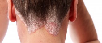
Ordinary or vulgar ichthyosis appears before the age of three years, but it is usually diagnosed before the third month of life. This is the most common form of ichthyosis, inherited in an autosomal dominant manner. First, the skin becomes dry and rough, then becomes covered with small whitish or gray-black scales tightly adjacent to each other. With ichthyosis, the area of the elbows, popliteal fossae, armpits and groin area are not affected.
Mucoid peeling appears on the palms, and the skin pattern becomes pronounced. The severity of ichthyosis depends on how deep the gene mutation is; an abortive course is possible, when the only manifestation of ichthyosis is dryness and slight flaking of the skin on the extensor surfaces.
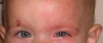
With ichthyosis, hair, teeth and nails undergo dystrophic changes. Dry, brittle hair is typical, nails break off and split, and multiple caries develops. Quite often, ichthyosis is accompanied by eye damage - chronic conjunctivitis and retinitis. Patients with ichthyosis have a hereditary predisposition to myopia, which begins to manifest itself in childhood. Since immunity is reduced, allergic diseases and purulent infections are permanent. Later, disturbances in the functioning of internal organs occur, most often cardiovascular failure and liver disease.
Recessive ichthyosis occurs only in males, although it is inherited on the X chromosome and differs in that the cause of the disease is a defect in placental enzymes. Clinical manifestations appear in the second week of life, less often immediately after birth. The horny layers of the skin look like large dense scales of black-brown color and resemble scutes. The skin between the scales is covered with cracks, so it looks like the skin of a crocodile or snake. Children with recessive ichthyosis often have mental retardation, abnormalities in the skeletal structure, and epilepsy. Juvenile cataracts and hypogonadism occur in 10-12% of cases.
Congenital ichthyosis develops in utero at 4-5 months of pregnancy. At birth, the baby's skin is covered with thick horny scutes of gray-black color. With congenital ichthyosis, the scales can reach up to 1 cm in thickness, the scales have different shapes, smooth or jagged, the skin between them is covered with grooves and cracks. Due to the dense, well-cohesive scales, the baby's mouth is either stretched or sharply narrowed so that the feeding tube barely fits. The ear openings are deformed and filled with horny scales, the eyelids are everted due to stretching. Almost all infants have skeletal anomalies - clubfoot, clubhandedness; many children with a congenital form of ichthyosis have interdigital bridges on the feet and palms, and sometimes missing nails. Pregnancy is often premature, and the rate of stillbirth is quite high. Since there are anomalies incompatible with life, most children with the congenital form of ichthyosis die in the first days of life.
Epidermolytic ichthyosis is a form of congenital ichthyosis. The baby's skin is bright red, as if scalded by boiling water. Nikolsky's syndrome is positive, as with pemphigus of newborns - with a slight touch, rejection of the epidermal scales is observed. The skin on the palms and soles is white and significantly thickened. In some cases, with the epidermolytic form of ichthyosis, there may be hemorrhages in the skin and mucous membranes. This is an unfavorable sign; if hemorrhages occur, children most often die. With milder clinical manifestations of ichthyosis, the blisters become smaller over time, but throughout life the disease recurs in the form of outbreaks, and during a relapse of ichthyosis the temperature often rises to high levels. By the fourth year of life, horny layers appear in certain areas of the body in the form of thick dirty gray scales, which are localized mainly in places of natural skin folds.
There are often defects in the nervous, endocrine and other body systems; many children with congenital ichthyosis are later diagnosed with mental retardation and spastic paralysis, which is caused by the accumulation of phytanic acid in the tissues. Polyneuropathy, anemia, and infantilism complicate the course of ichthyosis. The mortality rate is very high due to associated complications and associated diseases.
Forecast
The mortality rate from Harlequin ichthyosis is high, with rates approaching 50% worldwide. A review of 45 cases by Dr Rajpopat et al identified 25 survivors (56%) ranging in age from 10 months to 25 years. Twenty deaths (44%) occurred from days 1 to 52 and were as likely to cause respiratory failure as fulminant sepsis. A Japanese survey of 16 patients reported a survival rate of 81.3% (13 of 16 patients). Respiratory failure, fulminant sepsis, or a combination of both are the most common causes of death in newborns.
What does the development of ichthyosis include?
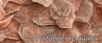
The biochemical properties of the disease have not been fully studied. However, keratinization of the skin occurs in the following order:
- An excessive accumulation of beneficial amino acids occurs in the blood of an infected person, which affects the disruption of the metabolism of fats and proteins in the body. As a result, cholesterol levels rise.
- In the patient's body, the thermoregulation of the skin is disrupted and metabolic processes are disrupted. Enzymes involved in the oxidative processes of the body, responsible for the “breathing” of the skin, on the contrary, are gaining activity.
- Vitamin A ceases to be absorbed by the body and the sweat glands cease to function normally. Keratin begins to be overproduced. All this leads to keratinization of the skin, which does not fall off, but remains on the skin. The scales are tightly connected due to a complex of amino acids located between them.
It is impossible to remove dead skin on your own, as it causes severe pain.
There are several forms of this pathology:
- ordinary (vulgar);
- recessive;
- congenital;
- epidermolytic;
- lamellar.