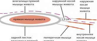
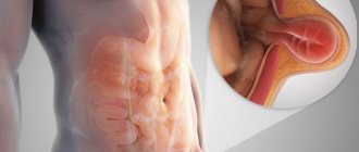
The linea alba is an anatomical structure that is formed by the fusion of the fascia and aponeuroses of the muscles of the anterior abdominal wall. The linea alba is located along the midline of the abdomen (dividing it into two symmetrical halves) and is named so because of its white color, which is caused by connective tissue.
Normally, the linea alba has small openings through which blood vessels and nerve endings pass. These gaps are a “weak” point, and under certain conditions, internal organs and fatty tissue located in the abdomen can prolapse through them. Such protrusions are called hernias. Most often they are localized in the upper third of the linea alba.
Types of hernias of the white line of the abdomen
Depending on the origin, a hernia of the white line of the abdomen can be congenital or acquired. A congenital disease manifests itself either immediately after the birth of a child, or a little later, but in early childhood. It occurs due to insufficient development of the tissues of the abdominal wall and can disappear on its own as the child grows. Acquired hernias of the white line of the abdomen occur with age. The reason for their development is the thinning and loss of tone of the aponeurotic tissues.
Hernias of the white line of the abdomen are divided into free (reducible) and irreducible. The contents of reducible hernias can move freely from the abdominal cavity to the hernial sac and back. With an irreducible hernia, such migration is impossible.
There is such a thing as a strangulated hernia. This condition occurs if the hernial contents are subjected to compression. As a rule, it occurs at the level of the hernial orifice, is life-threatening and requires immediate treatment.
In addition, complete hernias and incomplete hernias of the white line of the abdomen are distinguished. With a complete hernia, its contents extend beyond the abdominal wall. With an incomplete hernia, this does not yet happen.
Reliable signs of diastasis
Diastasis has one peculiarity. Even if you have a pumped-up and flat stomach, literally half a mug of water radically changes the situation, and your stomach looks like it would if someone was never friendly with sports. The anterior abdominal wall protrudes, the navel falls out.
The appearance of the abdomen is determined by the location and degree of diastasis: in some it manifests itself as a “bloated” abdomen, in others – an inverted navel.
Causes
The main reason for the development of acquired hernias of the white line of the abdomen is the loss of elasticity and thinning of the aponeurotic and fascial tissues. Various reasons can lead to this:
- Age-related changes.
- General exhaustion and cachexia.
- Traumatic damage, including during surgical interventions, when degenerative processes begin to occur in initially unchanged tissues along the incision line.
- Postoperative complications - inflammatory processes, seromas, hematomas. Most of all, the structure of the connective tissue of the aponeurosis is affected by purulent-necrotic processes that develop during infectious complications.
- Pregnancy.
- Obesity, in particular its abdominal form.
- Heavy physical activity.
- Prolonged constipation.
- Ascites.
- Problems with urination.
- Chronic lung diseases leading to persistent hacking cough (accompanied by tension).
Book a consultation around the clock +7+7+78
What to pay attention to during rehabilitation?
To ensure a sufficiently strong fusion during the rehabilitation period, restrictions are established. They concern:
- ability to carry heavy loads - 2 months, not more than two kg;
- playing sports and training - only from the third month with the permission of a doctor;
- food - only boiled, light dishes, milk porridges, juices, low-fat soups, kefir, cottage cheese (the rest is in accordance with the above dietary requirements).
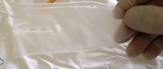
The photo shows a neat postoperative suture on days 2–3
Classification and stages
A hernia of the linea alba does not occur suddenly. One way or another, it is preceded by certain processes that lead to an increase in the size of the aponeurotic fissures. Preperitoneal fatty tissue begins to proliferate in them, leading to the formation of a lipoma. If you squeeze it, you will experience severe pain, which many patients and even doctors can mistake for an ulcer or other diseases of the gastrointestinal tract, accompanied by acute pain. Most often, the process ends at this stage and many people do not even suspect that they have such a pathology, especially if the disease is asymptomatic.
If the condition of the aponeurosis worsens, the situation may worsen. The peritoneum begins to stretch behind the fatty tissue, and when it gets into the aponeurotic fissures, a true hernia occurs. Following this, the greater omentum, a fragment of the intestine, or even the wall of the stomach may be pulled into the hernial sac.
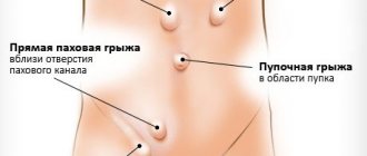
Based on location, the following types of hernias of the white line of the abdomen are distinguished:
- Supraumbilical. As the name suggests, they are located above the navel, in the so-called epigastric region. This is the most common pathology, found in 80% of hernias of the white line of the abdomen.
- Peri-umbilical hernias (not to be confused with umbilical hernias). They form around the umbilical ring and are very rare, occurring in less than 1% of patients.
- Subumbilical hernias, accordingly, are formed in the hypogastric region. They account for about 9% of all hernias.
A hernia of the linea alba is usually single. But there are also multiple formations located on top of each other.
Symptoms
At the stage of preperitoneal lipoma, the hernia can exist asymptomatically for a long time. If touched, acute abdominal pain develops, which can be confused with pathology of the gastrointestinal tract. It often happens that the hernia is in a stable state and exists at this stage for years without progression of the process.
If it increases, a protrusion forms along the midline of the abdomen. It causes painful sensations that intensify with palpation, after eating, when straining, etc. The pain can radiate under the shoulder blade, under the ribs or in the lower back. When the impact on the hernia stops, or intra-abdominal pressure normalizes, the pain subsides and may even stop for a while.
A serious complication of a hernia is strangulation, when the contents of the hernia are compressed by the hernial orifice. In this case, the patient experiences severe increasing pain. After some time, vomiting occurs, stool retention develops and symptoms of intestinal obstruction increase. In this case, the hernia cannot be repaired.
A strangulated hernia is a life-threatening condition and requires immediate hospitalization in a hospital followed by surgical care.
Diagnostics
The main symptom of hernia is the formation of a protrusion, accompanied by pain or discomfort at the site of its localization. In this case, the pain has a pulling or aching character and occurs or intensifies when pressure is applied to the hernia or when the abdominal muscles are tense. The intensity of pain does not depend on the size of the protrusion.
As for the protrusion itself, upon palpation you can determine that it has a soft consistency. When the position of the body changes, it can change its configuration and size. For example, with a vertical position of the body and straining, it increases. When lying down, it may decrease or disappear altogether.
Examination for hernias of the white line of the abdomen includes the following techniques:
- The linea alba of the abdomen is palpated along its entire length, starting from the xiphoid process and ending with the suprapubic region.
- To determine the hernial contents, palpation and percussion are performed. In the presence of intestinal loops, rumbling and a ringing tympanic sound will be detected. If there is a seal, the sound will be muffled.
- The reducibility of the hernia is determined with the patient lying on his back.
- The cough impulse symptom is examined. A finger is inserted into the hernial opening and the patient is asked to cough. If it is a hernia, then tremors will be detected during coughing.
- The patient is also asked to push. This will allow you to determine the maximum possibility of a hernial protrusion, if it is large, or to detect small hernias that may not yet appear.
Treatment methods
All hernias of the white line of the abdomen can be cured exclusively with surgery. Elective operations are performed for uncomplicated hernias. At the same time, it is possible to fully examine the patient and choose the optimal tactics for hernioplasty. Emergency operations are performed when complications develop (strangulated hernia), and in some cases several stages with delayed repair are required.
The choice of hernia repair method is determined based on the reasons for its formation, as well as the shape and size of the hernia. Sometimes it is impossible to carry out a full-fledged operation, in which case palliative interventions are limited. Such situations arise when the patient is old, in the presence of giant hernias, after closure of which suffocation may develop due to a sharp decrease in the volume of the abdominal cavity. Also, suturing is contraindicated in the early period after a strangulated hernia complicated by phlegmonous inflammation.
There are several ways to operate on hernias of the white line of the abdomen:
- Fascial-aponeurotic plastic surgery. This method implements to the maximum extent the principle of stitching homogeneous tissues, which, under certain conditions, ensures their reliable fusion and reproduction of the natural anatomical relationships of the tissues of the abdominal wall.
This method is implemented in two ways:
- Simple suturing of the edges of the aponeurosis. This method is considered unreliable because it often causes relapses. Can be used in the treatment of young people.
Duplication of aponeurosis. This strengthens the white line. Under certain conditions (good condition of aponeurotic tissues, small hernias), this method gives good results.
- The second method is suturing the aponeurosis with strengthening with muscle tissue . It is assumed that the muscle, due to its elasticity, will counteract the increase in intra-abdominal pressure. In practice, this method is also rarely used due to the technical difficulties of the operation. The main point is to preserve muscle function during muscle transplantation, and this is not always possible.
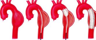
- And the last method is plastic surgery using artificial reinforcing materials that strengthen the white line. Such techniques are used for recurrent hernias, for large hernias, for atrophic changes in the structures of the abdominal wall, as well as for multiple hernias of the white line of the abdomen. Polymer meshes are used as reinforcement. They are chemically inert, non-toxic, elastic and durable. Their use has made it possible to sharply reduce the likelihood of relapses and achieve good treatment results.
In addition to the traditional method of performing surgery using skin incisions, endoscopic technologies are increasingly being used. They involve carrying out all surgical procedures through small punctures. The advantage of this technology is that the intervention is less traumatic, has a good cosmetic effect and a quick recovery period. The disadvantage of this method is the need for special equipment and trained personnel. To date, this is not available in every clinic.
Surgery for diastasis of the rectus abdominis muscles
An incision cannot be avoided if hernioplasty is necessary - surgery to remove diastasis and umbilical hernia, or abdominoplasty - removal of excess skin.
To carry out this procedure, we make a very small incision of 4-5 centimeters in the lower abdomen below the bikini line. It gives the surgeon freedom not only to remove hernias, but also to create a beautiful abdomen.
Expert comment:
“Despite the presence of a stitch, this is a very rewarding operation.
It allows not only crosslinking of diastase. This technique allows you to perform aesthetic abdominoplasty - remove excess skin, perform laser liposuction and remove fat, eliminate sagging abdomen and stretch marks, make the stomach flat, change the shape and position of the navel, emphasize the abdominal muscles and create the relief of a sports abdomen.
During the operation, I can create a sexy cleavage from the sternum to the navel, which looks very feminine. Or you can put special sutures on excessively wide muscles, limit the amplitude of their movement and form a pronounced waist.
The fat obtained during liposuction can be used as part of the same operation for liposculpture of the buttocks.
I make the cut in such a way that the seam can be covered with the smallest bikini. A special suturing technique reduces the time of surgery and rehabilitation, and makes scars and scratches almost invisible.”
Vladislava Gladysheva, plastic surgeon.
Typically, tummy tuck leads to breathing difficulties: the diastasis is stitched, and a feeling of a narrow corset appears in the abdominal area.
Over time, the body gets used to this condition, but the lack of oxygen affects both the general condition and recovery time. This is called compartment syndrome, or compartment syndrome. Before the operation, we carry out special, easy preparation, which almost completely eliminates the occurrence of compartment syndrome and ensures a quick and comfortable recovery after the procedure.
Prevention
In order to prevent the formation of a hernia, it is recommended to adhere to the following rules:
- Train your abdominal muscles.
- Organize a proper diet to avoid constipation and diarrhea.
- Maintain your weight at an optimal level.
- Try not to lift heavy objects.
- During pregnancy you need to wear a bandage.
As for the occurrence of recurrent hernias after their repair, the likelihood of relapse will be determined by the method of hernioplasty, the age of the patient and the condition of the tissues of the abdominal wall. The best results are achieved when strengthening the abdominal wall with synthetic materials in young patients.
Book a consultation 24 hours a day
+7+7+78
Features of the postoperative period
After the operation, the patient is observed and wound infection is prevented (a course of antibiotics, dressings with disinfectants). Men often have difficulty urinating. Walking is allowed on the second day. It is recommended to wear a bandage.
They are discharged home on days 8–10, immediately after the stitches are removed, but complete restoration of the tissue and wound will occur only after several months. At home, it is recommended to treat the wound with brilliant green. Dieting. Showering is allowed after two weeks. The doctor may prescribe physical therapy.