Trichophytosis is a fungal disease that affects the skin and hair, and sometimes the nails.
The causative agent is the fungus Trichophyton. Infection occurs through contact with a sick person, as well as his things (hats, combs, scissors, bedding, etc.). Possible transmission in hairdressers, kindergartens, boarding schools, schools. Rodents (mice, rats) and cattle (mainly calves) also serve as carriers of the fungus.
Infection of a person, as a rule, occurs through contact with hay, dust, contaminated hair affected by the fungus, and less often through direct contact with an animal. The disease is recorded more often in the autumn, which corresponds to periods of agricultural work.
Trichophytosis: symptoms of the disease
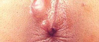
Trichophytosis of the scalp manifests itself as follows:
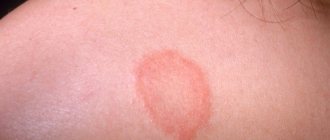
- small lesions appear (less than 2 cm in diameter);
- they have a reddish color, clearly defined boundaries; not prone to fusion;
- their surface may be covered with bubbles, which over time turn into crusts;
- slight itching is felt (however, it may be absent).
If there is a chronic form of trichophytosis of the scalp, the symptoms are less pronounced, only mild peeling is observed. The lesion is most often located on the back of the head. A careful examination reveals black dots of broken hair.
With the infiltrative-suppurative form of the disease, a long incubation period is observed (up to 2 months). This type of disease is characterized by the fusion of lesions, the appearance of pustules and crusts, and hair damage. In addition, dilated follicles can be seen on the surface of the lesions, from which pus pours out when pressed.
Pathogenesis
The pathogenesis of dermatophytosis is based on the virulence factors of fungi, due to their ability to secrete enzymes that destroy keratin (keratinolytic activity). The enzymes secreted by dermatomycetes and their metabolic products, as well as the contents of destroyed cells, cause an inflammatory process in the lesion, manifested by redness of the skin, itching and hair breaking. Keratinases have the ability to decompose not only keratin, but also other proteins (collagen and elastin), and it is the severity of keratinase activity that determines the characteristics of the manifestation of diseases when infected with various types of fungi. An important role in the spread of dermatomycetes is played by the presence of a specialized apparatus (perforator organs) for the directed growth of hyphae (the filamentous structure of the fungus). Thanks to this feature, the fungus grows in the direction of the intercellular junctions, which are the points of least resistance. Dermatomycetes, as a rule, do not penetrate deeper than the granular layer of the epidermis, since specific protective factors are activated at a deeper level.
The process of fungal invasion begins with the burrowing of hyphae into the skin and the release of keratinases (enzymes for the breakdown of keratins), which leads to irritation of nerve endings and, accordingly, the occurrence of itching. Dermatomycetes, loosening the cortex, cause acanthosis, parakeratosis, serous inflammation in the skin with the formation of blisters. Further, swelling occurs in the papillary/subpapillary layer, the vessels dilate, which leads to the development of polymorphic perivascular infiltration. The mycelium is usually located between the horny plates. Threads and spores of fungi can also be stuffed with hair follicles, which also undergo inflammatory changes.
Prevention
The basis of preventive measures is:
- Strict adherence to the rules of personal hygiene: do not use other people’s personal hygiene items, underwear, clothes, wash your hands thoroughly after contact with animals.
- Early (timely) identification of patients with trichophytosis with their isolation and quarantine of contact persons.
- Identification of animals with trichophytosis, carrying out deratization work.
- Carrying out current/final disinfection at infection sites (in schools, kindergartens, at home, bathhouses, hotels, hairdressers, medical institutions, dormitories, etc.).
Methods for eliminating fungal infections
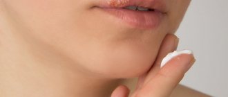
Treatment of trichophytosis requires the attention of a qualified specialist. In most cases, therapy is carried out at home. To successfully combat fungus, an integrated approach is essential: medications for external and internal use, and physiotherapeutic methods are used.
Treatment of trichophytosis with medications
Modern medicine offers the dermatologist a wide selection of antifungal agents. The pathogen is usually treated in a complex manner: ointments are applied topically, and drugs are taken orally in the form of tablets or capsules.
Local and general treatment of trichophytosis - table
| Preparations for external treatment of trichophytosis | Systemic treatment of trichophytosis | ||||
| Name of medicine | Active substance | Release form | Name of medicine | Active substance | Release form |
| Exoderil | Naftifin | Solution | Itrazole | Itraconazole | Capsules |
| Keto plus |
| Shampoo | Sangviritrin |
| Pills |
| Oflomil varnish | Amorolfine | Nail polish | Exifin | Terbinafine | Pills |
| Fungoterbin | Terbinafine |
| Mikosist | Fluconazole | Pills |
| Sangviritrin |
|
| Orungal | Itraconazole | Capsules |
| Mikoderil | Naftifin |
| Binafin | Terbinafine | Pills |
| Lamisil | Terbinafine | Cream | Rumicosis | Itraconazole | Capsules |
| Exifin | Terbinafine | Cream | Diflazon | Fluconazole | Capsules |
Antifungal agents - photo gallery
Exoderil is available in the form of a cream for external use.
Oflomil is available in the form of nail polish
Lamisil is an antifungal agent for external use.
Exifine contains terbinafine
Orungal is a drug for the systemic treatment of trichophytosis
Diflazone contains fluconazole
In addition to antifungal agents, a number of additional drugs are prescribed for the treatment of trichophytosis:
- antiallergic medications (Zodak, Loratadine, Suprastin);
- drugs that improve blood circulation (Troxevasin, Trental);
- antioxidants that improve the healing of damaged skin (Actovegin);
- immunomodulators that activate the body's defenses (Thymogen, T-activin).
Thymogen improves immunity
Physiotherapy
Physiotherapy is an effective method of treating many diseases, including superficial and deep trichophytosis. Specialists prescribe the following types of procedures:
- electrophoresis with iodine allows you to disinfect the lesion;
- iontophoresis with iodine can successfully replace the previous procedure;
- inductothermy (magnetic field treatment) improves blood circulation in problem areas of the skin;
- Ultrasound therapy in the area of the nearest lymph nodes leads to the activation of one’s own immune mechanisms to fight the fungus.
Household recommendations
Treatment of trichophytosis is often a long and difficult process. The patient must follow a number of recommendations that will speed up recovery and prevent the spread of infection:
- for trichophytosis of the head, the hair is shaved once a week until complete recovery;
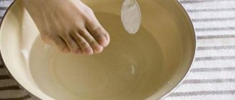
- if the feet are affected, it is necessary to disinfect the inner surface of the shoes once a week with a cotton swab soaked in a solution of Chlorhexidine;
Chlorhexidine solution perfectly disinfects shoes - it is necessary to use individual personal hygiene items - a comb, a towel and other linen, manicure accessories;
- shoes must be chosen that are comfortable, fit, made from natural materials, and well ventilated.
Treatment of trichophytosis in children is the task of an experienced specialist. The principles of therapy are similar to those in adults. Doses of antifungal agents used orally are calculated depending on the age and weight of the child.
Folk remedies are not used in the treatment of trichophytosis because they have not proven their effectiveness.
List of sources
- Leshchenko V. M. Fungal skin diseases. In the book: Skin and venereal diseases (a guide for doctors). Ed. Yu. K. Skripkina, V. N. Mordovtseva. M., 1999. T. 1. P. 257-311.
- Kubanova A.A., Potekaev N.S., Potekaev N.N. Guide to practical mycology. – Moscow, Financial Publishing House “Business Express”, 200 p.
- Bayazitova A.A., Bayazitova A.A., Kupriyanova-Ashina F.G., Khaldeeva E.V., Ilyinskaya O.N. Primary pathogens - Pathogens of superficial mycoses // International Journal of Applied and Basic Research. – 2015. – No. 11-2. – pp. 241-248.
- Medvedeva T.V. Trichophytosis: modern ideas about the etiology, clinical picture, features of diagnosis and therapy / T.V. Medvedeva, V.B. Antonov, L.M. Leina, T.S. Bogomolova // Klin. dermatology and venereology. - 2007. - No. 4. - P. 70–74.
- Rodionov A.N. Fungal skin diseases (Guide for doctors) St. Petersburg: Peter. 1998. – 288 p.
Clinical signs of lichen in cats and dogs
Spontaneous alopecia areata (baldness), sometimes with a red rim around the periphery of the baldness. The spot may peel off and is rarely accompanied by itching (scratching). Lesions are often located on the face and ears; young and immunocompromised animals are more susceptible to the disease (for example, with hyperadrenocorticism in dogs , diabetes mellitus in cats and dogs, immunodeficiency and feline viral leukemia, cancer).
A characteristic manifestation of lichen on the skin of the back of a Bambino cat.
Persian cats and Yorkshire terriers are at-risk breeds.
Cats can be carriers and sources of infection without showing clinical signs themselves. In long-haired cats, hair may not fall out, for example, in cats of Persian breeds, lichen can occur as single or multiple nodules in the thickness of the skin, felt like plaques under the skin with a diameter of 1–8 cm, with or without ulceration, on the back, sides or neck ( pseudomycetoma ). If you accidentally damage these areas of the skin, systemic mycosis may develop (spread of the fungus throughout the body, including into the internal organs).
In dogs, the fungus Microsporum canis causes a severe inflammatory reaction. Skin lesions look like a kerion ( a thickening of any shape that rises above the skin).
One of the manifestations of lichen in dogs is kerion. In this photo there is a round, hairless spot on the upper lip, rising above the surface of the skin.
Differences from microsporia
Quite often, trichophytosis is confused with microsporia.
But these diseases are characterized by the presence of certain distinctive features. Trichophytosis is popularly called lichen. During the pathological process, hair breaks off. This is why it feels like the hair has been cut. Trichophytosis and microsporia are fungal diseases. The distinctive features between them are fungi, which cause a pathological process . Ringworm occurs due to exposure to trichophyton fungi, and microsporia occurs due to microsporum. Since these diseases are characterized by almost the same symptoms, they are both called ringworm.
Trichophytosis and microsporia are quite difficult to distinguish. That is why, to determine ringworm, the patient is recommended to undergo appropriate laboratory tests.
Treatment of lichen in cats and dogs
In cats, cases of spontaneous recovery have been noted within 12–14 weeks. However, treatment should be carried out to prevent infection of other animals and people, to speed up recovery, and to reduce the spread of spores in the environment. The course of treatment is quite long, can take up to 4 months.
Successful treatment of lichen in cats and dogs is based on 3 areas:
- Systemic treatment . It is necessary to carry out this procedure, since it is mainly the growing hair that is affected; local products cannot penetrate into the hair in any way. For systemic treatment, antifungal agents (itraconazole, fluconazole, terbinafine) are used in tablet form. The course is long and well tolerated by animals. Some products accumulate in the skin and are contained in it for a long time in a therapeutic concentration; such products can be used in a pulse mode (the drug is taken only 2 days a week or every other day).
- Local treatment. It is used to prevent fungal spores from entering the environment and infecting other animals and people. The whole animal must be washed! Treating only the affected areas will not bring any effect, since spores can be found on the entire surface of the body. To wash animals with lichen, use Imaverol, Lime Sulfur, Liverazol, and shampoos with enilconazole. Treatment is carried out 2 times a week, some products do not need to be washed off, the therapeutic effect is achieved when the product dries on the coat and skin. For the best effect, long-haired animals are recommended to have their hair cut short or shaved.
- Environmental treatment . Carried out to destroy spores in the environment. Disinfectants with an antifungal effect are used (Imaverol, Clinafarm suppositories, sodium hypochlorite, 5–10% bleach, 0.2% enilconazole, 2% chlorhexidine). Items and clothes can be washed at 60 degrees in a washing machine, 2-3 cycles.
Evaluation of treatment effectiveness
Evaluation of treatment is carried out by culture on special media; culture is done after the disappearance of clinical signs every 2–3 weeks, until a negative result + 1 more control culture.
Only if there are no clinical signs and the culture is negative can the doctor say that the animal is healthy and not a carrier.
Vaccination is not used to treat or prevent lichen infection in cats and dogs!
Today there are no vaccines that can protect animals from infection, this is due to the fact that the vaccine creates humoral immunity (the production of antibodies in the blood and body fluids), and in the case of lichen, only cellular immunity (special cells in the body that react for antigens).