According to WHO criteria, the criterion of anemia is a decrease in hemoglobin level of less than 130 g/l in men and less than 120 g/l in women. The incidence of anemia ranges from 8% in individuals over 70 years of age to more than 40% in individuals over 85 years of age. Recent studies have established that poor age does not necessarily mean anemia, but rather a greater degree of weakness before its development. The clinical classification of anemia (Table 1) is based on the morphological principle - the average erythrocyte volume1 (MEV). In elderly patients, anemia may not be accompanied by a change in the size of erythrocytes. The most common etiological causes of anemia are listed in Table 3.
1 It is indicated using automatic laboratory analyzers or using the following formula: SEO = (hematocrit x 10) / number of erythrocytes (in million/l). (Note, switch)
Table 1. Classification of anemias
Macrocytic anemia (CEO>100 fl)
|
Microcytic anemia (CEO<80 fl)
|
| Normocytic anemia (CEO 80–100 fl) Increased consumption or destruction of erythrocytes (ICP2 > 2)
Reduced synthesis of erythrocytes (ICP < 2)
|
2 Index of reticulocyte synthesis (ISR) = number of reticulocytes (%) x hematocrit/45 x 1/2. The ISR is more reliable, the absolute number of reticulocytes is lower, which leads to the end stage of anemia and the level of life of reticulocytes. The ISR standard is set to 1. (Note, switch)
Table 2. Etiology of changes in the shape of erythrocytes (per M. E. Conrad, 2004)
| Red blood cell shape | Morphology | Characteristics of illness |
| Spherocyte | Loss of enlightenment at the center of the cell, more intensive preparation, often of microcytic size | Spasmodic spherocytosis, a type of full-blown hemolytic anemia |
| Klitina-target | Hypochromic cells with central accumulations of hemoglobin in the “target” shape | Liver disease, thalassemia, hemoglobin D, post-splenectomy condition |
| Leptocyte | Hypochromic tissue of normal diameter due to decreased SEO | Thalasemia |
| Ovalocyte | Oval or cigar-shaped | Congenital ovalocytosis, various types of anemia (especially in cases of deficiency of vitamin B12 and folic acid) |
| Schistocyte | Fragmented erythrocytes of tricutaneous shape or sholoma-like appearance | Microangiopathic anemia, artificial heart valve, uremia, malignant hypertension |
| Stomatocyte | Red blood cell with a slit-shaped central lucency | Liver disease, acute alcoholism, malignant disease, congenital stomatocytosis, artifact |
| Droplet-like | Speckled, often microcytic erythrocyte | Myelofibrosis, cerebrospinal fluid infiltration, thalassemia |
| Acanthocyte | Detection of 5–10 clusters of different species on the surface of the erythrocyte, distributed unevenly | Abetalipoproteinemia, insufficient food, liver disease. |
| Echinocyte | Evenly distributed sprouts on the surface of the erythrocyte (usually 10–30) | Uremia, schulci viral disease, schulcan cancer, pyruvate kinase deficiency, artifact |
| Crescent | Sickle-shaped clitina | Hemoglobin S, different types of hemoglobin C and I |
Table 3. The most common etiology of anemia
|
Diagnosis of anemia3
In especially elderly patients, typical symptoms (weakness, shortness of breath, weakness) often occur with less severe anemia than in patients of younger age. Anemia also causes an increase in other illnesses, changes in frailty, confusion, cognitive impairment and depression. Therefore, in patients with impaired cardiac or pulmonary disease, the presence of anemia should be turned off.
Nitrogen deficiency is an important etiological cause of anemia in patients of all ages. Although the function of the brain steadily decreases with age, in the presence of illness, the change is insignificant. Although anemia due to neurological causes occurs only in cases of impaired function of the nervous system, age-related changes are obviously not associated with episodes of significant anemia in the elderly.
Table 4. Indications for fasting due to anemia
|
Table 5. Tests for primary fasting due to anemia
Additional tests
|
3Div. also a statue
"Diagnostic algorithm for anemia."
— Medicine for the world. - 1996. part 1. p. 3–9.
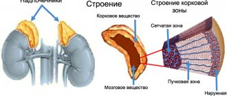
Normocytic anemia in endocrine diseases
Normocytic normochromic anemia most often accompanies the following pathologies:
- Cushing's disease.
- Hyperparathyroidism.
- Thyrotoxicosis.
- Addison's disease.
- Hypoandrogenism.
- Panhypopituitarism.
Endocrine glands produce hormones that are directly involved in the production of erythropoietin. Therefore, damage to the endocrine glands leads to a malfunction in the hematopoietic system.
As for the laboratory data of the blood picture, they are similar to the laboratory data obtained for renal failure.
Fluid deficiency anemia
Fluid deficiency causes anemia in 10–15% of elderly patients. The etiological factor, of course, is chronic blood loss through the blood vessels (see Table 6) and rarely, insufficient vascular drainage. Such bleeding is often asymptomatic and cannot be detected unless a negative stool blood test is present. Therefore, after identifying PDA in this category of patients, it is necessary to perform sterilization of the SCC, especially to exclude malignancy. The pathology of the upper branches of the SCC is most often affected, causing 40–50% of cases of HDA in summer people. It is important to cover the upper and lower sections of the SCC, as 10–15% of patients develop two pathologies at the same time. At the hour of esophagogastroduodenoscopy (EGD), a biopsy of the mucous membrane and duodenum is taken to exclude Helicobacter pylori
and celiac disease.
If patients cannot obtain an EGD, it is necessary to perform fluoroscopy of the upper lobes of the GCC with contrast barium, serological tests to detect H. pylori,
determine the level of antibodies against transglutaminase and immunosuppression globulins (to exclude celiac disease). If there is a contraindication, barium irigoscopy can be performed before colonoscopy. In 40% of patients with AD, it is not possible to establish the etiology of bleeding, regardless of the restraint. Fortunately, these patients are satisfied after replacement therapy and there is often no need for repeated quilting.
The most specific test for diagnosing ADHD is feritin
blood infusion, which indicates the amount of fluid reserves. Feritin level often decreases even before the decrease in mean erythrocyte volume, especially in the early stages of AD. As the saliva deficiency progresses, the value of SEO decreases along with hemoglobin. A feritin level <15 µg/l indicates a fertilization deficiency, and if its value is greater than 100 µg/l, the diagnosis of HDA can be turned off. The fragments of feritin are a protein of the acute phase of inflammation, and its rhubarb can be extremely high in the production of cytokines during inflammation, infection, or malignant illness, and can be significantly reduced during illness. Certainly, especially during the second and third trimester.
Since the level of feritin is doubtful (between 15–100 µg/l), the level of receptors for transferrin
(normal blood levels are 2–9 mg/l).
The transferrin receptor is a transmembrane glycoprotein that binds to circulating transferrin and transfers one or two atoms of release. Finally, the transferrin receptor is transported into the cytoplasm, where the membrane is formed, and the protein again rotates on the surface of the cell and is visible in the blood. A progressive decrease in salinity reserves is manifested by a decrease in the level of feritin in the blood serum, and a decrease in functional salinity in tissues is manifested by a decrease in the level of receptors for transferrin. The receptor for transferrin is not a protein in the acute phase of inflammation, so it is not affected by inflammation, infection, illness, or vaginosis. With ZDA, the number of receptors advances before transfer, even before changes occur in the CEO. The level of contamination in blood serum4,
as a result, has decreased, and
the level of serum concentration in blood serum5
has increased.
4The norm is 8.1–31.3 µmol/l in men and 5.4–31.3 µmol/l in women. (Note switch)
5The norm becomes 39.4–75.2 µmol/l. (Note switch)
Table 6. Etiology of bleeding from SCC
|
Treatment of the HDA involves the elimination of the source of blood loss (to the extent possible) and mixed treatment with salvage preparations.
Anemia caused by chronic illnesses
Anemia, caused by chronic diseases (ACD), is the most common cause of anemia in elderly patients and another common etiological cause of anemia in the world (after anemia). This anemia is due to the action of cytokines, which are seen in the response to many acute and chronic illnesses (see Table 8). ACD in typical episodes is manifested by normochromic normocytic anemia with CEO in the normal range (80–100 fL), although it can also be slightly microcytic (SEO <80 fL). If the hemoglobin level is lower than 100 g/l, then turn off the connection of ACD with another etiological cause of anemia. The response of the cerebrum to ACD is inadequate, so the index of reticulocyte synthesis is significantly lower 2. The number of reticulocytes, the rate of blood serum release, the secretive production and the saturation of transferrin6
є decreases, and the level
of feritin
as a protein in the acute phase of inflammation—movements (or may be normal). However, to confirm the diagnosis, it is necessary to perform a bone biopsy.
6The norm is 20–50%. (Note switch)
The most specific test for the differential diagnosis of salivary deficiency anemia (ZDA) and anemia caused by chronic diseases (CHD) is the level of feritin: a level greater than 100 μg/l is practical allows the indicator to turn off ZDA, and a value of less than 15 µg/l confirms the diagnosis of ZDA . In patients with chronic inflammatory diseases, the level of receptors for transferrin in the blood serum and the level of feritin is significantly less than 50, and in case of severe salinity deficiency, it increases above 50.
Treatment of ACD is used in the treatment of primary illness. Although there is no specific treatment, the results of the concentration of erythropoietin in certain patients with cancer or HIV infection with a similar hormone in blood plasma of less than 500 MO/ml are encouraging. Since the presence of feritin deficiency is suspected on the basis of the middle level of feritin, for differential diagnosis between HDA and AZH it may be necessary to treat ex juvantibus with feritin drugs.
Table 7. Differential diagnosis of salivary deficiency anemia (ZDA) and anemia caused by chronic illnesses (ACS)
| Laboratory test | ZDA | AHZ |
| Rhubarb in the blood serum | ↓ | ↓ |
| Zagalna zagalna zazvoyuchaya building | ↑ | ↓ |
| Transferrin saturation | ↓ | ↓ |
| The value of feritin in blood serum | ↓ | N or ↑ |
Table 8. Illnesses that can be caused by AZH
| The group is sick | Apply it |
| Gastroinfections | Bacterial, fungal, viral |
| Chronic infections | Tuberculosis, endocarditis, chickweed infection, coccidioidomycosis and other chronic fungal infections |
| Non-infectious chronic ignition illnesses | Arthrosis, rheumatoid polyarthritis, rheumatic polymyalgia, acute and chronic hepatitis |
| Evil illness | Metastatic carcinoma, hematological oncology |
| Gostri stani | Operations, significant trauma |
| Another illness | Blood diabetes, congestive heart failure, malnutrition, bedsores |
Table 9. Etiology of insufficiency of the function of the cerebrospinal fluid
Primary deficiency of the function of the cerebrospinal fluid
|
7Div. See also article Recommendations for screening and treatment of patients with chronic illnesses. —
Medicine for the world. - 2003. - T. 13, part 5. - P. 231-235.
Prerequisites for the emergence
There are several reasons for the development of pathology.
- Rheumatoid arthritis and anemia. What you need to know
- Mature age. Women over 85 years of age are more susceptible to the disease.
- Chronic inflammation, infections and malignant tumors.
- Iron deficiency due to constant blood loss caused by pathologies such as colon cancer or stomach ulcers.
- Heredity.
- Pathological condition of the kidneys.
Deficiency of vitamin B12 and folic acid
Deficiency of vitamin B12 and folic acid occurs in 5–15% of gerontological patients, resulting, as a result, in macrocytic anemia. Some CEO may be normal, so in case of normocytic anemia of unknown etiology, the deficiency of these vitamins should be included. Table 10 shows the main etiological causes of vitamin B12 deficiency in elderly patients. Hematological, neurological and psychiatric manifestations can occur simultaneously, sequentially or independently in one way or another. If, instead of B12, the blood does not exceed the third level of the norm, to confirm the diagnosis, measure the level of methylmalonic acid and homocysteine in the blood serum (if their concentration is normal, which does not include B1 deficiency 2). The increase in methylmalonic acid confirms the presence of vitamin B12 deficiency, and the increase in homocysteine will require further adjustment to exclude other causes of its increase.
8The norm is 162–835 pmol/l. (Note, switch)
Table 10. The main etiological causes of vitamin B12 deficiency in elderly patients
| Pathophysiological mechanism | Etiological officials |
| Insufficient care for the hedgehog | Chronic alcoholism Chronic malnutrition Suvora vegetarian diet |
| Impaired absorption | Atrophic gastritis Status after gastrectomy Abdominal proliferation of bacterial flora in the small intestine Malabsorption of Crohn's disease Chronic pancreatitis Gastritis, zoom Helicobacter pylori |
| Metabolism disruption | Medicines Transcobalamin II deficiency Analogues of antivitamin B12 |
| Reduced reserves of vitamin B12 in the body | Important liver disease |
| Interactions with medications | |
| Malabsorption | Metformin, antibiotics, diphenine, colchicine, para-aminosalicylic acid, cholestyramine |
| Malabsorption of grub products, which interferes with vitamin B12, inactivation of metabolism | Vitamin C, nitric oxide, proton pump inhibitors, H2 receptor antagonists |
The most common cause of folate deficiency is insufficient food. Hematological manifestations are similar to vitamin B12 deficiency, there are no neurological symptoms. Most summer patients have normocytic anemia. Instead of folic acid in the blood serum9 may be extremely normal after eating, the concentration of folates in erythrocytes10 more specifically reflects the tissue reserves of this vitamin. If the result reaches the lower value of the norm, then the level of homocysteine is measured - in 90% of cases of folate deficiency, its level increases. Rhubarb methylmalonic acid is, of course, normal.
9The norm is 7–28.1 imol/l. (Note, switch)
10 The normal level becomes 422–1464 nmol/l, (Note, switch)
Treatment for vitamin B12 deficiency is administered internally at a dose of 500–1000 mcg per day for 7–10 days, then change the dose twice and continue treatment for another 7–10 days , from 14–20 days switch to administering the drug every other day dose of 500 mcg over a period of 4–6 days, after which over a period of 2 months, supportive therapy of 500 mcg 1 time per week is required, then the patient must continue to take 500 mcg of vitamin B12 daily and for a month. In cases of folic acid deficiency, it is prescribed at a dose of 1 mg per day per os
.
Why does hypochromia occur?
In medicine, there are three main reasons for the development of hypochromia, depending on the type of anemia:
- Excessive loss of iron as a result of bleeding or excessive consumption of the useful element leads to the development of iron deficiency anemia.
- Disruption of the absorption of iron in the intestines, as a result of which iron ingested with food is excreted from the body. In this case, sideroachrestic or mixed types develop. Often such hypochromia develops against the background of drug treatment with certain drugs.
- Low intake of iron through food is a source of mixed type anemia.
Most often, hypochromia is provoked by the following factors:
- unbalanced diet, consumption of food that contains practically no vitamins, iron, deficiency of animal protein in the diet, vitamin deficiency;
- constant or periodic bleeding (nasal, intestinal, stomach), uterine bleeding, prolonged heavy menstruation, bleeding gums;
- acute massive blood loss;
- gastrointestinal pathologies that interfere with the absorption of iron (enteritis, helminthiases, acute/chronic infections, gastritis, dysbiosis, Crohn's disease, impaired intestinal absorption);
- poisoning with harmful toxic substances;
- surgical interventions;
- childbearing and breastfeeding;
- malignant blood diseases and other cancers;
- frequent stressful situations of a protracted nature;
- chronic infectious diseases (tuberculosis, pneumonia, liver/kidney pathologies).
In addition, hypochromic anemia is diagnosed due to the patient having autoimmune diseases (lupus, vasculitis, glomerulonephritis, rheumatoid arthritis, etc.), which form immune complexes containing red blood cells and at the same time producing antibodies against their own red blood cells.
Myelodysplastic syndrome
Myelodysplastic syndromes (MDS) is a pathology of the Stobur cells of the cerebrospinal fluid, which leads to ineffective hematopoiesis and, as a result, to various combinations of anemia, neutropenia and thrombocytopenia. These syndromes are sometimes called pre-leukemic due to their nature and sometimes transform into acute leukemia. MDS in patients with less than 50 years of age rarely occurs, but in patients with more than 65 years of age, the frequency is 5%. To confirm the diagnosis, a bone marrow biopsy is required. This disease is characterized by the presence of chronic cytopenia, hyperplasia of the cerebrospinal fluid and morphological pathology of the progenitor cells in the cerebrospinal fluid.
The response to treatment with chemotherapy, immunosuppressants, growth factors (granulocyte-stimulating hormone and erythropoietin) is limited, and the survival rate of patients does not increase. Therefore, in older patients, treatment is alternated with blood transfusions and other symptomatic therapy.
Prepared by Bogdan Boris
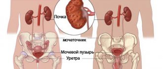
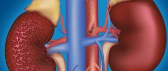
Normocytic anemia in renal failure
When the kidneys are unable to cope with their functions, the body's production of erythropoietin decreases. Chronic renal failure also contributes to the accumulation of uremic toxins in the body, which interfere with the production of erythropoietin. Chronic blood loss and other factors have a negative impact on the blood condition. This, in turn, leads to the fact that red blood cells quickly die and their level in the blood decreases.
Hemodialysis also contributes to the development of anemia. This procedure is often prescribed to treat chronic kidney failure.
Blood parameters in normocytic anemia against the background of kidney pathologies:
- Normocytic-normochromic anemia.
- Reticulocytosis with a slight increase.
- Thrombocytopenia.
- Decrease in erythropoietin levels.
- Decreased leukocyte levels.
In general, the pathogenesis of normocytic normochromic anemia in chronic renal failure is quite complex, but the leading role is played by the deficiency of endogenous erythropoietin, which causes accelerated apoptosis of erythroid cells in the bone marrow.