Adult respiratory distress syndrome (ARDS) is an acute respiratory failure, which can be caused by acute lung injuries of various natures, and which is characterized by non-cardiogenic pulmonary edema, disturbances in external respiration and hypoxia (lack of air in the body).
- Causes
- Pathogenesis
- Pathomorphology
- Symptoms
- Diagnostics
- Treatment
The syndrome was described in 1967 by researcher Esbach. Named similarly to neonatal respiratory distress syndrome, which is caused by a surfactant deficiency that affects babies from birth. In the case of the disease in question, this deficiency is secondary, that is, it does not arise from birth. Often other names are used for this syndrome: non-cardiogenic pulmonary edema, shock lung.
According to Marini data released in 1993, there are 150,000 cases of adult respiratory distress syndrome in the United States each year. That is, there are 0.6 patients per 1000 population.
Causes
Among the most common causes of the syndrome in question are:
- sepsis
- viral, bacterial, fungal and other pneumonias
- disseminated intravascular coagulation syndrome with subacute or acute course
- anaphylactic or septic shock, pronounced and long-lasting
- Chest compression syndrome and trauma
- aspiration of vomit, water (in cases of drowning)
- pulmonary embolism (air, fat, amniotic fluid)
- inhalation of irritating and toxic substances: nitrogen oxide, chlorine, ammonia oxides, phosgene, pure oxygen
- venous fluid overload (plasma, saline or colloid solutions, etc.)
- massive blood transfusions with the development of multiple microthromboembolisms in the vascular bed of the lungs
- severe metabolic disorders (eg, uremia)
- use of a heart-lung machine
- autoimmune diseases (Goodpasture syndrome, systemic lupus erythematosus, etc.)
- acute hemorrhagic pancreatic necrosis (enzyme intoxication is important in the pathogenesis, which provokes a violation of surfactant synthesis)
- long stay at high altitudes
Prevention
There is no specific prevention for ARDS. To prevent the development of pathology, it is necessary to beware of the effects of stressful damaging factors: promptly treat infectious diseases of the lungs, avoid chest injuries, and do not inhale toxic substances. Doctors should carefully monitor the blood transfusion process.
Prevention of respiratory distress syndrome in newborns consists of preventing premature birth, early detection and treatment of acute fetal hypoxia.
- For this, pregnant women are prescribed Dexamethasone and Betamethasone, which promote the maturation of surfactant in the lungs. The benefits of proper prophylactic use of drugs for a newborn should outweigh the potential risk and be expressed in a reduction in perinatal morbidity and mortality.
- To treat hypertension, pregnant women are prescribed Eufillin.
- The lungs mature faster in the fetus under the influence of Folliculin, Methionine, Essentiale, Lazolvan and Bromhexine.
Prevention of distress syndrome is not carried out for women beyond 34 weeks.
Timely diagnosis of ARDS and adequate therapy can cure even newborns. Otherwise, distress syndrome leads to the death of patients. With the development of multiple organ failure, the frequency of deaths increases many times. Mortality of patients depends on pulmonary dysfunction. After illness, the lungs can regain almost normal function.
Pathogenesis
Etiological (causal) factors lead to the fact that activated leukocytes and platelets accumulate in large quantities in the interstitial tissue of the lungs and in the capillaries of the organ. Researchers hypothesize that they release many biologically active substances:
- prostaglavdinov
- proteinases
- leukotrienes
- toxic oxygen radicals, etc.
They damage the alveolar epithelium and vascular endothelium, change the reactivity of blood vessels, the tone of the bronchial muscles, and contribute to the development of fibrosis. The above listed biological substances affect damage to the alveolar epithelium and endothelium of the capillaries of the lungs. In a short period of time, vascular permeability increases, spasm of the pulmonary capillaries occurs, and the pressure inside them increases. Pronounced sweating of plasma and red blood cells into the alveoli and interstitial tissue of the lungs is recorded, pulmonary edema and atelectasis develop. The development of atelectasis is also influenced by a secondary decrease in surfactant activity.
The processes described above affect the main pathophysiological mechanisms :
- shunting of venous blood into the arterial bed
- alveolar hypoventilation
- impaired diffusion of oxygen and carbon dioxide
- mismatch between ventilation and perfusion
Pathomorphology
Respiratory distress syndrome in adults develops at least 2-3 hours, maximum 3 days from the onset of the influence of the cause factor. There are 3 pathomorphological phases of the syndrome in question:
- acute
- subacute
- chronic
The duration of the acute phase of adult respiratory distress syndrome ranges from 2 to 5 days. Interstitial and then alveolar pulmonary edema develops first. Red blood cells, protein and leukocytes are found in the edematous fluid. In addition to edema, severe damage to the alveolar epithelium of types I and II and damage to the pulmonary capillaries is also detected. Damage to type 2 alveolocytes causes disruption of surfactant synthesis, which is why microatelectasis develops.
If the course of respiratory distress syndrome in adults is favorable, then after a few days the acute phenomena subside and the edematous fluid resolves. But the syndrome does not always proceed favorably. In some cases, it becomes subacute and then chronic. In the subacute phase, interstitial and broncho-alveolar inflammation occurs.
The chronic phase of adult respiratory distress syndrome is characterized by the development of fibrosing alveolitis. In the alveolar-capillary basement membrane, connective tissue grows, a sharp thickening of the membrane occurs, as well as its flattening. A pronounced proliferation of fibroblasts and increased synthesis of collagen, the amount of which increases two or even three times, are typical. Severe interstitial fibrosis may be present as early as 2-3 weeks after the onset of the disease. Also, the chronic phase is characterized by changes in the vascular bed of the lungs: the development of microthrombosis, emptying of blood vessels. As a result, chronic pulmonary hypertension and chronic respiratory failure develop.
Consequences and complications
Possible complications of ARDS in adults include ventilator-associated pneumonia , asphyxia , pulmonary barotrauma , bacterial pneumonia , left ventricular heart failure , and the development of disseminated intravascular coagulation syndrome .
The consequences of SDR in newborns are determined by gestational age, severity of respiratory failure, adequacy/timeliness of resuscitation/therapeutic measures, and associated complications.
Symptoms
In the clinical picture of respiratory distress syndrome in adults, doctors distinguish four periods. The first is hidden (when the influence of the factor-cause occurs). The period lasts a day after the body comes into contact with the causative factor. Pathophysiological and pathogenetic changes occur. But no symptoms appear, and there are no changes on the x-ray. But during this period, the patient can experience tachypnea, when he takes more than 20 breaths per minute.
The second period is called the period of initial changes. It is fixed 1-2 days from the onset of the etiological factor. Symptoms begin to appear, primarily tachycardia and severe shortness of breath. Auscultatory methods determine hard vesicular breathing and scattered dry rales. X-rays reveal an increase in the vascular pattern, especially in the peripheral parts. Such changes indicate that interstitial pulmonary edema begins. A blood gas study shows no abnormalities, or a slight decrease in PaO2 may be detected.
The third period is called the developed or period of pronounced clinical manifestations. The symptoms are pronounced and indicate acute respiratory failure. The person experiences severe shortness of breath. Accessory muscles take part in the act of breathing. There is swelling of the wings of the nose, retraction of the intercostal spaces. Diffuse cyanosis is also well expressed. Auscultation of the heart reveals dullness of heart sounds and tachycardia, and blood pressure (BP) drops significantly.
In this phase, percussion research methods reveal a dull percussion sound, mainly in the posterior lower parts, auscultation reveals hard breathing, sometimes dry rales. If moist rales and crepitus are detected, this indicates the appearance of fluid in the alveoli, which is called in medicine alveolar pulmonary edema, which can be either slight or severe. X-ray of the lungs shows pronounced interstitial pulmonary edema, as well as bilateral infiltrative shadows of irregular cloud-like shape, merging with the roots of the lungs and with each other. Often, focal-like shadows appear in the marginal parts of the middle and lower lobes against the background of an enhanced vascular pattern. During this period, PaO2 decreases significantly - 50 mm Hg, even if O2 inhalation is performed.
The fourth period is called terminal. Respiratory failure progresses greatly, severe arterial hypoxemia and hypercapnia, and metabolic acidosis develop. Acute cor pulmonale is formed due to increasing pulmonary hypertension. The fourth period of respiratory distress syndrome in adults is characterized by the following symptoms:
- profuse sweating
- severe shortness of breath and cyanosis
- a sharp drop in blood pressure until collapse
- deafness of heart sounds, tachycardia, often also various arrhythmias
- moist rales in large quantities (of different sizes) in the lungs, profuse crepitus
- cough with pink, foamy sputum
At this stage, signs of increasing pulmonary hypertension and acute pulmonary heart syndrome develop. The splitting and accent of the second tone are recorded on the pulmonary artery. Among the ECG signs, it is worth noting a pronounced deviation of the electrical axis of the heart to the right, high pointed P waves in leads II, III, avF, V1-2. An x-ray reveals signs of increased pressure in the pulmonary artery and bulging of its cone.
For the fourth stage of the syndrome, the development of multiple organ failure is typical. The functioning of the kidneys is impaired , therefore the following appear:
- proteinuria
- oligoanuria
- microhematuria
- cylindruria
- increased urea levels in the blood
- increase in blood creatinine levels
Liver function is also impaired, causing slight jaundice, and the amount of fructose-1-phosphate dolase, alanine aminotransferase and lactate dehydrogenase in the blood increases greatly. The function of the brain is impaired: the patient becomes lethargic, dizziness, headaches are observed, and there may be symptoms of cerebrovascular accident. When examining the blood gas composition, hypercapnia and deep arterial hypoxemia are recorded. A study of acid-base balance is carried out, which reveals metabolic acidosis.
Draft guidelines of the Russian Association of Perinatal Medicine Specialists (RASPM)
Baibarina Elena Nikolaevna, Moscow, Vereshchinsky Andrey Mironovich, Nizhnevartovsk, Gorelik Konstantin Davidovich, St. Petersburg, Grebennikov Vladimir Alekseevich, Moscow, Degtyarev Dmitry Nikolaevich, Moscow, Ivanov Sergey Lvovich, St. St. Petersburg, Ionov Oleg Vadimovich, Moscow, Lyubimenko Vyacheslav Andreevich, St. Petersburg, Mostovoy Alexey Valerievich, St. Petersburg, Mukhametshin Farid Galimovich, Yekaterinburg, Pankratov Leonid Gennadievich, St. Petersburg, Prutkin Mark Evgenievich, Ekaterinburg, Romanenko Konstantin Vladislavovich, Chelyabinsk, Fomichev Mikhail Vladimirovich, Nizhnevartovsk, Shvedov Konstantin Stanislavovich, Nizhnevartovsk
With the participation of:
Antonov Albert Grigorievich (Moscow), Babak Olga Alekseevna (Moscow), Vorontsova Yulia Nikolaevna (Moscow), Romanenko Vladislav Alexandrovich (Chelyabinsk), Rusanov Sergei Yuryevich (Ekaterinburg)
Diagnostics
Researchers Fisher and Foex in 1990 proposed the following criteria for diagnosing adult respiratory distress syndrome:
- greater work of breathing, increasing chest rigidity
- breathing disorder
- characteristic x-ray picture
- clinical picture (symptoms) of increasing pulmonary edema
- hypertension in the pulmonary circulation
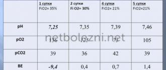
- arterial hypoxemia (usually PaO2 less than 50 mm Hg) and hypercapnia
- Arterial blood pH below 7.3
- normal pulmonary artery wedge pressure (<15 mm Hg) (this sign helps to distinguish the disease from cardiogenic pulmonary edema, in which pulmonary artery wedge pressure increases)
The examination program for respiratory distress syndrome in adults includes the following stages:
- General blood and urine tests
- Electrocardiography
- X-ray of the lungs
- Study of acid-base balance
- Study of blood gas composition: determination of PaO2, PaCO2
Treatment
The patient must be urgently hospitalized in the intensive care unit. Oxygen therapy is important for his survival to correct the decrease in O2 levels in the blood. If the oxygen supplied through the mask does not stabilize the patient’s condition, the person must be transferred to mechanical ventilation. A tube is inserted into the trachea through the mouth or nose and oxygen is pumped under pressure, which saturates the blood. The pressure must be adjusted so that the terminal sections of the bronchi (bronchioles) and alveoli remain open and the lungs do not receive excessive amounts of oxygen. This is important because too much O2 can have a negative impact on the lungs and lead to lung damage, which can lead to acute respiratory distress syndrome.
The patient is prescribed maintenance therapy, which means intravenous administration of fluids or nutrients, because with insufficient nutrition and lack of water in the body, the functions of many vital organs are impaired. Doctors call this condition multiple organ failure, as mentioned above.
The doctor, based on the cause, determines the types of treatment in each specific case. Sometimes it is necessary to prescribe antibiotics if the causative agent is an infectious agent. With a rapid effect of treatment, lung function is restored, long-term consequences are minimal or absent. With a long stay on a ventilator, there is a risk of developing fibrosis of the lung tissue. This means that normal lung tissue changes to coarse fibrous tissue. The degree of fibrosis may decrease several months after the patient begins to breathe on his own, without the help of a ventilator.