Cardiopulmonary failure is a decompensated stage of cor pulmonale, which occurs with acute or chronic right ventricular heart failure. The occurrence of pathology is associated with impaired blood circulation in the vessels of the lungs, which is accompanied by a decrease in blood oxygen saturation.
There are two main forms of cardiopulmonary failure - acute and chronic. The acute form in most cases occurs as a result of asthma, emphysema or massive thromboembolism. Clinical signs appear several hours after a sharp increase in arterial pressure. The chronic form is characterized by slow development with an increasing clinical picture (it can develop over several months or years).
There are several types of cardiac pulmonary failure, which differ in their characteristic symptoms. Among them: cerebral, anginal, respiratory, abdominal and collaptoid type of pathology. The cerebral type is characterized by signs of encephalopathy (for example, drowsiness and lethargy or, on the contrary, excitability and aggressiveness). Some patients complain of headaches and dizziness.
The anginal type is characterized by the appearance of severe pain in the area (with no irradiation) and a feeling of lack of oxygen. The progression of the respiratory type is accompanied by the appearance of shortness of breath, cough and cyanosis of the skin. The abdominal type is characterized by painful sensations and a feeling of nausea. Without further treatment, stomach ulcers may occur. The collaptoid type is accompanied by a persistent increase in blood pressure, tachycardia and general weakness. In this case, patients experience pallor of the skin.
Circulatory failure depends on two main factors: a decrease in the contractile force of the muscular membrane of peripheral vessels and a decrease in the contractility of the muscular organ. If the first factor significantly predominates, then we are talking about vascular circulatory failure, and if the second predominates, we are talking about chronic heart failure.
The left and right sections of the muscular organ are responsible for blood circulation. When one of these sections is damaged, isolated or predominant lesions appear, respectively, on the left or right half of the muscular organ. Based on this, left ventricular failure and right ventricular failure are distinguished.
From an anatomical and functional point of view, the lungs and heart have a close relationship. This explains the fact that when one organ is damaged, negative consequences subsequently affect the other. Depending on which of these organs is more affected, pulmonary-cardiac and cardiopulmonary failure are distinguished.
There are two main phases – compensation and decompensation. In the compensation stage, the muscular organ is able to cope with its load and work. But this does not always happen: over time, a period comes when its internal reserves are completely exhausted, resulting in a decompensation phase. In this phase, the muscular organ is no longer able to cope with the load.
First aid for pulmonary edema
Unlike the chronic form, the acute form of the pathology requires immediate assistance during an attack. A person is not able to help himself, so he needs help from the outside. First of all, you should call an emergency team. While waiting for her, the person should be seated on a chair and given a Nitroglycerin tablet. If the attack occurs indoors, be sure to open all windows to provide fresh air flow.
If a person gets worse, cardiopulmonary resuscitation methods are required. To do this, rhythmically press on the chest area or do 30 presses and mouth-to-mouth breathing (alternating these actions). The main goals of these measures are to normalize blood circulation and restore respiratory function. If you lack the skills, you can simply massage the sternum.
Diagnosis of heart failure
When diagnosing a disease, it is extremely important to identify the true cause of its development - only then can treatment be as successful as possible.
After collecting an anamnesis and physical examination, a cardiologist may prescribe:
- general clinical, biochemical blood and urine tests;
- electrocardiography (at rest);
- Ultrasound of the heart (Echocardiography);
- chest x-ray;
- coronary angiography;
- Holter ECG monitoring and other studies.
Causes of cardiopulmonary failure
Cardiopulmonary failure never occurs without a reason. As a rule, several reasons lead to its development. There are three main groups of causes that can provoke pathology:
1. Bronchopulmonary diseases. According to statistics, they act as a provoking factor in more than 70% of all cases. Among the most common diseases are bronchial asthma, pulmonary tuberculosis, chronic bronchitis and other severe disorders of the respiratory system (for example, cystic fibrosis).
2. Thoradiaphragmatic diseases. They are directly related to pronounced deformation of the chest and limited mobility of the diaphragm. The appearance is caused by severe curvatures of the spinal column (in particular, kyphoscoliosis), ankylosing spondylitis, and restrictions on the expansion of the lungs (pleuritis).
3. Vascular diseases. This group includes diseases that affect lesions of the pulmonary bed. These include sickle cell anemia, pulmonary vasculitis, compression of the pulmonary veins and arteries by neoplasms.
4. Vascular diseases. Often, the development of cardiopulmonary failure is provoked by aneurysms, atherosclerosis and blood clots in the arteries of the lungs.
What is heart failure?
Our heart is like a pump that pumps blood and supplies oxygen and nutrients to all parts of the body. Impaired ability of the heart to pump blood leads to stagnation and leads to the development of heart failure .
Poor blood supply puts other organs and tissues at risk. In terms of prevalence, heart failure rivals the most well-known infectious diseases.
According to statistics, around 28 million people suffer from heart failure throughout Europe. In our country, the number of patients with this disease exceeds 9 million (more than 25% of them are under 60 years of age)! At the same time, Russians die from it 10 times more often than from myocardial infarction.
1 Tests for heart failure
2 Tests for heart failure
3 Tests for heart failure
Diagnosis of pathology
The diagnosis of cardiopulmonary failure is established based on the results of a comprehensive examination. During the initial examination, the doctor collects anamnesis (listens about symptoms, chronic pathologies in the patient and immediate family), performs a visual examination (possible swelling of the feet and legs), conducts a physical examination and standard measures (measuring blood pressure, listening to breathing using a phonendoscope). Upon palpation, a cardiac impulse is detected, and upon percussion, an expansion of the boundaries of the region of the muscular organ, which is partially covered by the lungs, is detected.
Among the laboratory diagnostic methods for making a diagnosis, an analysis of the gas composition of arterial blood is required. If there are problems, patients experience a decrease in P02 and SAO2 P02, and an increase in PC02. Additionally, a general blood test is prescribed to detect an infectious process in the body (by increasing the level of leukocytes). The most informative ways to detect cardiopulmonary failure are instrumental diagnostic methods. Depending on the symptoms, the patient may be prescribed:
- study of respiratory function (external respiration function). With the help of this examination, it is possible to establish the nature and severity of pulmonary ventilation impairment;
- electrocardiography (ECG). This is an accessible method that does not require additional preparation from the patient. The essence of electrocardiography is to record the electrical potentials of a muscle organ, which makes it possible to detect changes in rhythm and electrolyte deficiency. If the disease occurs in an acute form, it is possible to detect signs of overload in the right parts of the muscular organ. If the disease is chronic, it is possible to detect markers (both direct and indirect) of right ventricular hypertrophy;
- echocardiography (EchoCG). It is often prescribed in addition to an ECG, since the information content of this examination regarding cardiopulmonary failure is very high. This is the main non-invasive method with which it is possible to assess intracardiac hemodynamics, determine the size of the cavities of the muscular organ and the degree of pulmonary hypertension. EchoCG allows you to detect a number of possible disturbances in the functioning of the heart, differentiate it from heart disease and other pathologies;
- chest x-ray. This is a simple technique with which it is possible to assess the condition of the respiratory system (detect damage to the internal organ and notice signs of pulmonary hypertension);
- transbronchial lung biopsy. These methods are rarely resorted to, in particularly advanced clinical cases. A puncture biopsy of the lung is performed under ultrasound or X-ray guidance using a local anesthetic.
Manifestations of acute form of insufficiency
Manifestations of the disease sometimes occur suddenly, develop quickly and give a clear clinical picture. This is an acute form of failure that requires emergency care and transportation to the intensive care unit. It occurs in the following cases:
- sharp spasm or thrombosis of the pulmonary artery trunk;
- inflammatory damage to a large volume of the lungs;
- status asthmaticus;
- pneumothorax, hydrothorax (accumulation of air or liquid in the pleural cavities);
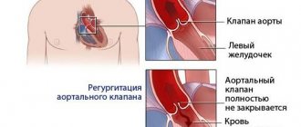
- severe degree of mitral valve incompetence;
- chest injuries;
- malfunction of the prosthetic valve.
As a result of the influence of unfavorable factors, a sharp disturbance of hemodynamics occurs in the form of insufficient blood circulation of the right ventricular type. The condition is characterized by the following symptoms:
- rapid shallow breathing;
- decreased blood pressure, in severe cases the development of collapse;
- shortness of breath with difficulty breathing;
- swelling of the veins in the neck;
- feeling of lack of air to the point of suffocation;
- cold extremities;
- blueness of the skin (cyanosis);
- sticky cold sweat on the skin;
- chest pain.
In the acute form of failure, pulsation can be detected in the epigastric region of the dilated right ventricle. The x-ray visualizes the expansion of the mediastinum upward and to the right, and the ECG shows the phenomenon of overload of the right atrium and ventricle. When listening (auscultation) of the heart, the gallop rhythm and muffled tones are clearly determined. With thromboembolism of large trunks of the pulmonary artery, pulmonary edema and painful shock develop quite quickly, which can lead to sudden death.
Disease prevention
In order to prevent pathology, all risk factors that can provoke it should be eliminated. If any negative symptoms appear, it is important to consult a doctor in order to promptly diagnose and treat lung or heart disease. Also, experts recommend leading a healthy lifestyle (not smoking or drinking alcohol), engaging in moderate physical activity and eating right. The diet should be balanced with a predominance of fresh vegetables and fruits.
In order to prevent the development of acute cor pulmonale, patients diagnosed with varicose veins and atrial fibrillation are prescribed indirect anticoagulants. If there are blood clots in the veins, a vena cava filter is often installed. It is a small device that is installed into the lumen of the inferior vena cava. With its help, detached blood clots do not enter parts of the heart muscle and the pulmonary artery system.
Symptoms
Acute pulmonary heart failure occurs suddenly. The patient's condition deteriorates sharply - within a few minutes or hours. The attack is accompanied by:
- pain in the heart area, rapid heartbeat;
- severe shortness of breath, suffocation;
- panic attack;
- blue nails, nasolabial triangle;
- a sharp drop in blood pressure.
Symptoms are especially severe in sitting and standing positions. Chronic pulmonary heart failure develops gradually, due to stagnation of blood in the venous system of the systemic circulation. This pathology provokes:
- constant shortness of breath;
- painful attacks in the heart and chest, for which nitroglycerin does not help;
- blue discoloration of the nasolabial triangle, tip of the nose, nails;
- fatigue, fatigue, drowsiness, fainting during physical activity;
- peripheral edema, edematous syndrome, ascites, exhaustion.
Establishment of clinical class and verification of diagnosis
Pulmonary function tests can identify obstructive or restrictive changes for the purpose of differential diagnosis of PH and clarify the severity of lung damage. Patients are characterized by a decrease in the diffusion capacity of the lungs for carbon monoxide (40-80% of normal), a slight or moderate decrease in lung volumes, a normal or slightly decreased PaO2, and usually a decreased PaCO2 due to alveolar hyperventilation.
Ventilation-perfusion lung scintigraphy is a screening method to exclude chronic thromboembolism as a cause of pulmonary hypertension. In patients after thromboembolism, perfusion defects are found in the lobar and segmental zones in the absence of ventilation disturbances. Perfusion scintigraphy has historically become one of the first methods for detecting perfusion defects in the pulmonary parenchyma in pulmonary embolism. The images obtained in acute PE and CTEPH differ significantly. Perfusion defects in acute PE are more clearly defined and contrast sharply with normally functioning tissue. In CTEPH, perfusion defects are not clearly defined and often do not correspond to the blood supply area of the large pulmonary artery.
Computed tomography and CT angiopulmonography
The CT picture of chronic thromboembolism can be represented by occlusions and stenoses of the pulmonary arteries, eccentric filling defects due to the presence of blood clots, including recanalized ones. CT angiopulmonography is performed on spiral computed tomographs during the phase of passage of the contrast agent through the pulmonary arterial bed. Among the methodological features, it should be noted that the study must be carried out using at least an 8-spiral tomograph, with a minimum step (no more than 3 mm) and a slice thickness (no more than 1 mm). A thorough scan should cover both lungs completely, from the apices to the phrenic sinuses. Contrast enhancement of the right chambers of the heart and pulmonary arteries should match or exceed the degree of contrast enhancement of the left chambers of the heart and aorta. The second, arterial phase of scanning is recommended for all patients over 40 years of age, especially if there is evidence of arterial thrombosis and coronary artery disease in the anamnesis. Modern software allows you to reconstruct images of the pulmonary arteries in any plane, construct maximum intensity projections and three-dimensional images. In most cases, to clarify the nature of the lesion, it is enough to analyze cross sections using an image viewer, which makes it possible to determine the presence of changes not only in the lobar and segmental branches, but also in a number of subsegmental arteries. Pathological changes, in addition to the presence of “old” thrombotic material, may include local thickening of the vessel wall, narrowing at the mouth of the vessels and along them, occlusions, intravascular structures in the form of membranes and bridges. If changes are detected in several branches of the pulmonary arteries, we can conclude that there is a high probability of thromboembolic nature of PH. It is important to note that the resolution of modern CT scanners is limited and does not allow the determination of very thin membranous and stringy structures in the lumen of the LA, especially if the size of the object does not exceed 2-3 mm. In some cases, calcification of the “old” thrombotic material develops, and CT can be invaluable in determining the location of the calcification. CT makes it possible to detect not only stenotic changes in the vessels of the lungs, but also disturbances in the perfusion of lung tissue by the nature of the contrast of the parenchyma. In some cases, parenchymal enhancement is so uneven that mosaic enhancement is detected on scans. A clearly defined mosaic pattern of segments usually indicates a good prognosis for surgical treatment. Contrasting exclusively in the root zones is not a true mosaic and is often observed in microvascular forms of PH. By providing detailed images of the lung parenchyma, CT allows the diagnosis of other lung diseases. In addition to the state of the arterial bed, CT can provide comprehensive information about all intrathoracic structures, which is important for confirming the diagnosis and developing a surgical treatment plan. Before performing the operation, the condition of the pulmonary parenchyma, bronchial tree, and pulmonary veins should be taken into account.
Angiographic diagnostics
The main objectives of angiographic diagnosis are to determine the severity of pulmonary hypertension, clarify the nature of the lesion in the pulmonary bed through angiopulmonography, and identify/exclude coronary disease. Carrying out catheterization in isolation without high-quality angiopulmonography in a patient with clear signs of CTEPH is inappropriate. This study should provide clear information to doctors to resolve the issue of the patient’s operability and the severity of his condition. Hemodynamic criteria for post-embolic pulmonary hypertension, identified during catheterization of the right heart, are: mean pulmonary artery pressure (PAP) above 25 mm Hg. Art., pulmonary artery wedge pressure (PAWP) ≤ 15 mm Hg. Art., PVR > 2 units. according to Wood (160 dyn. sec cm-5) in the presence of multiple stenotic and/or occlusive lesions of the branches of the pulmonary artery of various calibers.
Treatment and observation of a patient with SLN
Cardiopulmonary failure is a chronic disease that requires lifelong treatment. Under the influence of correctly prescribed medications, symptoms decrease, which significantly improves the quality of life. It is important to treat not only SLN, but also its cause. For most patients, treatment consists of dietary adjustments, medications, and surgical interventions.
The main medications used to treat SLN:
- ACE inhibitors - dilate blood vessels, lower blood pressure, improve blood flow and reduce stress on the heart
- Angiotensin receptor blockers - the principle of action is similar to previous drugs. Prescribed for intolerance to ACE inhibitors
- Beta blockers – slow down heart rate
- Thrombolytic drugs - for conservative treatment of pulmonary embolism
- Glucocorticoids and cytostatic drugs - for the treatment of connective tissue pathology
- Anti-tuberculosis drugs - for appropriate pathology
- Mucolytic drugs - optimize the viscosity of sputum and promote its discharge
- Diuretics – remove fluid that forms swelling, thereby lowering blood pressure and improving breathing
- Aldosterone antagonists – act like diuretics
- Digoxin – strengthens heart contractions, reducing their frequency
- Nitroglycerin – improves blood flow in the myocardium
- Statins – used to treat atherosclerosis
- Anticoagulants - used to reduce blood clotting properties
The drugs are selected individually and require several visits to the doctor to calibrate (titrate) the effective dosage. It is very important to use them regularly and not stop taking them on your own.
Surgery
Sometimes surgical treatment methods are necessary to treat the initial disease, such as:
- Thrombembolectomy for pulmonary embolism
- Sympathectomy
- Removal of a lung tissue tumor
- Heart and lung transplantation
- Balloon atrial septostomy
Palliative treatment
In severe and advanced cases, existing treatment methods are not effective, and cardiopulmonary failure progresses.
In this case, it is necessary to take active steps to maintain a decent quality of life for a person in a special clinic (hospice) or at home.
Recommendations for lifestyle changes:
- Stop smoking
- Weight control and maintaining a healthy diet
- Regularly checking your feet and ankles for swelling
- Limiting salt intake
- Vaccination against influenza and pneumonia
- Limiting alcohol intake
- Reducing stress levels
- Physical activity
- Monitoring the implementation of your prescriptions (creating a schedule, carrying the required amount of medications with you)
- Discuss taking additional medications, dietary supplements, vitamins with your doctor
- Keeping a diary to monitor weight, diuresis, blood pressure, fluids drunk and medications taken, and other important notes.
Mechanism of development of SLI
SLN often develops as a complication after diseases that lead to pulmonary hypertension and right-sided heart failure. The first version of the development of SLN begins with a pathology of the lungs, leading to an increase in the stiffness and density of the lung tissue, which is why the right ventricle requires more effort for systole. Subsequently, its wall hypertrophies, and the cavity expands, forming the so-called “pulmonary heart” (CP). Another variant of pathogenesis is associated with the progression of left ventricular heart failure to the last stage, as a result of which blood stagnates in the large and then small circle, increasing the pressure in the pulmonary vessels.
ACUTE HEART FAILURE Diagnosis and treatment at the prehospital stage
What are the main causes of AHF? What is the classification of OSN based on? What is the treatment algorithm at the prehospital stage?
Acute heart failure (AHF), which is a consequence of impaired myocardial contractility and a decrease in systolic and cardiac output, is manifested by extremely severe clinical syndromes: cardiogenic shock, pulmonary edema, acute cor pulmonale.
Main causes and pathogenesis
A decrease in myocardial contractility occurs either as a result of its overload, or due to a decrease in the functioning mass of the myocardium, a decrease in the contractile ability of myocytes, or a decrease in the compliance of chamber walls. These conditions develop in the following cases:
- in case of disturbance of diastolic and/or systolic function of the myocardium during infarction (the most common cause), inflammatory or dystrophic diseases of the myocardium, as well as tachy- and bradyarrhythmias;
- with the sudden occurrence of myocardial overload due to a rapid significant increase in resistance in the outflow tract (in the aorta - hypertensive crisis in patients with compromised myocardium; in the pulmonary artery - thromboembolism of the branches of the pulmonary artery, a prolonged attack of bronchial asthma with the development of acute emphysema, etc.) or due to stress volume (an increase in the mass of circulating blood, for example, with massive fluid infusions - a variant of the hyperkinetic type of hemodynamics);
- in case of acute disturbances of intracardiac hemodynamics due to rupture of the interventricular septum or the development of aortic, mitral or tricuspid insufficiency (septal infarction, infarction or avulsion of the papillary muscle, perforation of the valve leaflets in bacterial endocarditis, rupture of the chordae, trauma);
- with increasing load (physical or psycho-emotional stress, increased inflow in a horizontal position, etc.) on the decompensated myocardium in patients with chronic congestive heart failure.
Classification
Depending on the type of hemodynamics, which ventricle of the heart is affected, as well as on some features of the pathogenesis, the following clinical variants of AHF are distinguished.
- With a congestive type of hemodynamics: • right ventricular (venous congestion in the systemic circulation); • left ventricular (cardiac asthma, pulmonary edema).
- With hypokinetic1 type of hemodynamics (small output syndrome - cardiogenic shock): • arrhythmic shock; • reflex shock; • true shock.
Since myocardial infarction is one of the most common causes of AHF, the table provides a classification of acute heart failure in this disease.
Possible complications
Any of the variants of AHF is a life-threatening condition. Acute congestive right ventricular failure, not accompanied by small output syndrome, in itself is not as dangerous as diseases leading to right ventricular failure.
Clinical picture
- Acute congestive right ventricular failure is manifested by venous congestion in the systemic circulation with increased systemic venous pressure, swelling of the veins (most noticeable in the neck), enlarged liver, and tachycardia. Edema may appear in the lower parts of the body (with prolonged horizontal position - on the back or side). Clinically, it differs from chronic right ventricular failure by intense pain in the liver area, aggravated by palpation. Signs of dilatation and overload of the right heart are determined (expansion of the borders of the heart to the right, systolic murmur over the xiphoid process and protodiastolic gallop rhythm, emphasis of the second tone on the pulmonary artery and corresponding ECG changes). A decrease in left ventricular filling pressure due to right ventricular failure can lead to a drop in left ventricular minute volume and the development of arterial hypotension, up to a picture of cardiogenic shock.
With pericardial tamponade and constrictive pericarditis, the pattern of large circle congestion is not associated with insufficiency of myocardial contractile function, and treatment is aimed at restoring diastolic filling of the heart.
Biventricular failure, a variant where congestive right ventricular failure is combined with left ventricular failure, is not discussed in this section, since the treatment of this condition is not much different from the treatment of severe acute left ventricular failure.
- Acute congestive left ventricular failure clinically manifests itself as paroxysmal shortness of breath, painful suffocation and orthopnea, occurring more often at night; sometimes - Cheyne-Stokes breathing, cough (initially dry, and then with sputum, which does not bring relief), later - foamy sputum, often pink in color, pallor, acrocyanosis, hyperhidrosis and is accompanied by excitement, fear of death. In case of acute congestion, moist rales may not be heard at first, or a meager amount of fine bubbling rales is detected over the lower parts of the lungs; swelling of the mucous membrane of small bronchi can manifest itself as a moderate picture of bronchial obstruction with prolongation of exhalation, dry wheezing and signs of pulmonary emphysema. A differential diagnostic sign that allows one to differentiate this condition from bronchial asthma can be the dissociation between the severity of the patient’s condition and (in the absence of pronounced expiratory dyspnea, as well as “silent zones”) the paucity of the auscultatory picture. Loud, varied moist rales over all the lungs, which can be heard at a distance (bubbling breathing), are characteristic of a detailed picture of alveolar edema. Possible acute expansion of the heart to the left, the appearance of a systolic murmur at the apex of the heart, a proto-diastolic gallop rhythm, as well as an emphasis on the second tone on the pulmonary artery and other signs of load on the right heart, up to the picture of right ventricular failure. Blood pressure can be normal, high or low, tachycardia is typical.
The picture of acute congestion in the pulmonary circulation, which develops with stenosis of the left atrioventricular orifice, essentially represents left atrial failure, but is traditionally considered together with left ventricular failure.
- Cardiogenic shock is a clinical syndrome characterized by arterial hypotension and signs of a sharp deterioration in microcirculation and tissue perfusion, including blood supply to the brain and kidneys (lethargy or agitation, drop in urine output, cold skin covered with sticky sweat, pallor, marbled skin pattern); sinus tachycardia is compensatory in nature.
A decrease in cardiac output with a clinical picture of cardiogenic shock can be observed in a number of pathological conditions not associated with insufficiency of myocardial contractile function - with acute obstruction of the atrioventricular orifice by an atrial myxoma or a spherical thrombus/ball prosthesis thrombus, with pericardial tamponade, with massive pulmonary embolism. These conditions are often combined with the clinical picture of acute right ventricular failure. Pericardial tamponade and atrioventricular orifice obstruction require immediate surgical intervention; drug therapy in these cases can only worsen the situation. In addition, the picture of shock during myocardial infarction is sometimes imitated by dissecting aortic aneurysm; in this case, differential diagnosis is necessary, since this condition requires a fundamentally different therapeutic approach.
There are three main clinical variants of cardiogenic shock:
- arrhythmic shock develops as a result of a drop in cardiac output due to tachycardia/tachyarrhythmia or bradycardia/bradyarrhythmia; after stopping the rhythm disturbance, adequate hemodynamics are quickly restored;
- reflex shock (pain collapse) develops as a reaction to pain and/or sinus bradycardia resulting from a reflex increase in vagal tone and is characterized by a rapid response to therapy, primarily painkillers; observed with relatively small infarct sizes (often in the posterior wall), while there are no signs of congestive heart failure and deterioration of tissue perfusion; pulse pressure usually exceeds a critical level;
- true cardiogenic shock develops when the lesion volume exceeds 40-50% of the myocardial mass (more often with anterolateral and repeated infarctions, in people over 60 years of age, against the background of arterial hypertension and diabetes mellitus), is characterized by a detailed picture of shock, resistant to therapy, often combined with congestive left ventricular failure; depending on the selected diagnostic criteria, the mortality rate ranges from 80-100%.
In some cases, especially when it comes to myocardial infarction in patients receiving diuretics, the developing shock is hypovolemic in nature, and adequate hemodynamics are relatively easily restored due to replenishment of the circulating volume.
Diagnostic criteria
One of the most consistent signs of acute heart failure is sinus tachycardia (in the absence of sinus node weakness, complete AV block, or reflex sinus bradycardia); characterized by expansion of the borders of the heart to the left or right and the appearance of a third sound at the apex or above the xiphoid process.
- In acute congestive right ventricular failure, the following are of diagnostic value: swelling of the neck veins and liver;
- Kussmaul's sign (swelling of the jugular veins on inspiration);
- intense pain in the right hypochondrium;
- ECG signs of acute overload of the right ventricle (type SI-QIII, increasing R wave in leads V1,2 and formation of a deep S wave in leads V4-6, depression of STI, II, a VL and elevation of STIII, a VF, as well as in leads V1, 2; possible formation of right bundle branch block, negative T waves in leads III, aVF, V1-4) and signs of right atrium overload (high pointed waves PII, III).
- shortness of breath of varying severity, up to suffocation;
- a drop in systolic blood pressure of less than 90-80 mm Hg. Art. (or 30 mmHg below the “working” level in people with arterial hypertension);
| Figure 1. Algorithm for relieving pulmonary edema at the prehospital stage |
| Figure 2. Algorithm for relieving cardiogenic shock at the prehospital stage |
Treatment of acute heart failure
In any variant of AHF, in the presence of arrhythmias, it is necessary to restore an adequate heart rhythm.
If the cause of AHF is myocardial infarction, then one of the most effective methods of combating decompensation will be the rapid restoration of coronary blood flow through the affected artery, which can be achieved at the prehospital stage using systemic thrombolysis.
Inhalation of humidified oxygen through a nasal catheter at a rate of 6-8 l/min is indicated.
- Treatment of acute congestive right ventricular failure consists of correcting the conditions that caused it - pulmonary embolism, status asthmaticus, etc. This condition does not require independent therapy.
The combination of acute congestive right ventricular and congestive left ventricular failure serves as an indication for therapy in accordance with the principles of treatment of the latter.
When acute congestive right ventricular failure and small output syndrome (cardiogenic shock) are combined, the basis of therapy is inotropic agents from the group of pressor amines.
- Treatment of acute congestive left ventricular failure.
- Treatment of acute congestive heart failure begins with the administration of sublingual nitroglycerin in a dose of 0.5-1 mg (1-2 tablets) and giving the patient an elevated position (in case of an unexpressed picture of congestion - an elevated head end, in case of advanced pulmonary edema - a sitting position with legs down) ; these measures are not performed in cases of severe arterial hypotension.
- A universal pharmacological agent for acute congestive heart failure is furosemide, due to venous vasodilation already 5-15 minutes after administration, causing hemodynamic unloading of the myocardium, which increases over time due to the diuretic effect that develops later. Furosemide is administered intravenously as a bolus and is not diluted; the dose of the drug ranges from 20 mg for minimal signs of congestion to 200 mg for extremely severe pulmonary edema.
- The more pronounced the tachypnea and psychomotor agitation, the more indicated is the addition of a narcotic analgesic to therapy (morphine, which, in addition to venous vasodilation and reducing preload on the myocardium, already 5-10 minutes after administration reduces the work of the respiratory muscles, suppressing the respiratory center, which provides an additional reduction load on the heart. A certain role is also played by its ability to reduce psychomotor agitation and sympathoadrenal activity; the drug is administered intravenously in fractional doses of 2-5 mg (for which 1 ml of a 1% solution is taken, diluted with isotonic sodium chloride solution, bringing the dose to 20 ml and administered 4-10 ml) with repeated administration if necessary after 10-15 minutes Contraindications are respiratory rhythm disturbances (Cheyne-Stokes breathing), depression of the respiratory center, acute obstruction of the respiratory tract, chronic pulmonary heart disease, cerebral edema, poisoning with depressant substances breath.
- Severe congestion in the pulmonary circulation in the absence of arterial hypotension or any degree of acute congestive left ventricular failure during myocardial infarction, as well as pulmonary edema against the background of a hypertensive crisis without cerebral symptoms, are an indication for intravenous drip administration of nitroglycerin or isosorbide dinitrate. The use of nitrate drugs requires careful monitoring of blood pressure and heart rate. Nitroglycerin or isosorbide dinitrate is prescribed at an initial dose of 25 mcg/min, followed by an increase every 3-5 minutes by 10 mcg/min until the desired effect is achieved or side effects appear, in particular a decrease in blood pressure to 90 mm Hg. Art. For intravenous infusion, every 10 mg of the drug is dissolved in 100 ml of 0.9% sodium chloride solution, so one drop of the resulting solution contains 5 mcg of the drug. Contraindications to the use of nitrates are arterial hypotension and hypovolemia, pericardial constriction and cardiac tamponade, pulmonary artery obstruction, and inadequate cerebral perfusion.
- Modern methods of drug treatment have minimized the importance of bloodletting and the application of venous tourniquets to the extremities, however, if adequate drug therapy is impossible, these methods of hemodynamic unloading not only can, but should be used, especially with rapidly progressing pulmonary edema (bloodletting in a volume of 300- 500 ml).
- In case of acute congestive left ventricular failure, combined with cardiogenic shock, or with a decrease in blood pressure during therapy that has not given a positive effect, non-glycoside inotropic agents are additionally prescribed - intravenous drip administration of dobutamine (5-15 mcg/kg/min), dopamine (5- 25 mcg/kg/min), norepinephrine (0.5-16 mcg/min) or a combination thereof.
- A means of combating foaming during pulmonary edema are “defoamers” - substances that ensure the destruction of foam by reducing surface tension. The simplest of these means is alcohol vapor, which is poured into a humidifier, passing oxygen through it, supplied to the patient through a nasal catheter or breathing mask at an initial rate of 2-3 l/min, and after a few minutes - at a rate of 6-8 l/min.
- Persistent signs of pulmonary edema with stabilization of hemodynamics may indicate an increase in membrane permeability, which requires the administration of glucocorticoids to reduce permeability (4-12 mg of dexamethasone).
- In the absence of contraindications, in order to correct microcirculatory disorders, especially with long-term intractable pulmonary edema, the administration of sodium heparin is indicated - 5 thousand IU intravenously as a bolus, then drip at a rate of 800 - 1000 IU/hour.
- Treatment of cardiogenic shock involves increasing cardiac output, which is achieved in a variety of ways, the significance of which varies depending on the clinical type of shock.
- In the absence of signs of congestive heart failure (shortness of breath, moist rales in the posterior lower parts of the lungs), the patient must be placed in a horizontal position.
- Regardless of the clinical picture, it is necessary to provide complete analgesia.
- Stopping rhythm disturbances is the most important measure to normalize cardiac output, even if adequate hemodynamics are not observed after restoration of normosystole. Bradycardia, which may indicate increased vagal tone, requires immediate intravenous administration of 0.3-1 ml of 0.1% atropine solution.
- If the clinical picture of shock is extensive and there are no signs of congestive heart failure, therapy should begin with the administration of plasma expanders in a total dose of up to 400 ml under the control of blood pressure, heart rate, respiratory rate and auscultation of the lungs. If there is an indication that immediately before the onset of acute cardiac damage with the development of shock, there were large losses of fluid and electrolytes (long-term use of large doses of diuretics, uncontrollable vomiting, profuse diarrhea, etc.), then an isotonic solution is used to combat hypovolemia sodium chloride; the drug is administered in an amount of up to 200 ml over 10 minutes, repeated administration is also indicated.
- The combination of cardiogenic shock with congestive heart failure or the lack of effect from the entire complex of therapeutic measures is an indication for the use of inotropic agents from the group of pressor amines, which, in order to avoid local circulatory disorders accompanied by the development of tissue necrosis, should be injected into the central vein: dopamine in a dose of up to 2, 5 mg affects only dopamine receptors of the renal arteries, at a dose of 2.5-5 mcg/kg/min the drug has a vasodilating effect, at a dose of 5-15 mcg/kg/min it has vasodilating and positive inotropic (and chronotropic) effects, and in dose 15-25 mcg/kg/min - positive inotropic (and chronotropic), as well as peripheral vasoconstrictive effects; 400 mg of the drug is dissolved in 400 ml of a 5% glucose solution, while 1 ml of the resulting mixture contains 0.5 mg, and 1 drop - 25 mcg of dopamine. The initial dose is 3-5 mcg/kg/min with a gradual increase in the rate of administration until the effect is achieved, the maximum dose (25 mcg/kg/min, although the literature describes cases where the dose was up to 50 mcg/kg/min) or the development of complications (most often sinus tachycardia exceeding 140 beats per minute, or ventricular arrhythmias). Contraindications to its use are thyrotoxicosis, pheochromocytoma, cardiac arrhythmias, hypersensitivity to disulfide, previous use of MAO inhibitors; if the patient was taking tricyclic antidepressants before prescribing the drug, the dose should be reduced;
- the lack of effect from dopamine or the inability to use it due to tachycardia, arrhythmia or hypersensitivity is an indication for the addition or monotherapy with dobutamine, which, unlike dopamine, has a more pronounced vasodilatory effect and a less pronounced ability to cause an increase in heart rate and arrhythmia. 250 mg of the drug is diluted in 500 ml of a 5% glucose solution (1 ml of the mixture contains 0.5 mg, and 1 drop - 25 mcg of dobutamine); in monotherapy, it is prescribed at a dose of 2.5 mcg/kg/min, increasing every 15-30 minutes by 2.5 mcg/kg/min until an effect, side effect is obtained, or a dose of 15 mcg/kg/min is achieved, and with a combination of dobutamine with dopamine - in maximum tolerated doses; Contraindications to its use are idiopathic hypertrophic subaortic stenosis and stenosis of the aortic mouth. Dobutamine is not prescribed for systolic blood pressure <70 mmHg. Art.
- in the absence of effect from the administration of dopamine and/or a decrease in systolic blood pressure to 60 mm Hg. Art. norepinephrine can be used with a gradual increase in dosage (maximum dose - 16 mcg/min). Contraindications to its use are thyrotoxicosis, pheochromocytoma, previous use of MAO inhibitors; If you have previously taken tricyclic antidepressants, the dose should be reduced.
Indications for hospitalization
After relief of hemodynamic disturbances, all patients with acute heart failure are subject to hospitalization in cardiac intensive care units. In the torpid course of AHF, hospitalization is carried out by specialized cardiology or resuscitation teams. Patients with cardiogenic shock should, whenever possible, be hospitalized in hospitals where there is a cardiac surgery department.
A. L. Vertkin, Doctor of Medical Sciences, Professor V. V. Gorodetsky, Candidate of Medical Sciences O. B. Talibov, Candidate of Medical Sciences
1 The clinical picture of cardiogenic shock can develop with a hypovolemic type of hemodynamics: against the background of active diuretic therapy preceding a heart attack, profuse diarrhea, etc.
Clinical course of the disease
HF can have an acute onset and a chronic course. The signs are:
- Dyspnea (shortness of breath) during exercise or at rest, suffocation
- Weakness, lethargy
- Swelling of the feet, ankles and legs
- Swelling of the neck veins
- Pain in the right hypochondrium
- Cardiopalmus
- Inability to perform usual physical activities
- Persistent cough with light-colored or pinkish sputum
- Need to urinate at night
- Ascites
- Loss of appetite or nausea
- Inability to concentrate, absent-mindedness
- Chest pain relieved by taking aminophylline
- Sudden attacks of suffocation with coughing and discharge of foamy pink sputum
Emergency medical attention is necessary if the patient experiences:
- Chest pain
- Loss of consciousness or severe weakness
- Fast or irregular heartbeat associated with shortness of breath, dizziness, or chest pain
- Sudden cough with pink sputum or coughing up blood
- Bluishness around the mouth, tip of the nose and fingers
If you have already been diagnosed with heart failure or another cardiac disease and experience the symptoms described above, seek medical help.
Qualified care for heart failure at the CBCP clinic
At the slightest suspicion of this disease, undergo diagnostics of the functioning of the heart and blood vessels. Modern diagnostic methods make it possible to determine the true cause of the disease in order to promptly block its further development.
The CBCP clinic has the latest expert-class equipment for diagnosing all types of this disease. Experienced, qualified cardiologists will advise you and give recommendations on how to treat heart failure.
Make an appointment right now on the website or during business hours by phone.
Nutrition and daily routine
Meals should be fractional: 5-6 times a day in small portions. Limit the consumption of meat, salt, exclude smoked foods, chocolate, and alcohol. To replenish their strength, patients should eat foods high in potassium: buckwheat and oatmeal, bananas, dried apricots, Brussels sprouts, etc. A protein and vitamin diet is prescribed.
The daily routine depends on the form of the disease. In the acute form, only rest is necessary. In chronic cases, on the contrary, rest is contraindicated. The patient is advised to exercise in moderation, and a special system of exercises is developed to prevent the disease.