In this article
- Causes of exophthalmos
- Symptoms of exophthalmos
- Types of exophthalmos
- Stages of development of exophthalmos
- Diagnosis of exophthalmos
- Treatment of exophthalmos
- Orbital decompression for exophthalmos
- Prognosis and possible consequences of exophthalmos
Exophthalmos is a forward displacement of one or both eyes. Various factors can provoke it. The form of the course and the method of treatment depend on them. Often you need the help of several doctors: an ophthalmologist, an endocrinologist, a therapist. Exophthalmos can cause visual impairment. Let's take a closer look at this disease.
Exophthalmos is not always a pathology. It can be a symptom of a disease or a natural condition. But in all cases we are talking about the protrusion of one or both eyes. This syndrome is also called bulging eyes, protrusion and proptosis. It was first described in the 18th century by the Irish physician R.J. Graves, who considered exophthalmos as one of the signs of endocrine ophthalmopathy.
Causes of exophthalmos
Proptosis is caused by various factors. In women it usually occurs against the background of endocrine system disorders, in men - after head injuries. The causes can be both ophthalmological and systemic pathologies. Among the first:
- orbital inflammation;
- traumatic injury to the orbit;
- eye socket diseases;
- dysfunction of the ciliary muscle;
- tumor processes;
- intraocular hemorrhages;
- congenital glaucoma;
- high degree of myopia;
- paralysis of the eye muscles.
Inflammation of the orbit can occur due to panophthalmitis, infectious lesions of the lacrimal sac, purulent processes on the eyelids and nose, dental diseases, ear infections, etc. Complicated forms of these diseases can lead to exophthalmos.
If the primary disease develops on one eyeball, proptosis of one eye occurs. Bilateral pathological processes cause binocular proptosis. Mechanical injuries, tumors and damage to veins, as a rule, become the causes of unilateral exophthalmos. Congenital glaucoma most often affects both eyeballs, which is why bulging eyes occur bilaterally.
Proptosis can be a consequence of diseases and pathological conditions that are not initially associated with the organs of vision:
- thyroid dysfunction;
- circulatory disorders;
- blood diseases;
- incorrect structure of the skull;
- hydrocephalus, etc.
These causes cause bilateral exophthalmos, when both eyes protrude. Proptosis does not always develop evenly. At first, symptoms may appear in only one eyeball. This is even more noticeable than the binocular form of the syndrome.
The factors for the development of exophthalmos in children and adults may differ. In children, thyroid problems occur less frequently. Their proptosis may be a consequence of congenital glaucoma, abnormal skull structure, progressive myopia, or trauma.
Diagnosis and treatment of bulging eyes
In the initial stages, you can independently determine exophthalmos, for example:
- A white, often with blood vessels, gap between the upper eyelid and the iris is clearly visible
- If you look down, the tunica albuginea stands out strongly and the eyes protrude
- The skin on the eyelid becomes much darker than usual
Because bulging eyes are most often associated with other diseases, so a full diagnostic examination is prescribed to make a correct diagnosis. In this case, specialists of other profiles, for example, an endocrinologist, may be involved. Most often, when the general condition of the body improves, the bulging eyes disappear.
Therefore, you should not self-medicate, but rather consult a specialist. At the Eye Clinic of Dr. Belikova, the cause of the disease is diagnosed and treatment is prescribed in a timely manner. In our clinic, consultations are conducted by doctors and candidates of medical sciences, doctors of the highest and first categories.
Symptoms of exophthalmos
Clinical symptoms of bulging eyes depend on its etiology. One or another of its varieties is characterized by corresponding characteristics. A common symptom is bulging eyes. As a rule, this is noticeable from the outside. The eyeball can move not only forward, but also slightly to the side, as with strabismus. Some people complain of pain when moving their eyes.
Other ophthalmological signs are also observed:
- swelling of the mucous membrane;
- dry cornea;
- tearfulness;
- diplopia;
- photosensitivity;
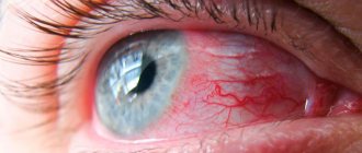
- redness of the sclera;
- failure to close eyelids;
- decreased visual acuity.
Any of these symptoms should be a reason to visit an ophthalmologist. A signal can also be systemic signs of a primary disease, against which exophthalmos develops: weakness, fatigue, insomnia, frequent dizziness, rapid weight loss, increased sweating, tachycardia, nervousness, anxiety, etc.
Types of exophthalmos
Proptosis can be true or false. The first is characterized by acute or chronic damage to the eye tissues. In this case, inflammation is not always observed. Mandatory treatment is required. Imaginary bulging eyes are a consequence of asymmetry of the eye sockets, congenital anomalies in the structure of the skull, or severe myopia. False exophthalmos does not cause an inflammatory process. The protrusion of the eyes is within normal limits. There is no need to treat such proptosis. But the patient is at risk, and therefore he needs to visit the ophthalmologist more often.
Based on the characteristics of the clinical picture of eye protrusion, the following types of exophthalmos are distinguished:
- Constant. Occurs with endocrine disorders and after removal of the thyroid gland. Characterized by rapid progression.
- Intermittent. Develops against the background of damage to the ocular vessels. Exophthalmos moves from one eye to the other when the body or head is tilted.
- Throbbing. It is a consequence of eye injury, aneurysm or thrombosis. The protrusion occurs in time with the pulse. Often accompanied by migraine.
Based on the causes of eye protrusion, there are:
- Edema exophthalmos. It occurs due to endocrine and autoimmune pathologies or due to increased production of thyroid-stimulating hormone in the body. Progresses rapidly. The patient's intraocular pressure rises, severe pain is felt in the orbits, and visual acuity is greatly reduced. There is a risk of ulcers forming on the cornea.
- Thyrotoxic exophthalmos. It develops due to excessive production of thyroid hormones, with hyperthyroidism and against the background of hormonal imbalances. Often, such bulging eyes are temporary, and they are detected in women. Exophthalmos in thyrotoxicosis is accompanied by tachycardia and tremor. Often the syndrome goes away on its own, once the hormonal levels are restored.
- Hypothalamic-pituitary exophthalmos. It occurs when the hypothalamic centers are irritated, which happens with autonomic, metabolic and endocrine disorders. The syndrome appears suddenly and progresses very quickly. It is characterized by symptoms such as conjunctival edema, optic nerve palsy and increased ophthalmotonus. There are disorders of the psyche, nervous and reproductive systems.
- Intermittent exophthalmos. Its main features are similar to pulsating. But it occurs when overexertion occurs and when the head is tilted forward and not to the side. The eyes bulge when the jugular vein in the neck is pinched. A pulsation is felt in the eyeball.
- Exophthalmos with diffuse toxic goiter (Graves' disease or Graves' disease). The bulge is moderate, develops slowly without any consequences. The patient retains eye mobility, there is no pain, diplopia or severe discomfort. But there are other signs: strabismus, lag of the upper eyelid when looking down, a decrease in the frequency of natural blinks.
Binocular proptosis often develops. Sometimes unilateral exophthalmos occurs, which subsequently becomes bilateral.
Stages of development of exophthalmos
The clinical picture is determined by the stage of development of the disease and depends on the degree of displacement of the eyeball in the orbit. There are three in total:
- First: eye diameter - 21-23 mm. There may be no significant symptoms. Upon external examination, the protrusion is not noticeable. It can only be detected using equipment.
- Second: eye diameter - 24-26 mm. The first signs of bulging eyes appear. A person is bothered by diplopia, strabismus develops, and the mobility of the eyeballs is limited.
- Third: eye diameter is more than 27 mm. The eyelids do not close completely. Part of the sclera is always open and is not wetted by tear fluid. Dryness, burning, irritation, and redness of the sclera appear. Compression of the optic nerve disc occurs. Visual acuity decreases.
Exophthalmos is congenital, in which the convexity of the eyeballs corresponds to the norm, that is, no more than 2 mm. The pathological process becomes noticeable already at the second stage, and sometimes at the first.
Diagnosis of exophthalmos
Protrusion of the eyes and the degree of displacement are detected using exophthalmometry, during which the distance between two parallel planes is measured: one passes through the apex of the cornea, the other through the lateral wall of the orbit. An exophthalmometer resembles a regular ruler, but has two frames. Two mirrors are attached to it at an angle of 45°. The ophthalmologist attaches the device to the outer sides of the eye sockets - closer to the temples, moves the frames and takes measurements.
The degree of exophthalmos is not the only indicator that needs to be identified. It is important to determine which parts of the eye are affected by the pathology. Typically, the following instrumental methods are prescribed for protrusion:
- Ophthalmoscopy. With the 3rd degree of bulging eyes, pathological changes in the fundus may occur. There is a risk of compression of the optic nerve head. He looks pale and puffy. Local hemorrhages are observed.
- Biomicroscopy. It can be used to detect ulcers and other damage to the cornea.
- Tonometry. Intraocular pressure does not increase in all types of exophthalmos. But this method is one of the mandatory ones in ophthalmology and is used during any examination. In addition, secondary complications of bulging eyes can be determined by ophthalmotonus.
- OCT. Allows you to visualize periorbital tissue, exclude or confirm swelling, neoplasms in the orbit and hemorrhages.
- Ultrasound of the eyeball in B-mode. Using this method, you can determine the degree of exophthalmos, evaluate the orbital tissue, and find out whether the disease is progressing or regressing.
The most important thing with exophthalmos is to find its cause, that is, the primary pathology. You need to undergo a comprehensive examination. The patient donates blood and urine for analysis, his hormone levels are determined, an ultrasound of the thyroid gland, MRI and other methods are prescribed. After the diagnosis is made and the type of exophthalmos is determined, treatment begins. As a rule, it is carried out not only by an ophthalmologist. You may need the help of an endocrinologist, otolaryngologist, neurologist and even a neurosurgeon.
Diagnosis of exophthalmos in thyrotoxicosis and other diseases
It is important not only to record the fact of bulging eyes, but also to determine its degree. To do this, exophthalmometry is performed - using special ophthalmic mirrors, the location of the eyeballs and the degree of their displacement are assessed. Diagnosis of exophthalmos in thyrotoxicosis or other diseases does not differ significantly.
In case of severe exophthalmos, the cornea is examined and assessed:
- surface (flat or not);
- presence of visible damage;
- degree of moisture.
In order to clarify changes in the orbit that could lead to exophthalmos, it is examined using methods such as:
- CT scan;
- Magnetic resonance imaging.
The most common cause of bulging eyes is endocrine disorders, in particular those of the thyroid gland. Therefore, in case of exophthalmos, laboratory determination of the amount of thyroid hormones is mandatory.
Treatment of exophthalmos
The treatment method is determined by the causes of the pathology. So, with thyrotoxicosis, exophthalmos is treated with steroid drugs. They relieve inflammation and stabilize hormone levels. If bulging eyes develop due to a diseased thyroid gland, for example, with hyperthyroidism, radioactive iodine is prescribed. Sometimes pulse therapy with Prednisolone is used for exophthalmos. Edema is eliminated with medications and radiotherapy.
Exophthalmos may result from inflammation in the eye or orbit. In such cases, antibacterial and anti-inflammatory drops are prescribed, which help reduce inflammation and its toxicity. In some cases, antibiotics are used, intravenously. Vitamins are used to maintain general immunity.
Edema exophthalmos, which often occurs due to hormonal imbalances, hypothyroidism and hyperthyroidism of the thyroid gland, is treated by a neurologist, endocrinologist and therapist. First you need to restore the functioning of the thyroid gland. This is done with the help of drug therapy. The choice of a specific drug is determined by the primary pathology.
Pulsatile exophthalmos is treated with orbital radiotherapy. In addition, a pressure bandage is placed on the eye to cause thrombosis of the ophthalmic vein. This helps stop the development of bulging eyes. In severe cases, the carotid artery is ligated. These procedures can only be performed by a doctor.
Intermittent exophthalmos requires surgical treatment. During the operation, the surgeon ligates the internal or external carotid artery or places a clip on it inside the skull. Oncological diseases in which the patient has to undergo a course of chemotherapy are also treated surgically. Surgery is necessary if the optic nerve is severely damaged. Excess fatty tissue is removed from the eye socket to reduce pressure on the fundus. Severe damage to the cornea may require stitching of the eyelids. Special ointments are prescribed that help restore tissue of the cornea.
Treatment
Therapy for exophthalmos of the eyes is selected taking into account the severity of the disease, the main reasons that provoked its development and the specifics of the clinical picture. In the most severe cases, surgery is necessary. Medicines used for treatment may include the following:
- Anti-inflammatory drugs for the eyes. The use of drugs with anti-inflammatory, immunomodulatory and antibacterial properties is mandatory in the treatment of exophthalmos if somatic inflammatory processes, as well as infectious eye lesions, contributed to its development.
- Glucocorticosteroids. The use of drugs in this group is necessary if the development of the disease is caused by damage to the thyroid gland. If there is no positive dynamics in the treatment of exophthalmos, the patient may be advised to remove the thyroid gland. In this situation, to restore the body, hormonal levels and prevent complications of true exophthalmos, it is necessary to take glucocorticosteroid drugs.
- Hormonal drugs. The use of medications in this group is necessary against the background of endocrine diseases, which include thyrotoxicosis.
- Surgical intervention. Severe exophthalmos, the development of which is caused by injuries, eye cancer, and damage to the nervous system, cannot be treated with conservative therapy; in this case, surgical intervention is necessary. The selection of the type of invasive manipulation depends on the characteristics of the type of disease. Congenital exophthalmos, which has a pronounced form, is treated through plastic surgery aimed at eliminating the defect. For mechanical damage associated with damage to the cornea of the eye, the method of stitching the eyelids is used. If the protrusion develops as a result of dysfunction of the endocrine system, surgical intervention is aimed at removing the thyroid gland.
Against the background of mild forms of imaginary or false protrusion, treatment is not mandatory, however, to prevent the progression of the pathology, it may be necessary to take drugs and medications that have an immunomodulatory effect.
Orbital decompression for exophthalmos
With exophthalmos, orbital decompression may be required, which helps reduce pressure on its walls. After the operation, the volume of space in the orbit increases, thereby reducing the degree of bulging eyes. Parts of one, two or three orbital walls are removed.
Orbital decompression is performed for compressive optic neuropathy, keratopathy, spontaneous eye prolapse, severe pain and to improve aesthetic appearance.
To relieve the symptoms of bulging eyes, it is recommended to adhere to the following rules:
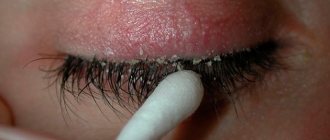
- use moisturizing drops, ointments and gels;
- when going outside, take sunglasses with you;
- eliminate salt, select food products more carefully;
- sleep with your head on a raised surface (no more than 15 cm).
Use anti-inflammatory eye drops only for the first three days. They help eliminate inflammation and redness of the conjunctiva. But subsequently the opposite effect may occur due to vasodilation. Buy all medications only with a prescription. Treatment of exophthalmos with so-called folk remedies is unacceptable. At best, they will not affect the condition of the eyes, at worst, they will cause complications.
Treatment of exophthalmos can take several years. Under no circumstances should you abandon therapy at the first signs of recovery. The risk of relapse is quite high.
Prognosis and possible consequences of exophthalmos
The prognosis depends on the severity of the disease, the timeliness of contacting a doctor, the correctness of the prescribed method of treatment and the individual characteristics of the patient. As a rule, it is possible to eliminate both the primary pathology and bulging eyes. Difficulties may arise with the malignant nature of exophthalmos, hydrocephalus and other serious illnesses.
As for complications of an ophthalmological nature, due to non-closure of the eyelids, increased intraocular pressure and insufficient nutrition of the eye with exophthalmos, there is a risk of:
- keratitis;
- neuritis;
- optic nerve atrophy;
- retinal hemorrhages.
Protruding eyes can lead to severe visual impairment, especially during atrophic processes. One or both eyes may lose the ability to move in their sockets. Complete loss of visual function is possible.
Forecast and prevention of exophthalmos
The prognosis for the development of eye protrusion depends on the stage of the disease and severity.
When treatment is started in the early stages of the disease, the chances of completely eliminating the pathology and restoring vision, as well as the patient’s appearance, are maximum. Against the background of complications and with advanced forms of pathology, the prognosis is not favorable; in such situations, complete loss of vision or proptosis of the eye is possible. If there is a predisposition to the development of pathology, as well as in the early stages of its development, preventive measures are required to prevent the progression of the disease. The most effective are the following:
- Rejection of bad habits. Consumption of alcohol and nicotine can provoke damage to the vascular system of the eyes, which in turn will lead to the development of complications of protrusion.
- Changing your diet. To enrich the body with vitamin complexes, it is recommended to avoid eating heavy foods in favor of fresh vegetables and fruits.
- Maintain eye hygiene.
- Prevention of stressful situations, which can also lead to protruding eyes.
- Prevention of injuries and mechanical damage to the eyeballs. Compliance with safety regulations at enterprises.
The most important factor in the prevention of eye disease is timely treatment of somatic diseases, the presence of which can provoke the development or complications of exophthalmos.