Anatomy
The anterior part is called the hard palate, it occupies two-thirds of the total volume, the posterior part, the soft palate, forms the remaining third.
The color of the soft palate is red with a pink tint, the color of the hard palate is pink and pale.
The hard palate is a bone formation that separates two cavities of the skull - the nasal and oral. For the oral cavity, the hard palate is the roof, and for the nasal cavity it is the base. The palatine processes of the upper jaws form its anterior part, and the horizontal plates of the palatine bones form the posterior part. The most common is a pronounced dome-shaped shape. The degree of curvature changes with age. In infancy, the dome is almost flat, towards adulthood its steepness becomes greater, and in old age the bend decreases again.
The mucous membrane contains glycogen in large quantities, so it is not susceptible to keratinization, as indicated by the pink color of the tissue. However, where the surface is subject to the greatest friction with food, the superficial layers of the epithelium are in various stages of keratinization.
The mucous membrane performs a protective function; it prevents microorganisms from penetrating into tissues. Receptors located in the mucous membrane perceive temperature, pain, and tactile stimulation and participate in the formation of taste sensations.
Rudimentary transverse folds of the hard palate are located in its anterior third, they are best expressed in children, and are practically smoothed out in old age.
On both sides of the midline, closer to the posterior edge of the hard palate, there are palatine dimples. Dentists use these grooves to determine the placement of removable dentures.
Injections for local anesthesia are made into the tissue surrounding the nasopalatine nerves, which pass through the incisive canals. The location of the canals is the incisive fossa of the hard palate.
The soft palate looks like a fold of mucous membrane that separates the pharynx from the oral cavity. At rest, it hangs freely down almost vertically, and during eating it regularly rises in response to swallowing efforts and blocks the upper part of the pharynx. The fold consists mainly of ligaments of tendons and muscles. The fibrous plate in front is attached to the hard palate; it is covered with mucous membrane on both sides.
The small uvula of the soft palate prevents food bolus or liquid from entering the nasal canals; in addition, it is involved in the formation of sounds, for example, the sound r. The uvula of the palate is located above the root of the tongue and has its own muscle fibers.
The two arches, which form a continuation of the ends of the velum palatine, are named in accordance with the organ to which they are closer - the pharyngeal and lingual.
Muscles of the soft palate
- the uvula muscle, it raises and shortens the uvula;
- palatoglossus muscle, lowers the palate and narrows the pharynx;
- the velopharyngeal muscle lifts the larynx, pharynx and tongue, narrows the swallowing space, pulls the soft palate to the back wall of the pharynx;
- The levator muscle is involved in lifting the velum palatine and separating the mouth and nasal cavity;
- The tensor muscle stretches the palatine aponeurosis and the soft palate.
Due to the action of the muscles, when swallowing, the lower palate blocks the posterior openings of the nose and the entire upper area from the rest of the pharynx.
Reasons for color change
A yellow tint may appear due to smoking. Tar and tobacco smoke stain the mucous membrane, settling on the surface. A yellow coating appears. Due to vascular contraction and changes in blood composition, blood supply is disrupted. With a lack of oxygen, the mucous membrane turns pale and yellow.
The causes of yellowing of the palate are diseases of the oral cavity and body. This is also caused by age-related changes due to metabolic disorders and blood microcirculation.
Diseases
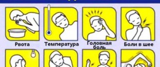
Pain in the palate is caused mainly by inflammatory processes.
Causes of inflammation
- Mechanical damage. Injury can occur from cuts from sharp edges of chipped teeth, improperly fitted dentures, or chewing hard food.
- Caries, pulpitis or osteomyelitis cause pain from cold or hot food or chewing it.
- An incision in the gum and removal of the dental nerve sometimes lead to disruption of the temporomandibular joint, which causes pain.
- Inflammation of the tonsil or trigeminal nerve.
- Stomatitis as a consequence of damage to the mucous membrane.
If the reasons described above are not corrected in a timely manner, the inflammatory processes will progress, which will only worsen the situation.
Signs and symptoms of inflammation of the palate
Depending on the causes of inflammation in the oral cavity, a person may experience the following unpleasant symptoms:
- A feeling of acute pain that makes it difficult to eat. Soon the pain increases, sometimes even swallowing becomes impossible.
- If the inflammation is caused by the action of a fungus, a white coating and erosion forms on the surface of the palate. The process is accompanied by an unpleasant putrid odor from the mouth.
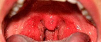
- When the cause of inflammation is an infectious lesion - sore throat or tonsillitis, the palate becomes red and swollen.
- In case of acute inflammation, an increase in body temperature and fever is possible.
- If the cause is dental disease, the patient will be bothered by toothache.
- With cancer, the patient complains of aching pain in the palate.
Treatment
The consequences of mechanical damage to the mucous membrane of the palate can be eliminated by rinsing the oral cavity with recommended compositions, for example, decoctions of various herbs; a solution based on iodine and salt, propolis works well. During treatment, you should refrain from eating solid food, there is a possibility of re-damage to the problem area. In such cases, doctors prescribe the use of targeted medications.
Treatment of tonsils does not involve independent actions. You should definitely consult a doctor and follow his instructions.
Ordinary stomatitis is a consequence of neglect of oral hygiene, so treatment of its mild form consists of following a diet, eliminating too cold and hot foods from the diet and regular rinsing with antiseptic agents.
Severe forms of stomatitis: aphthous, ulcerative, herpetic are characterized by massive damage to areas of the mucous membrane. In such cases, the help of dentists and therapists is required.
It is impossible to cure caries and pulpitis on your own. The sooner a patient seeks help from a dental clinic, the more successful the treatment will be and the less likely there are serious complications.
Functions of the sky and structural features
The palate is a vault that separates the oral cavity from the nasopharynx. The structure of the palate consists of two sections - hard and soft. This organ performs an important function - it prevents food from entering the nasopharynx from the oral cavity.
In addition, receptors on the surface of the palate are associated with the larynx and take part in articulation and influence the timbre of the voice and the pitch of sounds. Thus, the inflammatory process of the upper palate disrupts all the functions of this important organ, and therefore requires mandatory treatment.
The roof of the mouth hurts: causes and treatment in dental clinics
The services of highly qualified specialists are an opportunity to eliminate the problem quickly and safely for health. Already at the first appointment, the doctor conducts a comprehensive examination and diagnoses the problem. This approach allows us to determine the exact cause of the development of symptoms, as well as prescribe adequate treatment, taking into account the individual characteristics of the patient.
Are you experiencing discomfort and want to treat an inflamed palate in your mouth? Contact the specialized dental clinic "Alpha Dent" in Orenburg! Our specialists will conduct a comprehensive diagnosis, determine the cause of the problem and prescribe adequate treatment!
Diagnosis of oral cancer
Methods for diagnosing oral cancer are described below.
Biopsy
A biopsy takes a small piece of tissue from an area that may be affected by cancer. The tissue is sent to a pathologist (a doctor who examines body tissue to diagnose diseases). A pathologist will examine it under a microscope to look for cancer cells. Typically, biopsy results are ready no earlier than after 5 days.
If you have not yet had a biopsy, it may be done at your first appointment with your doctor at MSK. If you have already had a biopsy, a pathologist at MSK will review tissue samples taken during the biopsy to confirm the diagnosis.
Medical imaging
You may also have medical imaging tests, such as computed tomography (CT), magnetic resonance imaging (MRI), or panoramic X-rays of your upper and lower jaws. A panoramic x-ray shows the entire upper and lower jaw, including the sinuses. These images provide detailed information about the lesion (an area of diseased or damaged tissue). This can show how deep the tumor has gone and whether it has spread to other organs.
to come back to the beginning
If the child has a pathology
A diagnosis of “cleft palate” in a child is not a death sentence, and parents need to remember that the pathology responds well to treatment if it is started early. During the treatment period, the main role is given not only to surgical intervention, but also to the patience of parents, their responsibility and willingness to deal with the health of their own child. You must be prepared to carefully implement all recommendations.
Once a diagnosis of cleft palate is made, the child is taken to a surgeon who can create a treatment plan.
In our Clinic, such defects are dealt with by Sergey Nikolaevich Bessonov and Levan Avtandilovich Eremeyshvili, who are maxillofacial surgeons. Appointment with doctors S.N. Bessonov and L.A. Eremeyshvili takes place over the phone.
Thanks to the help of a charitable foundation, surgery to correct a cleft palate can be completely free. All you need to do is prepare the documents from the list and wait for an individual call to a specialist.
Pleomorphic adenoma of the minor salivary glands in the palate
Pleomorphic adenoma (PA) is the most common mixed benign tumor of the salivary glands. It is especially common in women in middle age. Approximately 80% of all cases involve the parotid gland, and about 7% involve the minor salivary glands. Histologically, this tumor is represented by both epithelial and mesenchymal elements, and is usually encapsulated in large salivary glands.
Mostly, tumors of the minor salivary glands are malignant (almost 50%). It has been established that the smaller the salivary gland, the greater the likelihood of tumor malignancy. The error of histological diagnosis varies from 1 to 14%. The difficulty of diagnosis lies not only in differentiating a benign neoplasm from a malignant one, but also in further, more detailed classification of the tumor.
The palate is the most common location of the minor salivary glands, followed by the lips and cheeks. PA appears as a painless firm formation and, in most cases, does not cause ulceration of the mucous membrane.
Ultrasound, CT and MRI may be used depending on the location and size of the tumor. The optimal treatment for PA is wide surgical excision of the formation along with the affected edges.
This clinical case examines a pleomorphic adenoma at the junction of the hard and soft palate.
Description of a clinical case
A 43-year-old woman was admitted to the clinic with complaints of a painless mass to the right of the junction of the soft and hard palate (Photo 1).
There was no somatic pathology in the medical history; the patient did not undergo drug treatment. Intraoral examination revealed a firm, approximately 2 x 2 cm, well-circumscribed, oval-shaped swelling to the right of the junction of the soft and hard palate (Figure 1).
Photo 1: Formation on the hard palate.
Also, palpation of the formation identified a tumor of soft consistency with well-defined boundaries.
Photo 2: Orthopantomogram
The orthopantomogram revealed changes in the bone structures and hard palate (Photo 2). MRI showed a well-defined mass on the palate (Figure 3). An incisional biopsy was prescribed. Histological examination revealed a keratinizing structure and myxoid degeneration (Figure 4). The cells were clustered into islands and layers separated from the myxoid matrix (Figure 4). Based on the data obtained, the final diagnosis was made: pleomorphic adenoma of the minor salivary glands of the soft and hard palate. The tumor was completely excised under local anesthesia, and the diagnosis was confirmed by repeat biopsy (Figure 5).
The postoperative period passed without complications and relapse.
Photo 3: MRI. Axial view showing a clearly visible mass.
Photo 4: Histological examination revealing keratinizing structure and myxoid degeneration, x40
Photo 5: Complete tumor removal
Photo 6: 3 weeks after surgery.
Discussion
PA occurs most frequently in the parotid salivary glands (56.7%), followed by the submandibular salivary glands (31.1%), palatine minor salivary glands (8.9%), and buccal minor salivary glands (3.3%).
A large number of tumors of the minor salivary glands are malignant (almost 50%). It has been established that the smaller the salivary gland, the greater the likelihood of malignancy of the tumor. Thus, differential diagnosis is vital.
Among all tumors of the salivary glands, malignant neoplasms such as mucoepidermoid carcinoma, adenoid cystic carcinoma and polymorphic poorly differentiated adenocarcinoma are distinguished. Mucoepidermoid carcinoma is the most common of all malignant tumors of the salivary glands.
The most vulnerable area after the parotid salivary gland is followed by the glands of the hard palate. Typically, such tumors are asymptomatic, grow slowly, have a different consistency, a bluish or red tint, and clinically resemble a mucous cyst. Adenoid cystic carcinoma most often occurs on the palate. It appears as a painful, slowly growing mass that affects surrounding structures and occurs in middle-aged people. Polymorphic poorly differentiated adenocarcinoma is also more often localized on the palate and usually presents as a slowly growing swelling. In the population, this disease predominates in older women.
Ultrasound, MRI and CT are used in diagnosis. In our case, MRI and CT were used. The CT scan was unremarkable, while the MRI revealed a mass on the palate.
Clinically and histopathologically, this formation can be mistaken for malignant; however, PA is a benign tumor that recurs quite rarely. The literature contains data on 2-44% of relapses. Wide surgical excision is the most preferred therapy. The occurrence of relapse may be associated with inadequate surgical treatment and removal of the tumor. In this clinical case, the tumor was completely enucleated.
Conclusion
The purpose of this report was to describe the diagnosis and treatment of a pleomorphic adenoma that was found at the junction of the soft and hard palate in a 43-year-old woman. The tumor was completely excised along with the affected edges. Recovery was uneventful with a short recovery period (Photo 6).
Authors: Firdevs AKPEK, Mustafa TEK, Orcun TOPTAS, Fat ih OZAN
The palate of the mouth is inflamed: how to treat it and where to go
Today, there are two main ways to treat pain and inflammation of the palate: at home and in a specialized dental clinic, with an ENT specialist or therapist. Treatment should be carried out only after determining the exact cause of the development of symptoms - improper treatment can only lead to aggravation of the problem.
If there are no prerequisites for serious diseases of the oral cavity: infectious, traumatic or burns, treatment can be carried out independently at home. First of all, you need to start by eliminating inflammation and pain. There are several folk and professional medical remedies that provide good effectiveness.
- To eliminate inflammation, use special herbal decoctions based on calendula and sage - they will soothe the mucous membrane and reduce swelling. Anti-inflammatory drugs can also be used. It is very important that in the first stages of treatment, inflammation is stopped and swelling is removed;
- If a pimple appears on the roof of the mouth, it is necessary to use special medications, such as: Chlorophyllipt, Chlorhexidine or Rotocan - they have a more powerful effect and limit the further spread of the inflammatory process to adjacent tissues of the oral cavity;
- If there is pain, it is necessary to use painkillers, as well as dental gels, such as Kamistad, Cholisal or Kalgel.
Particular attention should be paid to pain and inflammation of the palate caused by fungal etiology, which is accompanied by an unpleasant odor from the oral cavity. In this case, broad-spectrum antifungal agents are used; it is not recommended to treat this problem on your own.
If the cause of palate pain is aphthous stomatitis, it is recommended to use oils based on rosehip and sea buckthorn, and use propolis tincture for rinsing.
FREE CONSULTATIONS!
We are pleased to announce the start of free admission at the Konstanta Clinic for patients under 18 years of age by maxillofacial surgeon, pediatric surgeon L.A. Eremeyshvili. for questions:
- Congenital facial pathologies: cleft lip and palate;
- Benign neoplasms of the face and oral cavity;
- Diseases of the temporomandibular joints;
- Pathology of the frenulum of the oral cavity: lips and tongue.
Correction (plasty) of tongue frenulum is provided free of charge for children under 1 year of age.
*Valid for residents of Yaroslavl and the Yaroslavl region.
You can make an appointment by phone or through the website (4852) 37-00-85 Daily from 8:00 to 20:00
Sign up for a consultation
Congenital cleft palate or cleft palate is a fairly common malformation of the facial region. This pathology is ranked 4th among all developmental defects, and it is diagnosed with a frequency of 1:700 births. Often, a cleft palate is not an independent deviation, but part of a hereditary syndrome.
Vocal yawn
Let me give you a simple “exercise”. Try yawning: once with your mouth closed, and the second time with your mouth open. You felt like yawning wasn't that hard, right? I hope now you understand what this “thing” is - the sky. Just in case, I ask you not to take the “yawn” exercise literally.
The most important! When performing high notes using the soft palate, you need to keep your larynx relaxed and not lower it (as in academic vocals). In this case, the root of the tongue should be lowered.
By the way, the concept of “vocal yawn” refers more to academic vocals. This is the correct position in academic singing, which implies a raised soft palate and a lowered larynx. Not very suitable for pop performance! This is why the position of the larynx in stage performers should be average or neutral. We sing in a natural voice, so there is no need to study the “vocal yawn” in its academic version.
The situation is similar with the “raise the dome” technique. When you hear this command from your teacher, know that you should direct the sounds towards the hard palate, but not “yawn”. Do not take this literally, as a term from academic singing! Simply direct the sound to a point in the palate.
I also recommend that you study the short article “Surround Sound Secrets”.
How to feel the palate - the secret of surround sound! >>>