What is hydrocele?
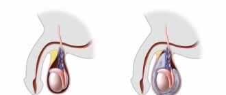
Hydrocele, also known as hydrocele or hydrocele, is an accumulation of fluid in the membranes of the testicle, which leads to enlargement of the scrotum, and sometimes swelling in the groin area.
There is isolated hydrocele of the testicular membranes, when the fluid surrounds the testicle and cannot flow into other cavities, and communicating hydrocele.
A communicating hydrocele differs in that hydrocele can flow into the abdominal cavity and back through a special duct - the vaginal process of the peritoneum. Hydrocele of the testicle is often combined with an inguinal hernia.
Lymphocele is a concept close to testicular hydrocele, meaning the accumulation of lymph in the membranes of the testicle, which occurs when the lymphatic vessels of the testicle are damaged or compressed. Typically, lymphocele is accompanied by stagnation of lymph in the testicle and its membranes - lymphostasis.
Prevention methods
It is necessary to take measures to prevent inflammation of the scrotum. Parents should regularly examine their child's genitals and contact a doctor immediately if they notice any swelling. Those children who have a congenital disease should be regularly monitored by a pediatric urologist.
Sources:
- https://www.ncbi.nlm.nih.gov/pubmed/12378019 Han CH, Kang SH. Epididymal anomalies associated with patent processus vaginalis in hydrocele and cryptorchidism // J Korean Med Sci. 2002 Oct;17(5):660-2.
- A.V. Grinev, S.I. Nikolaev, V.E. Serdyutsky, D. S. Efremenkov. New technologies in the treatment of hydrocele in adults and children // Bulletin of the Smolensk State Medical Academy, 2003, No. 5.
The information in this article is provided for reference purposes and does not replace advice from a qualified professional. Don't self-medicate! At the first signs of illness, you should consult a doctor.
Communicating hydrocele of the testicle in children. What is the mechanism of formation of a communicating hydrocele?
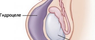
The term communicating hydrocele or communicating hydrocele means that between the cavity surrounding the testicle and the abdominal cavity there is a communication - an open vaginal process of the peritoneum, through which fluid from the abdominal cavity enters the scrotum and back.
During fetal development, the testicle descends into the scrotum through the inguinal canal. Together with it, the processus vaginalis descends into the scrotum - an outgrowth of the peritoneum that envelops the testicle and, thus, forms the two shells closest to the testicle.
By the time of birth or during the first months of life, normally the processus vaginalis of the peritoneum is overgrown, and the connection between the testicular membrane and the abdominal cavity disappears. Thus, neither peritoneal fluid nor abdominal organs can penetrate the cavity where the testicle is located. The lower part of the processus vaginalis of the peritoneum forms a slit-like cavity around the testicle, which, in case of dropsy, serves as a container for dropsy fluid.
The main cause of communicating hydrocele of the testicle is non-closure of the processus vaginalis of the peritoneum, which serves as a duct for moving peritoneal fluid from the abdominal cavity into the membranes of the testicle.
Complications of varicocele
As a rule, physiological dropsy disappears on its own during the first year of life. If surgical intervention was performed according to indications, then most often the disease goes away and does not occur again.
Chronic testicular hydrocele, which is a consequence of neglected treatment, poses a significant danger. It can cause spermatogenesis disorders and complete infertility in older age. The disease can also provoke a sensitivity disorder in the testicle and, as a result, its atrophy.
Causes of non-fusion of the peritoneal process.
Many theories explain non-fusion of the processus vaginalis of the peritoneum. Thus, in the open vaginal process of the peritoneum, smooth muscle fibers were found, which are not found in the normal peritoneum. Smooth muscles can prevent fusion of the peritoneal process.
According to our data, there is a higher incidence of reported hydrocele in children born after a pathological pregnancy with threatened miscarriage, as well as in premature children.
Another reason lies in the increase in intra-abdominal pressure, which is observed during resuscitation measures, with frequent restlessness of the child or during physical exercise.
How is the surgeon examined?
Diagnosis of an inguinal hernia begins with a questioning of the patient and his further examination. During the interview, the doctor finds out what exactly the patient is complaining about, how long he has been bothered by the listed phenomena, with what frequency, what precedes and causes them. Also, during the survey, factors contributing to the development of a hernia can be identified: conditions and characteristics of life, professional activity, leisure, the presence of injuries and surgical interventions in the past. The surgeon may ask whether any immediate relatives have suffered from a hernia, and this is not an idle question: the hernia itself, of course, is not inherited, but the specific structure of the ligaments, aponeuroses (connective tissue) and muscle tissue of the area can be passed on from the parents to the child groin Therefore, one can often see the “familial” nature of an inguinal hernia, which is explained by the elementary inheritance of the characteristic weakness of certain areas of the abdominal wall.
During the examination, the doctor assesses the size and shape of the hernia, and does this in different body positions: when the patient is standing and lying down. Pay attention to the skin above and around the formation: the presence of dilated veins, diaper rash, scratching and other damage. In obese patients, such an examination is difficult because, due to the large thickness of the fat layer on the abdomen, the hernia becomes invisible to examination. In addition, at the time of examination, the hernia may “slip” into the abdominal cavity. Therefore, after the examination, palpation (feeling) of the groin should be performed.
During palpation the following is determined:
- what is the shape of the hernia, its size, how does it change if the patient coughs or strains;
- is it possible to move the mass back into the abdominal cavity;
- does the hernia hurt when touched?
- whether the scrotum and testicles in it are enlarged, in what condition are the spermatic cords;
- what about the inguinal canal - with hernias it can increase significantly;
- condition of the lymph nodes in the groin area.
It is during palpation that, as a rule, the type of inguinal hernia is accurately determined, as well as its difference from other diseases in this area.
What does communicating hydrocele of the testicular membranes and an inguinal hernia have in common?
An inguinal or inguinal-scrotal hernia occurs in children with a wide, unclosed processus vaginalis of the peritoneum. Not only fluid from the abdominal cavity penetrates into the open vaginal process of the peritoneum, but also movable organs of the abdominal cavity (loop of intestine, strand of omentum, appendages in girls, etc.) can emerge, which characterizes an “oblique” inguinal or inguinoscrotal hernia.
In adults, inguinal hernias differ from those in children. They are associated with defects in the muscles and tendons of the anterior abdominal wall that occur during exercise. In childhood, such hernias are extremely rare. Therefore, operations for inguinal hernias in children and adults are performed using various methods.
How is hydrocele diagnosed?
For an experienced pediatric andrologist, diagnosing testicular hydrocele is a simple task. Especially in cases where parents’ anxiety due to the abnormal size of the child’s scrotum becomes the reason for contacting the clinic.
During the conversation, the doctor finds out whether this increase depends on the time of day, whether the boy is in a horizontal or vertical position, physical activity, and so on.
A preliminary diagnosis of dropsy can be made during an initial consultation with an andrologist. However, an enlargement of the scrotum can also be observed, for example, with an inguinoscrotal hernia, a large cyst of the spermatic cord and other pathological conditions. In order to dispel all doubts, ultrasound diagnostics is prescribed. Ultrasound allows you not only to make sure that there is a hydrocele, but also to understand the form of the disease, see whether the blood flow is suffering because of this, determine the amount of excess fluid and other nuances.
Tomography (both computed tomography and magnetic resonance imaging) is rarely used in cases of suspected dropsy, only if the doctor wants to exclude the presence of a malignant tumor.
Hydrocele of the testicular membranes in newborns and young children. Isolated hydrocele of the testicle.
In newborns and infants, hydrocele of the testicle in 80% of cases is (or becomes during the first months of life) isolated from the abdominal cavity and goes away on its own within 6-12 months. Isolated hydrocele of newborns is associated with birth trauma, peculiarities of hormonal status and the state of lymph outflow from the scrotum in children 1 year of age.
Isolated testicular hydrocele is often bilateral. Often the dropsy increases and becomes tense. In cases of intense hydrops, punctures are usually performed to remove fluid from the membranes of the testicles. Surgical treatment is usually not indicated.
Isolated hydrocele of the testicular membranes in boys over 3 years of age.
Isolated hydrocele of the testicle over the age of three often occurs after injury or inflammation. There are also cases of transformation of a communicating dropsy into a non-communicating one, due to the closure of the lumen of the peritoneal process from the inside, for example, by a strand of the omentum.
How to treat hydrocele? Operative and conservative methods
Whether a child requires surgery depends on the shape of the testicular hydrocele. If the pathology is congenital, then a wait-and-see approach is usually chosen under the supervision of a doctor. In 80-85% of cases, the disease goes away on its own in the first 1.5 years of life.
In the acute form of hydrocele, the underlying disease that caused the hydrocele is treated. In the case of a severe form of the disease, a puncture is performed and the fluid is removed, but this gives only temporary relief, because the cause of the accumulation of water must be eliminated. Source: A.V. Grinev, S.I. Nikolaev, V.E. Serdyutsky, D. S. Efremenkov New technologies in the treatment of hydrocele in adults and children // Bulletin of the Smolensk State Medical Academy, 2003, No. 5
How common is testicular hydrocele and how often is surgery required?
Hydrocele of the testicular membranes in newborns and boys in the first year of life occurs in 8-10% of cases. In 80% of cases it is isolated and goes away on its own. In 20% of children, surgery is performed after one year.
Communicating hydrocele of the testicle in children after 1 year 0.5-2.0%. In 95% of cases, surgical treatment is indicated.
Lymphocele and testicular lymphostasis in adolescents after operations for varicocele account for from 1% to 25% of all surgical interventions, depending on the type of operation and surgical technique (on average about 10-12%). In 80% it is amenable to conservative treatment. In the remaining 20%, surgical treatment is indicated.
Hydrocele and lymphocele after surgery for inguinal hernia in adolescents - statistics are the same as in adults 3-10%. Surgical treatment is often performed.
How is hydrocele treated?
Isolated hydrocele does not require surgical intervention in boys under two years of age. But, if this form of dropsy is found in an older child, a planned operation is mandatory, no matter what caused the disease.
A communicating hydrocele can be treated regardless of the boy's age. In this case, in order to choose the right treatment tactics, the pediatric andrologist must rely on examination data and the dynamics of the disease.
How to diagnose hydrocele?
The disease usually occurs with obvious external manifestations - swelling (increase in volume) of the scrotum on one or both sides. Scrotal enlargement may decrease or disappear at night when the child is in a horizontal position, and reappear when awake. This is evidence in favor of communicating hydrocele of the testicular membranes. Enlargement of the scrotum is sometimes also observed with tension or “inflating” of the abdomen.
Subjective sensations are insignificant. Complaints are rare. In case of acute, infected or tense dropsy, pain may be observed.
To establish the correct diagnosis, ultrasound is used - ultrasound examination of the inguinal canals and scrotal organs and duplex examination of testicular vessels.
Ultrasound often makes it possible to detect a problem from the other side - for example, an inguinal hernia or spermatic cord cyst that is invisible during examination.
Sometimes enlargement of the scrotum and groin area appears and disappears, and may be absent upon examination by a doctor. Then a photograph taken when a swelling appears in the scrotum or groin area, taken by the parents, helps resolve the issue of diagnosis.
Symptoms of hydrocele in boys
Pathology, as a rule, is discovered completely by accident by a pediatrician during preventive examinations or by parents during daily hygiene procedures. Visually, you can notice an enlargement of the scrotum on one or both sides. In an isolated form of the disease, the volume of tissue increases gradually; in the case of a communicating pathology, the size may vary depending on the position of the body, motor activity and the location of fluid accumulation. Hydrocele of the testicles itself does not cause pain, discomfort, itching or other unpleasant symptoms in the child.
Babies are often capricious, toss and turn, and restless, and older children may complain of discomfort when walking, running, a feeling of heaviness and fullness in the groin. In some cases, hydrocele is accompanied by the formation of a unilateral or bilateral inguinal hernia.
Diseases and circumstances that are often accompanied by the occurrence of hydrocele
- Cryptorchidism (undescended testicle)
- Hypospadias
- False hermaphroditism
- Epispadias and exstrophy
- Ventriculo-peritoneal shunt
- Prematurity
- Low birth weight
- Liver diseases with ascites
- Defects of the anterior abdominal wall
- Peritoneal dialysis
- Burdened heredity
- Cystic fibrosis
- Inflammatory diseases of the scrotum leading to the development of reactive hydrocele
- Testicular torsion
- Injury
- Infection
- Previous operations affecting the lymphatic system of the testicle
Prevention
At the stage of planning and maintaining pregnancy, the expectant mother needs to carefully monitor her health, promptly eliminating infectious and inflammatory diseases. To prevent a newborn child from developing hydrocele of one or both testicles, it is necessary:
- regularly examine the baby’s genitals and properly care for them;
- avoid hypothermia and overheating of the scrotum;
- teach children the rules of personal hygiene to avoid infection;
- prevent the development of obesity in children;
- stimulate adequate physical activity.
In the case of a congenital form of hydrocele, the child should be under regular supervision of a urologist or surgeon until complete recovery or surgery.
Hydrocele of the testicle is one of the diseases that can cause male infertility. Do not ignore a visit to a urologist-andrologist; at the slightest sign of a problem, make an appointment with an experienced specialist at the SM-Doctor clinic.
Treatment of hydrocele (hydrocele) and lymphocele without surgery. Duration of observation.
Hydrocele in children under 1 year of age requires observation by a pediatric urologist-andrologist. If fluid accumulates and tension appears in the membranes of the testicle, punctures are performed to remove hydrocele. Sometimes repeated punctures are required.
Communicating hydrops with a narrow peritoneal process is usually observed up to 2 years.
Observation is also required for traumatic dropsy, which occurs as a result of a bruise without compromising the integrity of the testicle. As a rule, 3 months are enough to assess the dynamics of the process and, if there is no improvement, prescribe surgical treatment. The same applies to hydrocele formed after inflammation.
The most difficult is the management of patients with lymphocele that forms after surgical treatment of an inguinal hernia and varicocele. In this case, prematurely performed surgery has little chance of success. For 6-12 months, it is necessary to monitor the condition of the testicle according to ultrasound and duplex examination of the scrotal organs in order to assess the dynamics of the process and the effectiveness of the therapy.
Methods for diagnosing the disease
A boy with such a problem should be shown to a pediatric urologist. At the initial consultation, the doctor collects an anamnesis of the child’s life and information about the course of his mother’s pregnancy. Then he examines the boy in a lying and upright position. This allows you to distinguish communicating dropsy from isolated dropsy. After this, the small patient needs to undergo an examination, which consists of the following procedures:
- Diaphanoscopy - transillumination of the scrotum with a special flashlight. If the disease is not complicated, the testicular membranes have a uniform color.
- Ultrasound of the groin area, which allows you to see the presence of a reported type of disease, determine the exact amount of fluid, and exclude tumors and other diseases of the scrotum.
- Scrotal biopsy – indicated when there is doubt about the diagnosis. With its help, the structure of the selected area is studied at the cellular level, malignant processes or benign formations are identified.
- A general blood test makes it possible to exclude infection and inflammation.
When is surgery performed for hydrocele?
- Operations for communicating hydrocele of the testicle are most often performed in children aged 2 years.
- From 1 to 2 years, operations for communicating hydrops are performed if:
- combined dropsy and inguinal hernia
- when the volume of the scrotum clearly changes with changes in body position
- dropsy increases, causing discomfort
- infection joins
- Surgeries for post-traumatic dropsy – 3-6 months after injury.
- Lymphocele that occurs after surgery for an inguinal hernia or varicocele is operated on 6 to 18 months after the appearance of fluid in the membranes of the testicle.
Treatment without surgery: clinical recommendations
An operation to remove testicular hydrocele is the only way to eliminate this pathology in a child. Doctors warn that folk remedies and dietary supplements can produce unpredictable effects. Only those children who have recovered by the time they reach 1.5 years of age can do without surgery.
Of course, many parents are afraid that the boy will remain infertile due to the presence of such a disease. Yes, overheating of the testicles can disrupt their hormonal function and reduce sperm quality. However, in early childhood (up to 3 years old) this is not critical, because all body systems are still just forming and starting to work correctly. If the doctor suggests observing the child and not performing surgery for now, do not be alarmed, because this is generally accepted practice.
Surgery for hydrocele (hydrocele). Surgical options.
The type of operation depends on the age of the patient and the characteristics of the dropsy.
Surgery for communicating hydrocele of the testicle. Operation Ross.
For communicating dropsy, as a rule, the Ross technique is used - isolation from the elements of the spermatic cord, excision and ligation of the internal inguinal ring of the peritoneal process, as well as the formation of a “window” in the membranes of the testicle. The operation is performed through a small incision in the groin area.
The operation is delicate, requiring good technique - careful and careful preparation while preserving all the anatomical formations of the spermatic cord - the vas deferens and testicular vessels, as well as the inguinal nerve.
Laparoscopic operations are sometimes used for testicular hydrocele, but the morbidity, risk of relapses and complications when using them are higher, and the duration of anesthesia is longer, so they are not widely used.
Operations for isolated hydrocele of the testicular membranes and lymphocele in children and adolescents.
Isolated hydrocele and lymphocele are indications for Bergman's operation - excision of the inner membranes of the testicle from the scrotal approach. In cases of large hydroceles and lymphoceles, drainage is often left in the wound and pressure bandages are applied.
Winkelmann's operation is a dissection of the testicular membranes in front and suturing the resulting edges of the membranes behind the epididymis. Currently used rarely due to changes in the appearance of the scrotum and testicular contours.
Among the complications, the most common is recurrence of dropsy (5-20%), which in case of lymphocele can reach 70%. A particularly high percentage of relapses is observed when operations are not performed on time.
Treatment of hydrocele
Treatment methods for hydrocele include therapeutic and surgical methods.
The choice of tactics depends on a number of factors: the boy’s age, general health, nature and form of pathology. In case of congenital disease, observation is usually indicated; if by 1.5–2 years the pathology does not go away on its own, surgical removal is resorted to.
Only surgical intervention can completely eliminate testicular hydrocele in infants and older children. There are several options for performing the operation:
- according to Bergman - this tactic is indicated for extensive accumulations of fluid in the scrotum area, but only if the boys have already formed testicles;
- according to Ross - an operation during which the duct between the peritoneum and the scrotum is sutured through a small incision in the groin area, prescribed for congenital communicating dropsy;
- according to Winkelmann - a similar technique is used in cases where the cause of the hydrocele is a violation of the outflow of lymph; after eliminating the pathology, excess fluid no longer accumulates.
Only the doctor decides how exactly to treat hydrocele, based on the information obtained during the examination of the child.
Hydrocele is quite easy to treat: in 98% of cases, the pathology completely disappears after surgery without any consequences. Re-development of testicular hydrocele is possible, but is rare, especially if treatment was carried out correctly and in a timely manner.
Complications of operations.
The overall risk of complications ranges from 2 to 8%.
Relapses of dropsy occur with a frequency of 0.5 to 6%. In adolescence, relapses of dropsy are more common.
The risk of infertility after such operations is due to surgical trauma and averages about 2-5% and mainly depends on the technique of performing the intervention.
Infertility is not always a manifestation of damage to the vas deferens. In 5-8% of patients, there are rudiments of the rudiments of the female genital organs, which indicate the presence of more or less pronounced defects of the reproductive system that arise in utero or are genetically predetermined.
One of the complications is high fixation of the testicle, when the testicle is pulled up to the inguinal canal and is subsequently fixed there with scar adhesions.
Testicular atrophy can be observed due to impaired blood circulation in the testicle, which occurs during mobilization of the peritoneal process from the elements of the spermatic cord.
Unpleasant or painful sensations in the area of the wound or scrotum on the side of the operation - hyperesthesia associated with pinching in the scar or damage to nerve endings. These phenomena usually disappear 6-12 months after surgery.
Prevention of complications.
The development of complications can be prevented by a high level of surgical technology and timely determination of indications for surgical treatment.
Winkelmann operation
Boys with isolated dropsy are operated on using the Winkelmann technique. This type of intervention is similar to the previous one. The fundamental difference is that after excision of the membrane, it is stitched, turning it inside out - behind the testicle. This operation has both its advantages - the absence of relapses, and disadvantages - trauma and a noticeable change in the external contours of the scrotum. Because of this, it is not very popular today.
The goal of any operation performed by the specialists of our center is not only to rid the child of the disease, but also to preserve his reproductive health - the opportunity to become a father in adulthood. Therefore, our highly qualified surgeons are armed not only with their own many years of experience, but also with the most modern medical equipment - microsurgical equipment and optical instruments, which ensure maximum precision of surgical intervention. And innovative suture material for plastic surgery provides the best cosmetic results.
Anesthesia, which meets the requirements of modern world medicine, completely relieves the child of pain not only during the operation, but also in the first days after it.
Over the 20 years of operation of our center, we have become friends of thousands of families not only in Moscow, but throughout Russia, returning children to health and the opportunity to live a full life. The effectiveness of all types of therapy, quick recovery and easy rehabilitation of young patients are the best indicators of the professionalism of our team.
You can make an appointment with specialists in the field of treatment of the reproductive system organs either on the website of the International Children's Andrology Center or by phone.
The health of our children is the key to their happy future! Take care of it today!
If it is not possible to come for an in-person consultation, then you can send photographs of the penis from different sides so that the external opening of the urethra is clearly visible via E-mail, WhatsApp or Viber
Postoperative period
Surgeries for dropsy are usually well tolerated by children and do not significantly interfere with their movements. However, with sudden movements or constipation as a result of increased intra-abdominal pressure or direct impacts, the formation of hematomas in the scrotum and groin area is possible. Therefore, children should limit their activity until the postoperative wound heals and follow a diet.
On the first day after surgery, non-narcotic painkillers (analgin, paracetamol, ibuprofen, Panadol and others) are usually prescribed. Laxatives are used for 4-5 days after surgery.
For 2 weeks after surgery, do not wear underwear that compresses the scrotum to avoid pushing the testicle up toward the inguinal canal, due to possible fixation of the testicle above the scrotum.
School-age children are exempt from physical education for 1 month.
Inguinal hernia: characteristic manifestations
The main complaint of a man suffering from an inguinal hernia is the appearance of a subcutaneous protrusion in the groin or in the iliac region. The patient feels some discomfort at the site of the protrusion, a feeling of stretching, tension or pain. The protrusion increases with straining, coughing or lifting a heavy object, after which it may completely disappear or decrease.
For an obliquely located inguinal hernia, a unilateral location is more typical; The protrusion itself is oblong, runs obliquely downwards, actually repeats the shape of the inguinal canal, and quite often penetrates the scrotum. With large oblique hernias, an increase in the volume of the scrotum may be observed. A direct inguinal hernia is round in shape and rarely reaches the scrotum. For direct hernias, symmetrical formation is the norm, on both sides at once.
The larger the hernia, the more discomfort it creates for the patient. Large protrusions can even interfere with walking, not to mention reduce the ability to work. The symptoms of a hernia partly depend on which internal organs are inside the hernial sac. If a loop of intestine gets into it, it can cause constipation and bloating. When there is a hernia of the urinary system organs, you may experience increased frequency of urination, pain, or even urinary retention. The listed phenomena disappear after the hernia is repaired, but return when it reappears.
The hernial mass may include the appendix; in this case, the pain and retention of bowel movements characteristic of a hernia may be accompanied by nausea and vomiting, an increase in general temperature and an increase in heart rate. Frequent penetration of the appendix into the hernia can cause chronic inflammation.
Get an online consultation
right now.
Get
Questions about the article
Ruslan
November 29, 2021 at 06:45 pm
Hello, Bergman's surgery was performed on November 23, 2021. Hydrocele on the left 75 ml. The stitches haven't been removed yet. It doesn’t hurt, but sometimes it’s a little painful. Is it possible to masturbate on the 7th day after surgery?
Anton Evgenievich Rotov
November 30, 2021 at 08:58
I think it's already possible
Konstantin
October 24, 2021 at 08:38
2 weeks ago I had a hydrocele operation according to Bergman. After the operation I had a fever for 2 weeks. On the 2nd day after the operation, the doctor examined the inside after removing the drainage, and said that there was nothing there, no infection either. The temperature was not high, up to 38. When taking painkillers, the temperature dropped. And every day the temperature became lower, and now it has passed. Is this a normal reaction of the body? And now when I touch the testicle, it is somehow large and very hard. There is also pain in the lower abdomen, on the side of the operated testicle, what could it be?
Anton Evgenievich Rotov
October 24, 2021 at 08:39
What you are describing is probably due to post-operative swelling that is more severe than normal due to inflammation. As a rule, the swelling completely disappears within 2-3 months. If in doubt, do an ultrasound of the scrotum
Vyacheslav
September 23, 2021 at 08:09 pm
Hello, on September 7, 2021, Bergman had an operation to remove a hydrocele. As of September 22, 2021, there are a number of problems that concern me, although I have been to the local doctor, I want to make sure that everything is in order. The first problem: somewhere around the 18th, the seam began to bleed from the edges, at the moment there is a “detachment” of the skin from above, but it does not cause any painful sensations (the local surgeon said that the seam had come apart, there was no need to stitch it up, he advised treating it twice a day with Chlorhexidine ) it’s already the 22nd, in my opinion, the seam divergence has not decreased, and I don’t understand how it will heal if it “peeled off”; there is still some liquid in the formed depression due to the seam divergence. Please tell me how normal this is and should I panic? Enough time has already passed, but the seam still doesn’t fit. The second question is how much is it acceptable to enlarge the scrotum after surgery, at the moment, compared to what was, of course, much less (there was 200 ml of water), but now there is swelling, I can hardly feel the testicle
Anton Evgenievich Rotov
September 23, 2021 at 08:18 pm
I am afraid that it will not be correct to assess the condition of the wound in absentia and draw any conclusions. Therefore, I will speak in general terms. Incomplete healing of the wound at this time is acceptable, although usually the wound is already closed by this time. Postoperative swelling of the soft tissues of the scrotum can persist for up to 3 months. If you still have doubts about the course of the postoperative period and you are not sure about the attending physician, then it is better to see another specialist
Peter
June 15, 2021 at 04:38 pm
A month ago I had a Winkelmann operation for hydrocele. The stitches have disappeared, the swelling is still there, but smaller, the wounds are also still present, but there is no bleeding from them. Can I already have sex or is it better to abstain for now?
Anton Evgenievich Rotov
June 15, 2021 at 04:39 pm
Sexual activity can be resumed
Eugene
March 4, 2021 at 12:18 pm
Thank you very much for the answer!