What is pneumonia
This is an inflammation of the lungs caused by bacteria, viruses or fungi. It may appear as an independent disease, but often becomes a complication of other diseases (influenza, coronavirus, AIDS and other immunodeficiencies, lung tumors, bronchiectasis, etc.). Inflammation affects the lower respiratory tract - cough, sputum appears, and if the process spreads widely, shortness of breath. Pneumonia is almost always accompanied by fever, weakness, and malaise. Pneumonia is divided into community-acquired and hospital-acquired. Community-acquired pneumonia will be discussed in more detail later in the article, but hospital-acquired pneumonia differs in that a person becomes ill with it while being treated in a hospital for another disease. Due to a weakened immune system, while taking medications, concomitant diseases, or if the patient is on artificial pulmonary ventilation (ALV), the body cannot cope with bacteria that have entered the respiratory tract (which in a healthy body would not have a chance to reproduce), inflammation begins, and develops pneumonia. Moreover, hospital-acquired pneumonia often becomes much more dangerous than community-acquired pneumonia: seriously ill, weakened patients, often older and elderly, become ill; hospital-acquired bacteria are often immune (resistant to antibacterial drugs, which leads to high mortality.
Diagnosis of pneumococcal pneumonia
Lobar pneumococcal pneumonia is characterized by certain physical manifestations, which are directly determined by the pathomorphological phase of the disease.
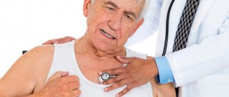
At the first stage of exudate accumulation, there is a dull tympanic sound above the source of inflammation, prolonged exhalation during hard breathing, and mild initial crepitus. In some cases, both types of wheezing are heard: wet and dry. During the second stage of compaction (also called hepatization), the trembling of the voice greatly increases, and bronchophony appears. During tapping, a dull sound is heard, vesicular breathing is not heard, there is no crepitus, and a noise from pleural friction is often heard. At the last stage, the trembling of the voice steadily returns to normal, bronchophony stops, redux crepitus occurs - sonorous, abundant and over a long period). In addition, the wheezing is sonorous, finely bubbled, and bronchial breathing over time turns into harsh, and then into vesicular. But it is important to take into account the fact that with pneumococcal pneumonia, these phases may not occur in the specified sequence, and in certain areas of the lungs different manifestations may be observed simultaneously. If pneumococcal pneumonia is focal, the symptoms are much less pronounced. So, in some cases, a dull percussion sound is heard above the lesion. And, as a consequence of concomitant focal bronchitis, crepitus and fine rales are heard.
Causes of pneumonia
- entry into the lungs of bacteria (pneumococci, staphylococci), mycoplasmas, fungi, viruses through breathing, less often - blood, lymph;
- decreased immunity (in a normal state, the immune system is able to cope with most pathogens);
- complication due to respiratory tract infections, diseases of the cardiovascular system; alcohol abuse. - previous operations (especially on the chest);
- exposure to chemical elements from the environment;
- respiratory tract burns.
Cardiovascular drugs
- Camphor oil is an important remedy needed in case of acute pneumonia. Camphor tones the cardiovascular and respiratory systems, enhances myocardial contraction, and produces an expectorant effect. The drug has a strong bactericidal effect. The literature talks about the property of camphor to improve ventilation of the alveoli. Camphor is indicated for use if the patient has severe pneumonia. Recommended subcutaneously 3 - 4 times / day, 2-4 ml. But it may have a side effect - the occurrence of infiltrates.
- Sulfocamphocaine is a drug obtained by combining sulfocamphoric acid with novocaine. A 1% solution is administered intramuscularly, subcutaneously or intravenously 2 or 3 times a day. The advantage of the drug is that it has the important therapeutic properties of camphor, but does not lead to infiltrates.
- Cordiamine – 25% solution of nicotinic acid diethylamide. It has a stimulating effect on the respiratory and vasomotor centers. Indicated 3 times a day subcutaneously, intramuscularly and intravenously, 2-4 ml, if the patient has been diagnosed with severe arterial hypotension or lobar pneumonia.
All these drugs normalize blood movement in the pulmonary circulation.
If the ability of the left ventricle to contract is greatly reduced, cardiac glycosides can be recommended, but taking into account the sensitivity of the inflamed myocardium. Administer small doses intravenously.
Symptoms of pneumonia
- pain in the side, especially when breathing deeply,
- dyspnea,
- dry cough or cough with sputum,
- increased fatigue, lethargy,
- headache,
- elevated temperature (up to 39-40°) or temperature below normal (especially in the elderly),
- nausea, vomiting.
If you do not contact a specialist when the first signs of the disease appear, the symptoms will begin to worsen and pneumonia will progress, affecting an increasing volume of the lungs. Therefore, you should not delay making an appointment with a doctor, especially if you or your loved one are at risk:
- children under 5 years old, adults over 65 years old,
- people with reduced immunity due to HIV infection or other serious diseases,
- diagnosed with asthma, COPD, diabetes mellitus or cardiovascular failure,
- heavy smokers,
Manifestations of the disease
In most cases, pneumococcal pneumonia is characterized by an acute onset and appears unexpectedly. Following a stunning single chill, a very noticeable increase in body temperature reaches 38-40°C. On the affected side, the patient feels pain while breathing. The cough is initially dry, very painful, and after a short period of time, the separation of purulent mucous sputum begins, in which blood is observed. Very often, patients notice quite a lot of such impurities - this is the so-called “rusty sputum”. Signs of intoxication in patients are very indicative, such as severe weakness, muscle and headache, almost complete lack of appetite, active tachycardia, the patient is suffocating.
Diagnostics
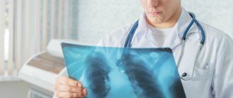
The main diagnostic method remains an X-ray examination of the chest - it will help the specialist understand the extent of the lesion. To select effective therapy, it is important to determine the nature of pneumonia: the disease is caused by the presence of bacteria, viruses or fungi - the results of a blood test, urine test and sputum secreted by coughing will help with this.
In the case of hospital-acquired pneumonia, diagnosis is complicated by concomitant diseases for which the patient is already being treated, as well as by a weakened and sometimes extremely weakened body. Therefore, it is fundamentally important for a doctor to quickly and accurately determine the bacteria that caused the disease and the affected area. Sometimes this requires bronchoscopy (examination of the lungs with an endoscope with collection of material for subsequent analysis); if there is no naturally produced sputum, then a liquid is taken for analysis, with which part of the lung is first washed - this procedure is called bronchial lavage.
Diagnostic criteria
Hospital pneumonia:
signs of newly emerging infiltrate of lung tissue in combination with signs of its infectious nature:
- development of fever;
- separation of purulent sputum;
- leukocytosis;
- decreased oxygenation.
The combination of data on a new site of infiltration of lung tissue with two of the three main signs (temperature > 38 º C, purulent sputum, leukocytosis) as a diagnostic criterion for pneumonia has 69% sensitivity and 75% specificity during pathological verification.
Auscultatory signs of pneumonia are an auxiliary diagnostic criterion.
The “gold standard” for radiological diagnosis of pneumonia is computed tomography. It is preferable to use the method in patients with high risk and complications of pneumonia.
Source:
Kalil AC, Metersky ML, Management of Adults With Hospital-acquired and Ventilator-associated Pneumonia: 2016 Clinical Practice Guidelines by the Infectious Diseases Society of America
and the American Thoracic Society. Clin Infect Dis. 2021 Sep 1;63(5):e61-e111.
PMCID: PMC4981759
Structure of the diagnosis
| Place of development | Localization | Respiratory failure**** | Determination of degrees of respiratory failure (not indicated in the diagnosis) | ||
| Community-acquired | focal, in the _ lobe __ of the lung, | pneumonia | etiology*** ± antibiotic- resistance | DN 0 tbsp. | no shortness of breath |
| Hospital* | focally - confluent, ... | DN I Art. | the appearance of shortness of breath with increased exertion, | ||
| Hospital, ventilator-associated** | …third party…..equity | DN II Art. | the appearance of shortness of breath with normal exertion, | ||
| subtotal, total | DN III Art. | the appearance of shortness of breath at rest. |
* Onset ≥ 48 hours after hospital admission in patients who were not intubated
** Developed ≥ 48 - 72 hours after endotracheal intubation.
*** Abbreviations for the names of microorganisms on the website https://www.antibiotic.ru
**** The degree of DN is not indicated in the diagnosis of ventilator-associated pneumonia.
Examples of diagnosis formulation
Example 1: Community-acquired focal pneumonia in the lower lobe of the left lung. DN 0.
Example 2: Community-acquired right-sided lower lobe pneumonia caused by S. pneumoniae. Right-sided exudative pleurisy. DN 1 tbsp.
Example 3: Bilateral focal confluent pneumonia in the lower lobes of both lungs, the upper lobe of the right lung, on the right - abscess (abscess in S3), caused by K. pneumoniae, S. pneumoniae; DN – 2 tbsp. Chronic alcoholism.
Treatment of pneumonia
To treat pneumonia, the most accurate diagnosis indicating the causative agent of the disease is necessary. If the cause of the disease is a virus, the doctor will prescribe antiviral therapy, and if fungi, antifungal therapy. If the bacteriological nature of pneumonia is confirmed, the patient will be prescribed antibacterial therapy, taking into account a large selection of modern medications that can destroy the pathogen and stop inflammation.
In the case of prescribing antibacterial drugs, i.e. antibiotics, there are two possible ways to take the medicine: in the form of tablets for mild pneumonia (often the patient is allowed treatment at home with unconditional regular monitoring by the attending physician) and intramuscularly/intravenously in a hospital in case of severe disease. Medicines will also be prescribed to combat symptoms and generally strengthen the body: expectorants, vitamins, drink plenty of fluids; Additionally, it is advisable for the patient to follow a certain diet to restore the body’s strength and support the immune system.
Pneumonia is considered a well-studied disease and, despite its sometimes severe course (if it is a hospital-acquired form, or if the patient has not sought help for a long time in a community-acquired condition), in the hands of competent specialists it can be easily cured.
The pulmonology department of our center is rightfully considered one of the leading in the country, thanks to modern diagnostic and treatment equipment and the composition of the team of specialists: our doctors develop unique treatment and rehabilitation programs, train colleagues from other centers, and publish the results of their research in leading international publications.
The department has all possible means of diagnosing and treating lung diseases: both modern and time-tested. Moreover, in our center, the only one in Russia, clinical trials are conducted, and it is our specialists who confirm the effectiveness of new diagnostic and therapeutic equipment, which is being registered for future use in all pulmonology departments of the country. This is a great trust in the center’s qualifications, which we justify year after year, first of all, to our patients.
Diagnosis criteria
The diagnosis of pneumococcal pneumonia is made if the following symptoms are present.
- The disease occurs very acutely, chills are felt; fever is pronounced. The patient experiences chest pain, suffocates, and coughs heavily.
- Indicative data from physical examination and pulmonary radiology.
- Gram-stained sputum preparations contain gram-positive lanceolate diplococci - they create short chains. In this case, it is necessary to identify 10 or more diplococci or typical pneumococci. The latest evidence that the identified streptococci belong to pneumococcus is the swelling reaction of its capsule. This reaction occurs when polyvalent pneumococcal antiserum is added.
- Antipneumococcal antibody titers increase in the patient's paired blood sera, which were taken on the first day of illness and on days 10-14.
Prevention
It consists of practicing a healthy lifestyle, proper nutrition, and, if desired and there are no contraindications, hardening, i.e. Maintaining a strong immune system in general can protect you from the risk of this disease.
In addition, it is important to be attentive to your own health - pneumonia often acts as a serious complication against the background of a common ARVI or flu, if they are not treated in time.
If, however, you or your loved ones experience symptoms of pneumonia, we recommend that you consult a specialist without delay. Do not forget that in some forms, pneumonia can be dangerous not only for the patient, but also for others with reduced immunity due to the possibility of infection.
You can sign up for a consultation with a specialist through a special form on the website or by phone.
Radiology data
During the stage of compaction or hepatization, the most striking changes in the lung tissue are determined. Lobar pneumonia is characterized by very intense darkening of the lobe of the lung. During a tomographic examination, inflammatory infiltration is observed, and the bronchi are clearly visible against its background. This symptom reliably distinguishes between pneumonia and pulmonary atelectasis. Local consolidation or focal shadow reveals focal pneumococcal pneumonia.