General information
The problem of mycotic diseases in general, including fungal diseases of the throat (pharynx and larynx), becomes important due to the wide distribution and severity of the disease. The risk of dissemination of the process and even the development of fungal sepsis increases. Thus, over the past 10 years, the incidence of candidal pharyngitis ( pharyngomycosis ), laryngeal mycoses ( laryngomycosis ) and candidal tonsillitis have increased sharply - they account for 30 to 45% in the structure of all infectious lesions of the tonsils and pharynx.
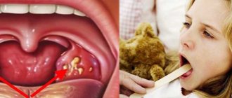
Below you can see what a fungus in the throat looks like (photo). The growth of fungal infections of the throat is facilitated by massive antibiotic therapy, taking immunosuppressive and glucocorticoid drugs over a long period of time.
These and a number of other factors create favorable conditions for the development and spread of deep mycoses caused by opportunistic fungal flora, which is widespread in the external environment, can saprophyte the mucous membrane of the throat and was previously considered non-pathogenic.
Pharyngomycosis / laryngomycosis is not a highly contagious disease. In the vast majority of cases, inflammation of the mucous membranes of the throat caused by fungi of the genus Candida is secondary, developing against the background of predisposing factors. Fungal diseases of the throat are easily chronic (especially in cases of unreasonable/inadequate/adequate, but not aggressive enough therapy) and are prone to frequent recurrence. In most cases, they become stubbornly persistent and therapeutically resistant, and in cases of absence/incorrect treatment can lead to severe complications. The symptoms and treatment of fungal diseases of the throat are practically the same when the throat is affected by one or another type of fungus.
Risk group and causes
In order for a fungus of the genus Candida to trigger symptoms of inflammation, it is necessary to reduce the protective properties of the body. Therefore, children with reduced immunity are at risk.
- Premature newborns.
- Children with intrauterine growth retardation and congenital defects.
- Birth trauma helps to reduce defense mechanisms.
- Severe infectious diseases.
- An unbalanced, monotonous diet leads to hypovitaminosis and decreased immune status.
- Long-term antibiotic therapy and chemotherapy directly suppress the immune system.
- Metabolic disorders.
- Endocrine diseases.
- Poor hygiene of newborns. The fungus reaches the baby from the mother through dirty nipples, hands, and care items.
The listed reasons for decreased immunity are the most studied. In reality, there are many more such factors. That is why the diagnosis of oral thrush is very common in pediatric practice.
Pathogenesis
The pathogenetic mechanisms of fungal infection of the pharynx are based on a restructuring of the body’s immune reactivity (accumulation in the blood and circulation of fungal antibodies), which causes immediate/delayed reactions. The development of throat lesions by fungi is also facilitated by changes in cellular immunity . In the pathogenesis of pharyngomycosis, an important link is specific/nonspecific sensitization of the body and allergy . The factor of traumatization of the mucous membrane of the oropharynx, which contributes to the development of the fungal process, also plays an important role. Tissue reactions are manifested by the formation of granulomas, atypical hyperplasia of the epithelium, necrotic changes of varying severity. Fungal elements in the tissues of the mucous membrane of the throat and the stroma of the tonsils are located predominantly subepithelial. Most often, mycotic foci in the pharynx are localized on the palatine tonsils, posterior wall, arches, velum and tongue.
Causes
The main causative agents of fungal infections of the oropharynx are fungi of the genus Candida (97%) and molds of the genus Aspergillus (in 3% of cases). Among the fungi of the genus Candida, the yeast-like Candida albicans (39%), C. tropicalis (12%), and C. krusei (9%) were most often isolated in patients. Almost all of these types of listed fungi belong to saprophytes (opportunistic microflora), which are activated when the body’s reactivity decreases and acquire pathogenic properties.
The leading reasons for the decrease in the body's resistance are immunodeficiency states caused by frequent and long-term administration of immunosuppressive/glucocorticoid drugs and massive antibacterial therapy. The development of pharyngomycosis is promoted by systemic diseases of the gastrointestinal tract and blood, diabetes mellitus , bronchial asthma and especially often intestinal dysbiosis , in which the lack of bifidobacteria leads to a decrease in the synthesis of B vitamins and, as a result, the unhindered and rapid colonization of fungal flora not only in the intestines, but also in all other cavities organisms in contact with the external environment (oral cavity, nose).
Occupational hazards include constant contact with gases, dust, and elevated temperatures. Often, due to diagnostic errors and inadequate treatment, an acute mycotic process in the throat becomes chronic.
Why is the formation of fungus in the maxillary sinus dangerous?
How can the growth of a fungal colony be dangerous? In any case, it’s not very pleasant when some kind of parasite, mold, or fungus lives in you. This is really a parasite that lives in the cavity of the maxillary sinus and feels great there. In addition, mycetoma is dangerous because the blood supply and oxygen supply to the brain deteriorates, since the function of nasal breathing is disrupted. A person simply begins to experience partial oxygen starvation due to mycetoma.
Plus, the waste products of the fungus in the maxillary sinus flow into the nasopharynx, which can additionally lead to additional complications, including the development of allergies and provoking respiratory diseases. And, of course, chronic sinusitis.
The role of CT in the study of mycetoma
Of course, a good computed tomogram gives a complete picture of the maxillary sinuses and is the main tool in diagnosing “mycetoma” when examining a patient. A CT scan of the maxillary sinus shows the location, size, and volume of mycetoma damage (local volume or total fungal infection of the cavity).
In fact, computed tomography is the gold standard for diagnosing mycetoma today.
Symptoms of fungus in the throat
Symptoms of fungal diseases of the throat (pharyngomycosis) are variable and are determined primarily by the general/antimycotic activity of the body, and then by the type of fungus that caused the disease. The clinical manifestations of candidiasis , aspergillosis and penicilliosis are practically the same. The acute form of the disease is characterized by a rise in temperature to a subfebrile level, a feeling of dryness/scratching and pain in the throat, hyperemia of the oropharyngeal mucosa, the appearance of necrosis of the epithelium, first white small and then extensive plaques that arise under the influence of a fungal infection, which persists when treated with conservative traditional methods .
Upon examination, multiple, difficult to remove, grayish-white plaques of irregular shape are detected on the mucous membrane, which are located on the palate, tonsils, arches and other places. After their removal, as a rule, erosion remains on the mucous membrane.
In the chronic form, patients' complaints appear cyclically every 2-3 weeks, that is, with frequent alternation of new exacerbations and unstable remissions, depending on the current immune status and other determining factors.
In cases of superficial damage, unexpressed hyperemia of the mucous membrane is determined with areas of translucent/dense deposits of a grayish-white color, having a predominantly lumpy, curd-like character, which are easily removed, exposing the hyperemic smooth mucous membrane. Less often, plaques become denser and merge, and when they are removed, the mucous membrane that bleeds with any damage is exposed (like diphtheria).
With the ulcerative-membranous variant of the lesion, localization of the pathological process is characteristic of the tuberous infiltrated tonsils and adjacent formations. At the same time, the ulcers are covered with an easily removable white coating. The submandibular and submandibular regional lymph nodes are in most cases slightly enlarged and slightly painful. Often lesions are found in other parts of the digestive/respiratory tract ( candidiasis , cheilitis , glossitis ).
Tests and diagnostics
The diagnosis of mycotic lesions of the oropharynx is based on complaints, examination of the patient’s oral cavity and laboratory data. If upon examination there are changes in the mucous membrane and a curdled white coating is detected, then a diagnosis can be made at this stage. If the picture of the pharynx (with an erosive-ulcerative/catarrhal process) is not specific, then additional studies are prescribed:
- Microscopy of the native preparation (examination of plaque under a microscope in a smear from the oropharynx allows you to quickly visualize fungal cells, spores, pseudomycelium).
- Bacteriological method (inoculation of the contents of the mask on a nutrient medium). Allows you to identify a specific type of fungus and determine its sensitivity to antifungal drugs.
- Immunological examination (detection of pathogen antigens in the blood).
Fungus in a child's throat
The incidence of FM in childhood is significantly higher. Candidiasis of the mucous membrane of the mouth and throat is especially common in newborns (thrush) and in children under 1 year of age. This is due to the incomplete formation of the child’s immune system from exposure to a mycotic infection. Pharyngomycosis also often affects older children, in whom the onset of the disease is caused by fungal infection at an early age and the lack of complete elimination of the source of infection in the oropharynx. Treatment of fungus in the throat in a child is identical to the treatment of adults, taking into account the age-specific dosage of drugs. It is also necessary to take into account the complexity of local treatment in children.
Treatment of throat fungus
Treatment of the disease is based on three basic principles:
- Treatment of throat fungus will require the use of both local and systemic antifungal drugs. All previously used antibiotics should be discontinued.
- It is necessary to restore disturbances in the intestinal microbiocenosis. This is achieved through dietary nutrition, through the use of antibacterial drugs (Mexaform, Intestopan) and products containing live bacteria (Bifidumbacterin, Lactobacterin, etc.).
- The interferon status is corrected, for which patients are prescribed Viferon for a period of 30 days.
Therapy for uncomplicated mycosis of the throat begins with the use of local antimycotics; only if they are ineffective, the patient is transferred to oral administration.
For this purpose, drugs from three medicinal groups are used:
- Group of polyenes, drugs: Nystatin, Amphotericin, Levorin.
- Azole group: Ketoconazole, Fluconazole, Itraconazole, Diflucan, Mycoflucan.
- Allylamine group: Terbinafine.
If a fungus in the throat is diagnosed in the acute stage, the course of treatment most often ranges from 1 to 2 weeks. When it is possible to achieve a relapse, it is possible to use drugs in a prophylactic dosage. In case of a complicated course of the throat fungus, the patient is hospitalized and treated in a hospital setting.
Most doctors, when treating throat fungus, prefer Fluconazole (if it is confirmed that the disease is caused by yeast fungi of the genus Candida). Most often, this drug is well tolerated by patients and rarely causes side effects. It is enough to take it once a day for 7-14 days. The dosage is selected individually by the doctor. It can vary from 50 to 200 mg per dose. If there is no effect, then the drug is replaced with another.
When standard antifungal therapy is ineffective and the fungus has developed resistance to it, intravenous administration of Amphotericin is indicated. When administered intravenously, monitoring of liver and kidney function is required, since the drug has pronounced toxic properties. Molds are treated with Terbinafine or Intraconazole.
As for local treatment, it is carried out with the help of local antiseptics, this can be: Miramistin, Oxyquinoline, Clotrimazole, Natamycin suspension, Chlorhexidine. The back wall of the pharynx is treated, endopharyngeal installations are performed and the inflamed tonsils are washed. It is important to alternate antiseptic agents every week.
At the same time, it is necessary to treat concomitant diseases that provoked mycosis of the throat. After performing an immunogram, the patient is prescribed therapy using immunomodulators, if necessary.
If the patient seeks medical help in a timely manner, the disease is correctly diagnosed and appropriate treatment is carried out, then most often the fungus in the throat can be successfully treated. The prognosis for complete recovery with chronic disease is less favorable.
Prevention
Prevention of pharyngomycosis is aimed at increasing immunity and involves the rational and justified use of antibiotics and glucocorticoid drugs. The duration of antibiotic therapy should not exceed the period sufficient to eradicate the pathogen. Antibiotics should not be prescribed for prophylactic purposes, especially for viral respiratory infections. If repeated courses of antibiotic therapy are indicated, mandatory antifungal therapy is required.
Adequate treatment of endocrine pathology ( thyroiditis , diabetes , obesity ) is necessary. Hardening, good nutrition, healthy lifestyle.
How can mycetoma be cured?
Is it possible to do without surgery? It is impossible to get rid of fungus in the maxillary sinus without surgical intervention, I will say this right away. No pills, no drops of “dancing with a tambourine” and everything else will give the desired therapeutic effect. First of all, it is necessary to surgically remove the fungal body, remove this mycelium, remove all the fungus from the maxillary sinus.
This can be done either through a nasal surgical approach or an intraoral approach.
How to remove fungus from the maxillary sinus
With intraoral access, a perforation is made in the vestibular wall of the maxillary sinus - access, and evacuation occurs through this hole, i.e. removal of the fungal body, mushroom mycelium. Then the maxillary sinus is well washed, treated with antifungal and antimicrobial drugs and sutured. Subsequently, the patient is prescribed antifungal therapy.
Surgical removal of mycetoma shows good results today, relapses are extremely rare.
List of sources
- Kryukov A.I., Kunelskaya V.Ya., Shadrin G.B. Epidemiology of fungal diseases of the upper respiratory tract and ear. Problems of medical mycology. St. Petersburg, 2011. T. 13, No. 1. — P. 28-31.
- Sergeev A.Yu., Sergeev Yu.V. Fungal infections. Guide for doctors. 2nd ed. -M.: BINOM Publishing House, 2008. - 408 p.
- Kunelskaya N.L., Izotova G.N., Kunelskaya V.Ya., Shadrin G.B., Krasnikova D.I., Andreenkova O.A. Pharyngomycosis. Diagnosis, prevention and treatment. Medical advice. 2013. No. 2. P. 42-45/
- Kunelskaya V.Ya., Romanenko S.G., Shadrin G.B., Krasnikova D.I. Laryngomycosis. Modern approach to diagnosis and treatment. Materials of the XIV Scientific and Practical Conference “Pharmacological and physical methods of treatment in otorhinolaryngology”, Russian Otorhinolaryngology. 2021. 3(82). pp. 38-39.
- Kunelskaya V.Ya., Shadrin G.B., Krasnikova D.I., Andreenkova O.A. Rational methods of treating UDP candidiasis. Advances in medical mycology. 2013. 11. P.99-102.