The diagnosis of leukoplakia from the mouth of a doctor sounds quite terrifying, and most patients begin to panic after such a verdict. This article contains the basic information necessary for a person who has been diagnosed with this disease: what kind of disease it is, what its manifestations look like, what are the main types, causes and signs of leukoplakia, how this disease is diagnosed in a clinical setting, what are the consequences of the lack of timely treatment, and what preventative measures exist to prevent the occurrence of this disease in the future.
What it is
It is believed that leukoplakia occurs against the background of chronic irritation of the oral mucosa and is a kind of protective reaction of the body. This is a local reaction and is not transmitted through contact with a sick person.
The disease is not so harmless, since it most often occurs shortly before the development of oral cancer. Many people mistakenly believe that leukoplakia itself is cancer, however, this opinion is incorrect. This disease affects the epithelial covering of the oral cavity in response to constant external irritants; vitamin deficiency, a decrease in the level of immunity and the presence of chronic foci of inflammation in the oral mucosa serve as an additional impetus for its development.
Leukoplakia of the oral cavity is a significant lesion of the mucous membrane of the lips, tongue, cheeks, upper and lower palate, manifested by keratinization (hardening) of the epithelial cover to varying degrees. Doctors usually classify leukoplakia as a disease that precedes the development of a tumor.
The greatest risk of developing this disease are men in the age group from 30 to 70 years, who abuse alcohol, smoke and wear dentures. Leukoplakia does not occur overnight, and its development can last for years.
Etiology of oral leukoplakia
The etiology of oral leukoplakia includes some external (exogenous) irritants that act for a very long time, that is, they have already entered the chronic stage.
- Mechanical factors:
- Rough food;
- Poorly fitted removable dentures;
- Poor quality fillings;
- Anomalies of teeth position;
- Bite abnormalities;
- Destroyed tooth crowns;
- Galvaniz;
- Bad habits
- Chemical irritants:
- Household factors (passion for spices, smoking, ethyl alcohol);
- Production factors (exposure to bromine, iodine, acids);
- Temperature irritants;
- Meteorological factors;
- Special attention is also paid to the pathology of the gastrointestinal tract. Pathological conditions such as gastritis, ulcers, enteritis and colitis lead to a decrease in the resistance of the mucous membrane to the action of pathogenic factors
- Anemia;
- Genetic predisposition;
- Anatomy - physiological factors;
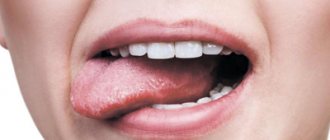
What does leukoplakia look like?
The disease can affect not only the oral cavity, but also other organs (esophagus, bladder, genitourinary organs, etc.) that have a mucous membrane, however, it is in the mouth that the lesion can be diagnosed earlier than in other places.
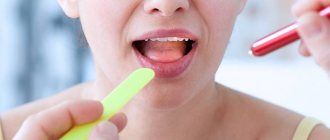
Foci of damage to the mucous membrane are characterized by plaques and growths of whitish or gray color, ranging in size from a few millimeters to three to four centimeters. During a quick examination of the oral cavity, plaques located under the tongue and on the walls of the cheeks can be quite difficult to notice. At the initial stage of the disease, the plaques practically do not rise above the rest of the surface of the mucous membrane, but later they can become hard, heterogeneous, ulcerated, and acquire sharp edges that cause discomfort.
Focal leukoplakia can form over months and even years, however, most often the lesions are visually determined within a couple of weeks after the onset of the disease. In this case, in most patients there is no pain, only a slight increase in the sensitivity of the affected area to temperature effects.
Causes of oral leukoplakia
- taking strong medications (in particular, antibiotics, as well as constant use of drugs of various spectrums of action in large quantities);
- smoking, as harmful components penetrate through cigarettes and deplete the mucous membrane;
- poor nutrition and abuse of spicy, salty and sour foods;
- chronic gum disease in an advanced stage;
- abundant carious lesions of teeth;
- incorrectly placed fillings;
- incorrectly selected dentures that cause an allergic reaction;
- alcoholism;
- hormonal changes;
- bite problems;
- diabetes;
- HIV infection;
- avitaminosis;
- diseases of the stomach and intestines;
- stress;
- hereditary predisposition;
- unfavorable environmental conditions;
- harmful production activities.
Typically, at the initial stage, oral leukoplakia can be completely asymptomatic. That is why it is very important for people who are susceptible to risk factors to undergo regular dental examinations for prevention. The 32 Dent dental network will help you, even if the disease is in an advanced stage.
Types and forms
The classification of typical signs of the disease is divided into the following types of leukoplakia:
| Verrucous | In the absence of timely treatment and elimination of external irritants, verrucous leukoplakia develops from flat leukoplakia. Due to the acceleration of the process of keratinization of epithelial cells, the affected dense area begins to stand out strongly above the level of healthy tissue and acquires a characteristic white color. In this case, the patient may complain of constant discomfort in the oral cavity, since the plaques have a hard, warty surface. This type of leukoplakia often develops into a malignant form. |
| Erosive | Often, patients develop erosion on the lateral surface of the tongue or in the corners of the mouth; leukoplakia in such cases passes from the verrucous to the erosive stage. In this case, the patient has difficulty opening his mouth, and eating becomes very difficult and painful for him. The erosive form of the disease ends in the formation of cancerous tumors more often than other forms. |
| Smoker's leukoplakia (Tappeiner) | It occurs in people who are prone to regular smoking. A characteristic manifestation is a white coating with red dots. Such formations rarely develop into a malignant tumor; the best method of treatment is complete cessation of cigarettes. |
| Soft leukoplakia Pashkova | It is characterized by a benign lesion of the epithelial surface of the oral cavity (most often in the lips and cheeks), associated with heredity or with frequent neuropathic biting of the affected areas. Most often, Pashkov's soft leukoplakia is observed in children and young people under 30 years of age and is not accompanied by unpleasant or painful sensations. It is diagnosed during a simple routine examination at the dentist. If the seals are frequently bitten, roughness and peeling appear, the damaged area may have a grayish color, and erosions may occur. |
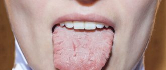
| Flat | Flat leukoplakia is the most common. In most cases, it does not cause a person any inconvenience or pain, so it is diagnosed only during a routine preventive examination by a doctor. Sometimes the patient may complain of constant dryness of the oral mucosa, which leads to increased fluid consumption. This type of mucosal lesion is determined by one or several white spots with clearly defined edges, and the disease itself can accompany a person for many years without causing any inconvenience or developing. |
| Simple leukoplakia without atypia | It is diagnosed by the presence on the oral mucosa of a thin whitish film or dense plaques with pronounced contours. By removing the keratin layer, you can see the actual size of the lesion in simple leukoplakia. In most cases, the simple form is asymptomatic and does not lead to tumor formation, however, patients are recommended to be observed by a specialist and periodically undergo histological tests to exclude cellular activity and atypia of the affected area. |
Kinds
- The most common is simple or, as it is also called, flat leukoplakia . It is usually discovered by chance during an examination by a dentist, since the patient does not experience any subjective sensations. A burning sensation occurs extremely rarely, and the appearance of the mucous membrane may change. If the disease affects the tongue, loss of taste may occur.
- Hairy leukoplakia of the tongue resembles stomatitis. The shape of the spot that appears, as well as its size, can be different, the color - from pale gray to white. The surface of the mucous membrane at the site of the lesion becomes slightly rough, which can be felt to the touch. On the cheeks it appears as solid or broken lines. It can also be found on the lips, where it looks like thin paper pasted on.
- Verrucous leukoplakia is the second stage of the development of the disease. The keratinization thickens, the affected area seems to rise above the nearby tissues. When you touch it with your fingers, you feel a compaction.
- Erosive form . Untimely diagnosis of the two previous stages of the disease leads to a worsening of the situation - the person feels pain when exposed to any irritants, erosions or ulcers are visible in the mouth.
- Soft leukoplakia is a type of cancer. Its distinctive feature is peeling of tissue in the area of the lesion. To clarify the diagnosis, a histological method of studying cells is required.
- Tappeiner's leukoplakia . This form of the disease affects people who abuse smoking. According to studies, daily smoking 10 cigarettes a day increases the chance of developing the disease by 50 times (as the number of cigarettes increases, the risk also increases)
The disease begins with the formation of lesions on the roof of the mouth (sometimes they appear on the gums). The mucous membrane changes its color to a pronounced gray or bluish, which is noticeable to the naked eye, folds appear on it. Reddish nodules may begin to appear, which is accompanied by infectious inflammation of the oral cavity (caused by the accumulation of salivary gland secretions in the tissues).
Causes
Most often, the causes of leukoplakia are trivial, and the culprit for the active development of the disease is the patient himself. The fact is that this disease is provoked by frequent irritating effects on the mucous membrane, and irritation is caused by hot smoke and tar from cigarettes and strong alcoholic drinks.
In people who do not smoke or abuse alcohol, the reasons for the development of leukoplakia may lie in other bad habits. For example, the disease is provoked by the constant consumption of too spicy or too hot food and drinks. In patients with reduced immunity, mucosal damage may occur due to long-term use of potent medications. Local irritation of the epithelium by sharp edges of teeth, crowns and dentures is also not uncommon.
Since leukoplakia is not an isolated disease, but rather a consequence of the main one, the cause of its appearance can also be associated with the presence or development of diabetes, hypovitaminosis, gastrointestinal dysfunction, anemia, papilloma virus and HIV. So, if obvious damage to the epithelial layer appears, it is recommended to undergo tests to identify hidden diseases.
How to treat hairy leukoplakia
Before starting treatment, you need to conduct a number of studies. The patient is referred to a therapist at the clinic. There, the doctor can prescribe laboratory tests: biochemical blood test, testing for sexually transmitted diseases, etc.
If a person is diagnosed with HIV infection, antiretroviral therapy is administered. With long-term use of drugs, the patient’s well-being improves, and the disease in the mouth goes away.
The dental clinic provides oral examinations, dental diagnostics, and professional hygiene. If sharp edges of fillings or failed prostheses are detected, they are replaced. Treatment of caries and its complications is carried out. Recommendations are given for home dental care: personal hygiene products (brushes, pastes, dental floss, rinses) are selected.
Keratolytic drugs are suitable for local treatment. In advanced cases, they resort to surgical manipulations: excision of the affected areas with a laser.
Symptoms
In oral leukoplakia, symptoms are most often typical for this disease, namely:
- Localization on the mucous membrane of the lips (in the corners of the mouth or on the inside of the lower lip) and cheeks, less often on the back and side surface of the tongue.
- The affected area is a whitish or grayish epithelium desquamated in the form of a plaque with clearly defined edges, sometimes its surface is covered with cracks and ulcers.
- The number of lesions can vary from one to several pieces, the sizes sometimes reach three to four centimeters, the shape is arbitrary.
- Most often, the presence of the disease does not cause discomfort to the patient and is diagnosed only at a dentist’s appointment. However, as the condition worsens, the plaques can thicken, burst and become covered with sharp crusts that cut the surrounding mucosa. This process clearly indicates the onset of oral cancer and requires urgent treatment. If leukoplakia symptoms have acquired a similar character, you should not delay visiting a specialist; the disease will definitely not reverse.
Chemical coagulation
For leukoplakia of the tongue, more gentle therapy should be chosen than for lesions of the cheeks or lips, since the soft tissues of the tongue are much easier to damage and take longer to heal. In such cases, the affected area is exposed to a special chemical composition . It does not affect healthy cells, but has a detrimental effect on atypical ones, which leads to their death and the gradual restoration of healthy epithelial cells.
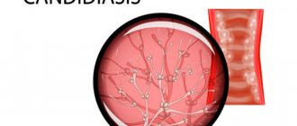
This method also has a significant drawback: the chemical composition kills not only the affected cells, but also beneficial bacteria, as a result of which candidiasis can begin after or even during treatment. To combat this complication, the doctor usually prescribes concurrent use of antifungal drugs.
Nutrition
Since in most cases of disease the patient does not experience any complications or pain symptoms, there is no need to follow a diet for leukoplakia. However, most often the cause of the formation of plaques is a low level of immunity, and therefore, nutrition should be complete, healthy and rich in vitamins.
In advanced and severe forms of the disease, when it is difficult for the patient to open his mouth and chew food due to the formation of erosions and ulcers in places where the mucous membrane is damaged, it is recommended to switch to a porridge-like and puree-like consistency of food. It is also necessary to avoid eating too spicy and too hot foods.
Treatment of patients with chronic diseases of the oral mucosa (ORM) and the red border of the lips is one of the serious problems of modern dentistry. The question of choosing treatment tactics for such patients remains relevant [3, 8, 67].
In recent years, the frequency of these diseases has increased under the influence of external and internal factors, and a variety of their clinical forms has been observed. These diseases are characterized by rapid development and a risk of malignancy [11, 14, 32, 65, 66, 111].
Leukoplakia is one of the types of keratoses characterized by a chronic course and affecting the mucous membrane and red border of the lips [38, 86]. It is a keratinization of the oral cavity, accompanied by inflammation of the stroma and occurring, as a rule, in response to chronic exo- and endogenous irritations [38, 74, 86]. Leukoplakia is characterized by the presence of foci of hyperkeratosis with symptoms of chronic inflammation in areas that are not normally subject to keratinization [71]. Most clinical forms of leukoplakia are characterized by pathological keratinization of the surface epithelium, which is a risk factor for the development of squamous cell carcinoma [10, 39, 106].
Despite experimental and clinical laboratory studies, the etiology and pathogenesis of leukoplakia have not been fully elucidated.
It is assumed that the occurrence of damage to the oral cavity in leukoplakia is based on such causative factors as smoking, injuries of mechanical, chemical and thermal origin, as well as a genetic predisposition to hyperkeratosis processes [71, 89]. It has been established that the appearance of areas of leukoplakia on the oral cavity is provoked by weak but long-acting irritants [94]: hot or spicy food, strong alcoholic drinks, tobacco and betel, meteorological (cold, wind) and occupational factors (aniline paints and varnishes, pitch, coal resins, phenol, some benzene compounds, formaldehyde, products of dry distillation of coal, gasoline vapors, bromine, etc.) [28, 61, 62]. The combined effect of thermal and chemical irritants on the oral organs (ammonia and phenolic compounds, nicotine, tar derivatives) is observed during smoking [27, 28, 70, 72, 75, 77, 95, 99, 105, 109].
It has been shown that hyperkeratosis of the oral cavity also occurs with pathology of the gastrointestinal tract [30, 50], stressful situations, lack of vitamin A in the body [15, 76, 80, 98, 102,103], hormonal disorders [33], under the influence of genetic factors [85, 104].
Recently, a relationship has been identified between the presence of a viral infection and the onset of a pathological process in the tissues of the oral cavity, which contributes to the development of this disease. A number of authors have established a relationship between the occurrence of leukoplakia SOP and chronic candidiasis infection [12, 82, 88, 97, 110].
The existence of a connection between the occurrence of leukoplakia SOP and the detection of human papillomavirus is still questionable [100, 101, 108].
Domestic physician researchers have proposed many classifications of leukoplakia. According to the modern WHO classification of the 10th revision (1999), homogeneous and inhomogeneous leukoplakia are distinguished. Non-homogeneous leukoplakia, in turn, is divided into erythroplakia, nodular, macular and verrucous leukoplakia [71, 74, 81, 90, 107]. Evidence has accumulated that inhomogeneous leukoplakia, especially macular leukoplakia, is epithelial dysplasia in 50% of cases and is characterized by a high incidence of malignancy [29].
The following forms of leukoplakia are distinguished according to the classification of A.L. Mashkilleyson: Tappeiner smokers’ leukoplakia, flat (simple), verrucous and erosive-ulcerative [9]. In different areas of the oral cavity, a combination of these forms of leukoplakia in the same patient is possible.
Some authors [18] believe that flat, verrucous and erosive-ulcerative forms of leukoplakia can be not only independent forms of the disease, but also successive stages of its development. This ranking order of individual forms of leukoplakia is also due to a gradual increase from form to form in the frequency of the potential for the development of dysplasia, followed by malignancy of the process and the development of local or invasive cancer in the final stage. Leukoplakia, localized on the floor of the mouth, the lower surface of the tongue and its edges, the hard palate and the inner surface of the mucous membrane of the lip, is especially prone to malignant degeneration [84]. Flat or simple leukoplakia never becomes malignant, but it can be a component of other pathological conditions, and then malignancy is possible [75].
Verrucous leukoplakia is the next stage in the development of flat leukoplakia. Its main symptom is more pronounced keratinization than with flat leukoplakia; in this case, a significant thickening of the stratum corneum is detected. The area of leukoplakia rises significantly above the level of the SOR and differs sharply from it in color. There are plaque and warty forms of verrucous leukoplakia [87]. In the plaque form, limited milky-white, sometimes straw-yellow, plaques with clear contours are identified, rising above the surrounding mucous membrane. The most pronounced tendency to malignant transformation is proliferative verrucous leukoplakia with warty growths [3, 29, 73, 79].
The warty variety of verrucous leukoplakia is characterized by dense grayish-white bumpy or warty formations, rising 2-3 mm above the level of the mucous membrane and has a greater potential for malignancy than plaque. A number of foreign and domestic authors note that it is verrucous leukoplakia that in 70% of cases transforms into squamous cell carcinoma [19, 29, 34, 78, 96]. The results of biochemical studies show that with verrucous leukoplakia SOP, membrane damage and cell apoptosis are observed, which determines the need for complete excision of the affected areas.
Currently, more and more attention is paid to timely, adequate treatment of patients with precancerous diseases of the oral cavity and red border of the lips, in particular with leukoplakia [4, 53, 60].
The effectiveness of care for patients with this form of pathology is determined by the timeliness and accuracy of diagnosis, followed by the choice of the optimal treatment method [18, 66]. Treatment tactics always depend on the nature of the process, the duration of the disease, the state of local and general immunity, the microflora present, the timeliness of eliminating irritating or traumatic factors, and the effectiveness of previously carried out conservative treatment [17].
In complex conservative treatment, local drugs with analgesic, anti-inflammatory, antimicrobial and keratoplasty properties are used. However, it is not always possible to obtain a positive result in the form of complete structural and functional restoration of the oral cavity by using only therapeutic methods [95]. When the disease becomes protracted, long-term or recurrent, often complicated by secondary infection or malignancy, the only treatment method is surgical [67].
Conservative methods are acceptable only for flat leukoplakia. In the case of verrucous leukoplakia, treatment should be surgical, but even with traditional surgical treatment, relapses of the disease are possible and, due to the traumatic nature of the operation, disruption of the regeneration process in the postoperative period [11, 31]. The surgical method of treatment traditionally plays a leading role in the treatment of this pathology. Surgical intervention is aimed at removing the pathological focus within apparently unchanged tissues. Removal of the lesion must be radical to avoid complications such as relapses or malignancy [67].
Therefore, it is necessary to look for methods that provide a more gentle excision of the affected areas of the oral cavity and promote the activation of reparative processes in the postoperative wound. If conservative treatment does not produce results within 3 weeks, surgical removal of the lesion is indicated by excision with a conventional scalpel, radioelectric scalpel, electrocoagulation, cryodestruction, or laser evaporation. Indications for the use of one or another of the listed methods are determined by the nature and extent of the pathological process [54].
Any surgical treatment is characterized by the development of a wound process, the course and outcome of which depend on many factors: wound location; state of general and local immunity; degree of microbial contamination; virulence of the microflora present in the outbreak; therapeutic measures; and also on the nature and severity of the injury [67].
Excision of the pathological focus with a scalpel
is the most common way to perform such operations. The advantages of the method include the fact that its use does not require special equipment, and the entire tissue removed can be sent for pathohistological examination. If a small area of tissue is removed and it is possible to bring the edges of the wound together without tension with sutures, it usually heals quickly with the formation of an inconspicuous scar. If tissue excision is carried out over a large area and it is not possible to bring the edges of the wound closer together, it heals by secondary intention with the formation of a rather rough scar, which can limit the mobility of the lower jaw and tongue and often ulcerates.
In recent decades, alternative methods to standard ones have been increasingly used in surgery. Excision of the affected area is performed using low temperatures, radio wave coagulator, electrocoagulator and laser radiation [2, 5, 9]. Most often, pathological areas affected by leukoplakia are excised using a surgical scalpel, cryotherapy, CO2 laser radiation, etc. [7, 9, 21, 41, 83].
The action of high-energy lasers on biological tissue is based on the transformation of light radiation energy into heat with the occurrence of an exceptionally high temperature, which results in the evaporation of intercellular and intracellular fluids of tissues with the subsequent formation of gaseous products of evaporation and combustion and the formation of a surgical incision, the basis of which is coagulation thermal necrosis of tissues [13, 26]. When using surgical lasers, 2 basic principles are implemented: 1) high-intensity laser radiation acts like a scalpel - a multidisciplinary surgical instrument; 2) a physical factor with a wide range of biological effects. The main technical characteristics of a laser device are wavelength, mode and radiation power. The depth of impact on biological tissue depends on the wavelength. The higher the radiation power, the higher the temperature effect [1].
To ensure optimal laser radiation modes, the correct ratio of its parameters is necessary: power, duration and pulse repetition rate, while observing the fundamental principle of laser radiation: at higher power, the depth and speed of tissue dissection and ablation are ensured; a longer pulse provides a higher thermal input of light energy into the tissue. The correlation of these parameters ultimately gives an integral effect: effective surgical operations with minimal thermal changes in the tissue adjacent to the incision line (or surface) [25].
Currently, highly efficient pulsed lasers of a new generation have been developed that provide accessibility and visualization of the operated area, anesthesia, hemostasis, lymphostasis, excision of a large area without subsequent application of an iodoform tampon. In the postoperative period, a decrease in pain, a decrease in swelling, and an acceleration of tissue regeneration are observed [51, 64]. It has been shown that with the help of laser technologies, the most complete excision of pathologically changed tissues, a good overview of the surgical field is achieved, and antibiotic therapy and anti-inflammatory drugs are not required in the postoperative period. Wound healing occurs without the formation of rough scars with earlier epithelization [64].
According to A.S. Kasparova (2009), optimal healing of incisions during operations using a laser scalpel was obtained when laser radiation was used in continuous mode at a power of 4 W and in a pulse-periodic mode of 2000/100 ms at a power of 5 W. In 5 (10%) cases, a relapse of the process was observed.
After laser exposure in a pulse-periodic mode, the postoperative wound heals with minimal complications; a virtually painless postoperative course is achieved, with minor or no postoperative swelling and mild hyperemia around the wound or its absence. With the pulse-periodic mode of diode laser radiation, there is less risk of unwanted thermal damage to tissue, which makes it possible to perform atraumatic operations on the soft tissues of the oral cavity and periodontium. The postoperative period proceeds more smoothly than in patients operated on using continuous laser radiation. A more favorable course of reparative processes is observed. The operations are bloodless, with minimal damage to surrounding and underlying tissues, less swelling and no pain in the postoperative period, the formation of a thin, delicate scar at the site of laser exposure and good long-term results. The coagulation film that appears on the wound surface after exposure to a laser beam protects the wound from infection, the macerating effect of saliva, and ensures uncomplicated healing of postoperative wounds without their plastic closure.
The use of laser diode radiation in a pulse-periodic mode in the treatment of various diseases of the oral cavity does not cause severe thermal damage to tissues, reduces their inflammatory response, which accelerates regeneration processes.
The more fluid the tissues contain, the slower they dissect, i.e. the depth and speed of dissection are inversely proportional to the water content in the tissues. The high degree of absorption of CO2 laser light by water makes it one of the most suitable for treating lesions of the mucous membranes and skin. The CO2 laser beam can easily cut and remove tissue without fear of burning the underlying tissue [16, 92, 93].
Some authors have indicated that the radiation of semiconductor lasers with a wavelength of 0.97–0.98 μm occurs at a local absorption maximum in biological tissues and is absorbed at a depth of 1–2 mm, due to which it provides an optimal combination of cutting and coagulating properties for surgery.
In the treatment of long-term non-healing erosions of the mucous membrane of the cheek and tongue, limited para- and hyperkeratosis, erosive-ulcerative forms of lichen planus and leukoplakia.
A.S. Kasparov (2008) used a CO2 laser with a power of 3.5–5.5 W with a pulse duration of 500–2000 ms and a pause between pulses from 100 to 1000 ms. The essence of the method was layer-by-layer ablation (evaporation) of pathologically changed tissues or their removal using laser excision. In this case, a coagulation film was formed, which reliably protected the wound surface from the macerating effect of saliva and microflora and ensured tissue epithelization.
According to A.O. Evgrafova (2011), the use of laser technologies contributed to the rapid return of patients to normal life and a reduction in the length of stay on sick leave. Thus, when using laser technologies, the average stay on sick leave was 1.5 days, and after excision with a scalpel - 6.5 days. The operation of excision of the area of the oral cavity affected by verrucous leukoplakia using CO2 laser radiation was bloodless with the formation of a deep coagulation layer covering the entire wound surface, with areas of black carbonization. The postoperative period was uneventful; final epithelization of the wound occurred after 14±0.5 days.
According to J. Ishii et al. (2003), the use of standard surgical instruments led to cancerous degeneration in 38.1% of cases, and in the case of the use of laser radiation - only in 9% of cases, from which it was concluded that the use of laser technologies in the treatment of leukoplakia SOP is more justified than the standard surgical.
Previously, positive results were obtained when excision of soft tissues of the oral cavity using erbium laser radiation. It has been established that the energy of an erbium laser is absorbed by water more efficiently than, for example, the energy of a CO2 laser, which provides a greater range of coagulation depths and, therefore, a more complete range of procedures. After excision of neoplasms using erbium laser radiation during the formation of a coagulation film on the surface of the wound, there is no need to close it with sutures or an iodoform tampon. In the process of wound epithelization, there is no phase of granulation formation [48, 64, 68]. This was the basis for the use of this type of radiation to remove the pathological focus on the oral cavity in the event of the development of leukoplakia. According to the method of A.O. Evgrafova (2011), excision of the area of the oral cavity with leukoplakia was performed with a focused beam of an ErYAG laser with a light spot diameter of 0.05 cm (wavelength - 2940 nm, power - 4 W, pulse duration - 700 μs in the "long" mode, energy - 300 mJ, frequency - 20 Hz, exposure time - 15 s per 1 cm with a water-air spray), retreating from the edge of the lesion by 0.5 cm and gradually going deeper into the thickness of the mucous membrane by 0.5 cm until it is completely excised. The sapphire fiber was located at a distance of no more than 0.5 cm from the surface of the fabric. Then the wound surface was treated with a defocused laser beam at a distance of 1.5-2 cm in coagulation mode without water-air aerosol (power - 4 W, pulse duration - 700 μs in the "long" mode, energy - 300 mJ, frequency - 20 Hz, exposure time - 15 s per 1 cm). Circular movements of the fiber formed a coagulation film over the entire wound surface. In addition, the wound healing rate was 2 times greater than during surgery performed with CO2 laser radiation and a scalpel. In the postoperative period, pain and collateral swelling were less pronounced or absent altogether. During the process of epithelization of the wound, there was no phase of granulation formation and directed regeneration of the surface layer of the epithelium under the fibrinous coating occurred without scar formation.
When using ErYAG laser radiation in the surgical treatment of patients with verrucous leukoplakia, postoperative wound healing occurred 2 times faster than when using CO2 laser radiation [20].
The combined use of radiation from erbium and carbon dioxide lasers made it possible to excise the affected layer of the COP with an erbium laser and form an even deeper coagulation layer covering the entire wound surface, with areas of black carbonization, which made this result different from the result of exposure to only a CO2 laser. This combination of laser radiation with different penetration depths helped improve intraoperative hemostasis, reduce the healing time of a postoperative wound to 7 days, and reduce the risk of inflammatory complications and disease relapse.
After excision of the affected area of the oral cavity using erbium laser radiation, wound healing under fibrinous plaque was observed after 7.0±0.5 days, and when using a CO2 laser - after 14.0±0.5 days. When excision of the mucous membrane with a scalpel, healing occurred under an iodoform swab through the granulation phase within 14.0 ± 1.5 days [52].
In patients after surgical excision of an area of verrucous leukoplakia with laser radiation without antimicrobial and anti-inflammatory therapy on the 3rd day, in 95% of cases there was no pain and collateral swelling in the postoperative area. After traditional treatment, despite taking antibiotics and non-steroidal anti-inflammatory drugs, 56% of patients complained of pain, and collateral edema persisted in 62%.
However, during surgery using laser technologies it is impossible to obtain biopsy material for histological examination, which is a significant drawback of the technique.
Cryogenic interventions have virtually no contraindications or side effects, but are not widely used in outpatient dental practice [49]. The cryogenic treatment method is based on the destructive effect of low temperatures on biological tissues [24, 47].
Typically, a jet or application cryotherapy method is used, with liquid nitrogen as a cryoagent. The cryotherapy regime depends on the nature and extent of the pathological process and the characteristics of the equipment used. In case of pronounced phenomena of hyperkeratosis and hyperplasia, the tissues of the pathological focus are frozen twice for 30-60 seconds with a break for thawing. In case of erosive and ulcerative processes, they are limited to a single cryo-exposure. The tissue freezing zone should overlap the pathological focus area by 0.5 cm. With increasing exposure to cryotherapy, the degree of cryodamage to the layers of the SOP becomes more pronounced, and the regeneration period lengthens. These processes depend on the morphology of the areas of the frozen COP [46].
The use of cryodestruction to remove lesions of the oral cavity has a number of advantages over the use of other methods. If you have the appropriate equipment and coolant, the cryotherapy procedure is relatively simple, painless, and bloodless. The scar formed after cryodestruction of large areas of the altered mucous membrane is more delicate than after excision of the same large areas of the oral mucosa and electrocoagulation. According to some authors [46], a scar does not form at all. The cryogenic method is one of the simplest and safest and provides such important effects as absence of bleeding, painlessness, the ability to clearly determine the focus of cryotherapy, tumor regression not only at the site of direct contact with the applicator, but also in deeper layers of tissue. During cryotherapy, nerves, muscles, and tissue contours are preserved intact [23, 37, 69].
The disadvantages of the method include the duration of periods of rejection of necrotic tissue, compensation of the resulting defect with a scar and its epithelization. To accelerate these processes in the postoperative period, the wound surface is exposed to radiation from a low-energy helium-neon laser. For the same purpose, patients are prescribed rinsing, irrigation, application of antiseptic solutions, proteolytic enzymes, and after rejection of necrotic tissue - keratolytic agents [54].
The cryogenic method also has another significant drawback - the cryogenic zone is limited to 2-5 mm in depth. Clinical observations have shown that some cells, for a number of reasons (life cycle phases, etc.), have different tolerance to cold exposure, which allows them to survive after cryoexposure. Surviving cells become a source of relapse [47, 56, 59].
Radio scalpel.
The use of a radioelectric scalpel makes it possible to remove a pathological focus with virtually no bleeding and in compliance with the requirements of ablastics. The use of a radiosurgical scalpel ensures a low degree of tissue damage due to the atraumatic effects of radio waves and the evaporation of necrotic tissue and microbial bodies. Smooth edges of the wound are formed, and simultaneous coagulation of small vessels occurs. After radio wave treatment, the edges of the wound remain smooth, the tissue remains viable, without foci of necrosis, i.e., conditions are created for wound healing by primary intention [35].
The hemostatic effect of a radiofrequency scalpel is significantly less pronounced than that of a semiconductor laser in contact mode; while the incision using a radio scalpel is much faster [44].
The radiosurgical method of excision of lesions of the oral cavity has a number of significant advantages, the main one of which is the absence of the damaging effect of radio waves on the tissues and vessels of the mucous membrane; this is manifested by a lesser severity of hemodynamic changes already on the 1st day after surgery, an earlier onset of regenerative processes, and a reduction in the area of necrosis, which is confirmed by data from a morphological study [67].
The use of radio wave surgery gives positive results in the treatment of many diseases in general surgery and in its special branches - otorhinolaryngology, maxillofacial surgery, dermatovenerology, gynecology, etc. [6, 22, 36, 40, 42, 45, 55, 57, 58 ]. According to histological analysis of tissue after laser exposure, coagulation necrosis predominated in it, which spread into the deep areas of the submucosal layer, while after radiofrequency exposure, liquefaction necrosis was predominantly observed, mainly only within the mucous membrane [44].
A.S. Kasparov (2009), when exposed to a radio scalpel, observed manifestations of dry necrosis at the edges of a linear defect, i.e., mummification of tissue structures adjacent to the defect.
A.V. Nagikh (2007) used high-frequency electric current to excise hyperplastic formations on the oral cavity in the “cut-coagulation” mode with an output power of 4 to 6 W and pulsed current supply to the active loop-shaped electrode for no more than 1 s, using the technique of planar electrical excision with a predominance of scraping movements in the plane of the lesion. The author argues that if radiosurgery is used, suturing and additional steps can be avoided during operations on the oral cavity.
In a comparative morphological study of the condition of the surgical wound and the characteristics of its healing when using simple and radio wave scalpels, O.S. Shvyleva (2008) revealed the advantages of the latter: traumatic tissue damage is less pronounced; weak inflammatory reaction in the wound area; formation of a delicate connective tissue scar.
ON THE. Yanova (2009), when using the radiosurgical method of treatment, already on the 1st day after surgery, observed the absence of pain in 56.54% of patients with verrucous form of leukoplakia, in 76.22% - with erosive-ulcerative form of lichen planus, in 95. 32% + operated on for papillomas SOP.
Analysis of literature data on surgical methods for treating leukoplakia indicates the possibility of using different surgical methods; their advantages and disadvantages are indicated in the literature.
However, information is not provided about the features of surgical treatment for leukoplakia SOP depending on the location of the lesion; the volumes of tissue excision in different parts of the mouth with possible suturing of the wound are not specified; Indications for the use of different methods have not been determined depending on the localization of the process in the area of attached and mobile gums, as well as the mucous membrane of the tongue.
Diagnostics
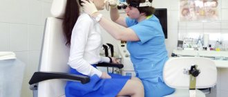
Even with the current high level of medicine, it is impossible to determine leukoplakia by eye with one hundred percent accuracy. To make an accurate diagnosis, it is necessary to perform a biopsy under local anesthesia. After a histological examination of the damaged mucosa and determination of the type of leukoplakia, the diagnosis is considered successfully completed.
Based on the results obtained, the doctor prescribes treatment, which may include removing the source of irritation, local treatment of the damaged areas with retinol acetate or other oil solutions, and taking immunostimulating and vitamin-mineral preparations. Or, in case of a serious precancerous form and the presence of cellular atypia, the doctor will recommend cryodisruption of the lesion.
Diagnosis of the disease
The diagnosis is made based on examination and questioning of the patient. Mandatory diagnostic studies confirming the diagnosis include:
- Cytology is the collection of epithelial cells to identify cancerous lesions.
- Histology is taken to perform a biopsy, which allows examination of the affected epithelial tissue to detect cancer cells.
- Blood from a vein.
Additional clarifying studies make it possible to distinguish leukoplakia from other pathologies.
Consequences
Many patients, having received a similar diagnosis from a doctor, are interested in whether leukoplakia is dangerous. Most doctors consider simple forms of this disease to be safe and do not require special treatment. Leukoplakia in the initial stage has a favorable prognosis, especially if it is caused only by external irritants, and not by a functional disorder of the internal organs.
However, there are also frequent cases of precancerous leukoplakia - the danger of this form is clear to everyone. If atypical cancer cells are detected in the histological analysis of the obtained material, this type of disease requires immediate surgery. If left untreated, the consequences of leukoplakia at the cancer stage can be disappointing. After the operation and the end of the rehabilitation period, the patient is recommended to undergo periodic examinations and take a course of medications aimed at a speedy recovery.
Radio wave therapy
A new, more effective and gentle method is radio wave treatment . The impact of radio waves on the affected areas is non-contact. It occurs through an electrode that acts on leukoplakia of the mucous membrane of the cheek, tongue or lips with high-frequency discharges that heat the foci of the disease to such high temperatures that the atypical cells simply evaporate.
This method is excellent for verrucous leukoplakia, the treatment of which cannot be delayed for long, since there is a high risk of it developing into a malignant tumor.
Unlike electrocoagulation, the radio wave method exposes tissue to much less trauma and does not leave behind scars and necrotic plaques. For this reason, it is more preferable for the treatment of leukoplakia of the lips, especially their visible part.
Simpler forms of the disease, such as Tappeiner's leukoplakia or mild leukoplakia, require less aggressive therapy aimed at eliminating local symptoms. To do this, it is quite enough to identify the cause of the lesions, cure it (if the damage to the mucosa is caused by diseases of the internal organs) or eliminate it (smoking, dentures, fillings with sharp edges), and lubricate the damaged areas of the mucosa with oil solutions of vitamin preparations for several days. To treat leukoplakia of the red border of the lips, the doctor may also recommend an oil-based chapstick with vitamins A and E.
The duration of the disease and the form of its course are individual for each patient. This disease is completely curable; the main thing is not to delay seeing a doctor, since it can very quickly turn from a mild form into a malignant one.
Prevention
The best means of preventing any disease associated with the oral cavity are careful hygiene, a healthy lifestyle and giving up bad habits. In addition, to prevent leukoplakia from developing, prevention should consist of the manufacture and installation of high-quality dentures in a trusted clinic. Regular sanitation of the oral cavity, treatment of damaged teeth and preventive examinations at the dentist will prevent the development of the disease or at least detect it in the early stages.
Prognosis and complications
The disease progresses rather slowly, i.e. represents a sluggish pathology, with periods of subsidence and increase in symptoms. But it is important that in the absence of treatment for oral leukoplakia , the disease is constantly growing and irreversible (that is, it does not go away on its own). The gradual transition (degeneration) into inhomogeneous forms is a transition to a precancerous state and even the onset of oral cancer.
The simple, most common form of leukoplakia (homogeneous form) degenerates quite rarely and practically disappears with treatment and elimination of provoking factors (for example, permanent damage to the mucous membrane by orthopedic structures). But other forms of the disease, such as oral verrucous leukoplakia, have a high risk of becoming cancerous. Therefore, even after successful treatment of oral leukoplakia, certain precautions and prevention must be taken, since this disease has a tendency to return.