Tongue cancer is a fairly common pathology in the structure of malignant neoplasms of the head and neck organs (about 55% of cases), and in the general structure of malignant neoplasms it accounts for 0.45%. The average age of those affected is 60 years, men suffer from it 3 times more often than women. But the tumor can also occur in young people and even children.
- Causes of tongue cancer
- Symptoms of tongue cancer
- Methods for diagnosing tongue cancer
- Treatment of tongue cancer
- Prevention
Most often, cancer is localized on the lateral surface, a little less often on the root of the tongue, and very rarely in the area of its back and tip. [1,2]
Anatomical structure of the tongue
General information
A malignant neoplasm in the tongue comes from the mucosal epithelium.
Despite the fact that tongue tumors are uncommon, they have an aggressive course, so early detection is important. Distinctive features of this disease are early metastasis (including distant), rapid growth, resistance to treatment, high recurrence rate and mortality. At the time of diagnosis, half of the patients had affected regional lymph nodes. Recurrence is observed in 30-50% of patients. If we consider the localization, then most often a tumor of the tongue occurs on the lateral surface, much less often there is cancer of the root of the tongue, the back and the tip. A neoplasm of the root of the tongue is classified as oropharyngeal cancer. The asymptomatic course of the disease makes early diagnosis difficult. Tumors are either not visible in the early stages or initial changes are regarded as a consequence of trauma to the tongue. Due to the lack of alertness of patients and doctors regarding malignant neoplasms, most cases are diagnosed with a full clinical picture or already in an advanced stage.
The highest incidence is observed after 50 years of age, but this disease also occurs in people under 30 years of age. In men, this pathology is diagnosed 5 times more often. In 95% of cases, tongue tumors are squamous cell carcinoma. Squamous cell carcinoma of the tongue is an aggressive tumor and insensitive to treatment ( chemotherapy ), but radiation treatment .
Methods of treating the disease
Treatments for this type of cancer include surgery, radiation therapy, and chemotherapy. Let's consider these methods separately.
Surgery
The main method of treatment is resection (removal) of the tumor. The principle of the surgical technique is to remove a malignant tumor from the patient. Surgery is always performed, except in cases where the tumor is inoperable.
Operation options:
- Gentle resection, in which the tongue or most of it is preserved.
- Radical removal (the operation is called glossectomy, this is the total removal of the tongue).
- Together with glossectomy or after it, reconstruction of the tongue is often performed - one-stage plastic surgery with a skin flap. This intervention allows you to preserve the functions of the removed organ.
Radiation therapy
Radiation is designed to destroy cancer cells or slow their growth. For small tumors this is the main treatment method. Radiation can also be given before surgery (to reduce the tumor) and after it (to avoid relapse). In later stages, the technique is used as palliative therapy, that is, it alleviates symptoms.
Chemotherapy
This is a systemic therapy that aims to destroy malignant cells throughout the body. Chemotherapy is given before surgery (to shrink the tumor) or after it (to avoid relapse).
Classification
Depending on the location, cancer is distinguished:
- Bodies. These include tumors of the tip, dorsum and lateral surface (most often the middle of the lateral surface). This localization occurs in 70% of cases.
- Root (accounts for 20% of all cases).
- Lower surface (sublingual area).
By growth pattern:
- Exophytic form (this includes papillary and ulcerative forms of tumors growing outward).
- Endophytic (growing inside the tissue of the tongue - infiltrative and ulcerative).
Papillary form a is a dense papillary growth of a mushroom shape. There may also be raised plaque-like growths that have clear boundaries.
Ulcerative (occurs in 50% of cases). Characterized by a superficial ulcer surrounded by a ridge. The ulcer is constantly increasing in size. At first, the ulcer does not bother the patient, but as it grows, pain and bleeding appear. The ulcer can become infected and inflamed, making diagnosis difficult.
Infiltrative form . The tumor grows into the thickness of the tissues, and they become denser. With diffuse growth, the compaction spreads to the entire tongue, which impairs its mobility.
The infiltrative-ulcerative form is characterized by thickening of the tongue with the presence of deep ulcers.
According to histological composition:
- Adenocarcinoma.
- Squamous cell carcinoma.
Classification according to the TNM system.
- Tis "cancer in place."
- T1 Tumor no more than 1 cm. Not accompanied by complaints, discovered by chance during examination.
- T2 ≤2 cm. There are ulcers and areas of compaction.
- T3 >4 cm. The tumor is equal in size to half the tongue. Metastases to the occipital lymph nodes, postauricular.
- T4 Locally advanced cancer that occupies the entire tongue and invades the tissues of the mouth and face. Metastases to internal organs (brain, liver, heart) and bones.
Symbol N - the presence of metastases in the lymph nodes:
- N0 - No lymph node involvement.
- N1 One node on the side of the tumor is affected.
- N2 Metastasis: one metastasis no more than 6 cm, several up to 6 cm on one side or both sides up to 6 cm.
- N3 Metastases larger than 6 cm.
With T1 tumors, lymph nodes are affected in 40% of cases, with T4 stage in 85%. A reliable factor for metastasis is the depth of invasion - 4 mm is considered a critical value. Most often, metastases are found in the submental, submandibular and cervical nodes (upper third).
Histopathological differentiation:
- G1 High degree.
- G2 Medium.
- G3 Low.
Kinds
In 70% of cases, cancer of the body of the tongue is detected, in 20% - damage to the root, and in 10% - to the lower surface of the organ. If we divide diseases according to cell characteristics, we can distinguish the following forms:
- Papillary. It looks like a dense growth with papillary outgrowths and plaque-like formations.
- Ulcerative. It is observed in approximately 50% of cases. Ulcers develop over time, can bleed, and often become infected, thereby masking the root cause of the disease.
- Infiltrative. Cancer grows inside the tongue and hardens its tissues. The form can be diffuse or spread throughout the entire organ.
If we talk about microscopic analysis, then in 95% of cases we are talking about a squamous cell form of tongue cancer, other options are much less common.
Causes
- Bad habits. Among which, smoking is of particular importance. The influence of carcinogenic substances in tobacco in the development of cancer has long been proven. The risk increases with duration and intensity of smoking. The risk increases further with alcohol consumption.
- Occupational hazards (contact with radiation, work in the oil industry).
- Constant traumatization of the mucous membrane (poor-quality dentures and damaged teeth, poor treatment of fillings, and constant biting of the tongue are important).
- Chewing mixtures (betel nut, non-smoking tobacco), consuming spices that have a carcinogenic effect.
- Oral infection.
- Pre-tumor conditions ( leukoplakia , Bowen's disease , ulcerative-erosive form of lupus erythematosus , papillomatosis , erythroplakia , leukokeratosis ).
- Action of oncogenic viruses. The connection of this pathology with chronic human papillomavirus infection and HIV . The oncogenic effect of viruses is associated with blocking or the influence of tumor suppressor genes.
- Long-term use of immunosuppressive drugs.
Treatment results for cancer of the oral cavity and oropharynx
In patients with stage 0 cancer of the oral cavity and oropharynx, long-term survival is 95-100%, but if the disease recurs (returns), more serious surgery or radiation therapy may be required. These patients need to be aware that smoking can contribute to the development of another malignant tumor.
The use of surgery or radiation therapy in patients with stage I cancer allows long-term survival to be achieved in 80-85% of cases, and in stage II patients - in 60-80%.
In patients with stages III-IV cancer of the oral cavity and oropharynx, 2 or all 3 treatment methods are used. Long-term survival rates range from 20 to 50%.
Symptoms of tongue cancer
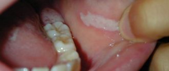
During the course of the disease, the initial stage, developed and advanced, is distinguished. It is important to know and identify the symptoms of the initial stage. Tongue cancer initially has an asymptomatic course. What does the neoplasm look like?
It may manifest itself:
- whitish spots similar to plaque;
- compaction and slight redness;
- painless nodules;
- superficial ulcers or cracks.
Most often, such changes are located on the lateral surface and are mistaken for glossitis (inflammation of the tongue). It is important that conservative treatment (treatment with antiseptics, sea buckthorn oil) does not help. It is rare, but it still happens that already at an early stage discomfort (burning or tingling) appears when eating and pain.
Photo - initial stage of cancer of the lateral surface of the tongue
Signs of tongue cancer at an advanced stage are characterized by pain, which is observed in 100% of cases. It has different intensities, can be local and diffuse, and also radiate to the temple, various areas of the mouth and ear. Patients are bothered by a feeling of numbness. When an infection occurs, bad breath appears. If the tumor begins to disintegrate, necrosis products irritate the mucous membrane and cause salivation. Bleeding of the tongue may also occur.
The advanced stage is characterized by an aggressive course and rapid invasive growth. Signs in an advanced stage are massive destruction of the tumor and surrounding tissues, including bone structures, and metastasis to local lymph nodes (occipital, submandibular, cervical and mental). With this disease, distant metastases occur in the lungs, bones, brain and liver. As the tumor disintegrates, the smell from the mouth increases, difficulty swallowing, difficulty speaking, numbness of the tongue and bleeding are observed. Common symptoms include weakness, weight loss and exhaustion.
Symptoms of tongue root cancer
This localization, which is observed in 20% of cases, is characterized by a rapid and aggressive course. There is a sore throat and discomfort when swallowing as the tumor reaches a large size. It often spreads to the auditory nerve. Patients have impaired tongue mobility.
Stages of the disease
Stages of tongue cancer
We offer you a table of correspondence between tongue tumor stages and clinical TNM classification, which uses the following designations:
- T – indicates the primary tumor: Tx – the primary tumor cannot be assessed;
- T0 – no data on the primary tumor;
- Тis – cancer in situ (pre-invasive stage);
- T1-T4 – the primary tumor is enlarged and/or widespread.
- Nx – regional lymph nodes cannot be assessed;
- M0 – no distant metastases;
Correspondence table for stages of tongue cancer TNM classification
| TNM stage | |
| 0 (carcinoma in situ) | Tis, N0, M0 |
| I | T1, N0, M0 |
| II | T2, N0, M0 |
| III | T3, N0, M0 or T1-T3, N1, M0 |
| IV | 4a: T4a, N0 M0 or T4aN1, M0 or T1-T4a, N2, M0 4b: T4b, any N, M0 or any T, N3, M0 4c: any T, any N, M1 |
Tests and diagnostics
To clarify the diagnosis, patients are prescribed:
- Biopsy of a tumor formation (or scraping and impression smears from the surface of erosions).
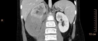
- CT and MRI of the affected area with intravenous contrast. This study makes it possible to assess the extent of the tumor and the depth of invasion into the tongue tissue.
- CT scan of the facial bones with contrast if there is a suspicion of tumor spread to the bones of the jaw and skull.
- Ultrasound of the lymph nodes of the neck.
- Puncture of the affected cervical nodes with cytological examination.
- Ultrasound of the abdominal cavity to exclude distant metastatic process.
- Chest X-ray to exclude metastases.
- Osteoscintigraphy for metastatic lesions of skeletal bones.
Diet
Diet for tongue cancer
- Efficacy: therapeutic effect for a month
- Timing: constantly
- Cost of products: 1300-1500 rubles per week
Difficulties with swallowing in the patient before and after surgery force the installation of a nasoesophageal tube. Through it, broth with meat and eggs beaten in a blender, sour cream, cream and other high-calorie liquid products are introduced. A nutritional mixture must be prescribed in addition to the main diet . This is a liquid high-calorie and high-protein mixture enriched with micronutrients. This can be Nutrizon Advance , Nutrizon Protein Advance , Nutrizon Energy , Nutrizon Protein Intense , Supportan Mixture for enteral nutrition, Fresubin original , Fresubin Energy , Nutricomp . With the transition to natural nutrition, a diet table with liquid dishes is organized - puree soup, boiled fish and meat (veal), poultry (turkey, chicken), whipped in a blender, meat and fish soufflé, liquid omelet, yoghurt, milk, cauliflower, broccoli in the form of mashed potatoes, mashed potatoes.
Prevention
- Quitting bad habits (smoking, alcohol).
- Elimination of tongue injury (high-quality treatment of fillings after their installation, correct selection and correct installation of dentures, timely treatment of dental chips).
- Regular oral hygiene, including professional.
- Timely detection of precancerous diseases of the tongue ( leukoplakia , dyskeratosis , papillomatosis ).
- Diet rich in vitamins . It has been proven that vitamin A and β-carotene are important in the prevention of neoplasms. With a deficiency of carotenoids, there is a risk of squamous cell carcinoma .
Prevention of cancer of the oral cavity and oropharynx
Most oral and oropharyngeal cancers can be prevented by avoiding known risk factors.
Tobacco and smoking are the most important risk factors in the development of cancer of the oral cavity and oropharynx. The best solution for all people is not to start smoking, not to drink alcohol, or to sharply limit their consumption.
If you smoke and drink alcohol, even for a long time, then giving up these habits will significantly reduce the risk of cancer in these locations.
Avoiding sun exposure during the middle of the day, when exposure to ultraviolet radiation is greatest, will reduce your risk of developing lip and skin cancer.
A nutritious diet including plenty of vegetables and fruits several times a day and whole grain products will help reduce the incidence of cancer of the oral cavity and oropharynx.
Forecast
With early diagnosis and radical treatment, the prognosis for recovery from tongue cancer is more favorable. It is believed that complete recovery from cancer is impossible and the goal of treatment is to achieve long-term remission of 5-8 years. Thus, with this pathology, the five-year survival rate ranges from 65 to 85%. Complete remission within 5 years after surgery and radiation therapy with T1 is achieved in 80% of patients, with T2 in 60% of patients, and with T3-4 does not exceed 35%. Metastases in the lymph nodes are an important prognostic factor and, if present, survival rate is halved.
How long do people live with stage 4 tongue cancer or with relapse after chemotherapy? Such patients have a poor prognosis: they live no more than 3.5 months while on maintenance treatment. The addition of cetuximab to chemotherapy increases survival by 2 times.
Treatment of cancer of the oral cavity and oropharynx
When treating patients with cancer of the oral cavity and oropharynx, surgical, radiation and medicinal methods are used. In this case, depending on the stage of the tumor, one or more methods of therapy are used.
For the surgical treatment of tumors of the oral cavity and oropharynx, various operations can be used, taking into account the location of the tumor and the stage of the process, as well as the need to perform reconstructive (restorative) interventions in order to restore lost functions.
In patients with a mobile tumor in the oral cavity, the tumor is removed without excision of bone tissue. If the mobility of the tumor is limited and there are no changes in the bone (on x-rays), the tumor is removed along with part of the jaw. Clear damage to the jaw, visible on radiographs, requires wider excision of bone tissue.
If the lip is affected, in some cases a special surgical micrographic method can be used, in which the tumor is removed in layers and examined under a microscope. This allows you to completely remove the tumor and preserve as much normal lip tissue as possible.
Malignant tumors of the oral cavity and oropharynx often spread to the lymph nodes of the neck. In these cases, surgery to remove them and suspicious lymph nodes is indicated. The extent of the operation depends on the extent of the tumor and can be significant, including the removal of muscles, nerves and blood vessels.
Side effects and complications during surgical removal of lymph nodes are associated with nerve damage: numbness of the ear, difficulty raising the arm above the head, drooping of the lower lip. These phenomena may gradually subside, but may remain permanently if the nerve has been completely removed.
In some cases, for large tumors of the oropharynx that lead to difficulty breathing, a tracheotomy (cutting the trachea) is performed and a tube is inserted into the trachea to restore breathing. After the tumor is removed, the tube is removed and normal breathing is restored.
Radiation therapy may be the mainstay of treatment for patients with small tumors of the oral cavity and oropharynx. In patients with large tumors, radiation is used along with surgery to destroy remaining tumor cells. Radiation therapy is also used to relieve pain, stop bleeding, and eliminate difficulty swallowing.
For tumors of the oral cavity and oropharynx, external irradiation is most often used. Treatment is carried out 5 times a week for 5-7 weeks.
In some patients, brachytherapy (internal radiation) may be used. In this case, metal rods containing radioactive material are inserted into or near the tumor for a certain period of time. These rods are removed before discharge home.
In some cases, both external and internal irradiation are used.
Side effects of radiation therapy: redness of the skin, dry mouth, sore throat, hoarseness, partial loss of taste, weakness. Treatment complications may include: damage to the thyroid gland and blood vessels supplying the brain.
Chemotherapy refers to the use of anticancer drugs. It can be used before surgery or radiation therapy to shrink the tumor. In some cases, chemotherapy is used in combination with radiation or surgery.
The most commonly used chemotherapy for oral and oropharyngeal cancer is cisplatin and 5-fluorouracil. In addition, other drugs can be used: methotrexate, bleomycin, carboplatin. The drugs are used both individually and in combination to enhance the antitumor effect.
Side effects of chemotherapy may include nausea, vomiting, loss of appetite, baldness, mouth ulcers, fatigue, increased susceptibility to infection, and bleeding. Most side effects go away over time, but some, such as hearing loss with cisplatin, may be persistent.
List of sources
- Alieva. S.B., Alymov Yu.V., Kropotov M.A., Mudunov A.M., Podvyaznikov S.O. Cancer of the oral mucosa. Oncology. Clinical recommendations / Ed. M. I. Davydova. – M.: Publishing group RONC, 2015, pp. 27-37.
- Romanov, I.S. Features of regional metastasis of squamous cell carcinoma of the oral cavity, detected during preventive lymph node dissections. / Romanov, I.S., Yakovleva L.P., Udintsov D.B., Dzhumaev M.G., Tsiklauri V.T. // Dentistry. – 2012. – T.91, No. 4. — P. 28-31.
- Romanov I. S., Yakovleva L. P. Issues in the treatment of oral cancer. Farmateka 2013; (8):59–63.
- Paches A.I. Tumors of the head and neck. 5th ed., supplemented and revised M.: Practical Medicine, 2013. P. 119‒146.
- Romanov I. S. Prospects for the use of cetuximab in the treatment of squamous cell carcinoma of the head and neck / Oncology. Hematology. Chemotherapy. - 2015. - No. 17/1.
Leukoplakia, erythroplakia and dysplasia
Leukoplakia and erythroplakia are terms denoting various changes in the mucous membrane of the oral cavity and pharynx due to smoking, chewing tobacco, trauma to the oral mucosa with a denture
The appearance of a changed white mucous membrane may indicate the presence of leukoplakia in the patient. With erythroplakia, the altered mucous membrane is red in color, may protrude somewhat above the surface and bleed easily.
The severity of the resulting changes in the mucous membrane of the oral cavity and pharynx can only be clarified with the help of a biopsy (taking a piece of tissue for microscopic examination) or scraping of individual cells.
These changes may be harmless and disappear after the cessation of exposure to the causative factor, but may precede the onset of cancer.
This precancerous condition is called dysplasia.
There are minor, moderate and severe degrees of dysplasia. Knowing the degree of dysplasia, it is possible to predict (predict) the likelihood of self-healing, disappearing after treatment, or turning into a malignant tumor.
Without proper treatment, 5% of leukoplakia develop into cancer over a 10-year period.
Erythroplakia is a more serious condition in which almost 50% of cases are diagnosed as cancer after biopsy.
More than 90% of tumors of the oral cavity and oropharynx are squamous cell carcinoma, developing from integumentary (epithelial) cells.
Verrucous (warty) carcinoma is a type of squamous cell carcinoma and accounts for 5% of all oral tumors. This type of cancer is a low-grade tumor that rarely metastasizes, but can spread deeply into surrounding tissues. In this regard, wide removal of the tumor within healthy tissue is recommended.
The minor salivary glands, located in the oral and pharyngeal mucosa, can give rise to various types of cancer, such as adenoid cystic carcinoma, mucoepidermoid carcinoma and low-grade polymorphic adenocarcinoma.
The tonsils and base of the tongue contain lymphoid tissue from which lymphomas (non-Hodgkin's lymphoma/lymphosarcoma and Hodgkin's disease/lymphogranulomatosis) can develop.