Everyone knows what a runny nose is or, as doctors call it, rhinitis. A runny nose almost always accompanies colds, flu and other respiratory diseases. Most often, rhinitis has an acute form, that is, it develops quickly and quickly subsides.
If acute rhinitis is not treated in time, it may develop into a chronic form. Rhinitis begins to torment a person all year round. Sometimes it seems that it has completely passed, but at the slightest weakening of the immune system it makes itself felt again. Allergy sufferers are also very familiar with the chronic manifestation of rhinitis, which instantly wakes up when the body is exposed to an intolerable allergen.
Like most diseases, chronic rhinitis has its own forms, from familiar and simple to complex and dangerous. Among this diversity, a rather rare form of chronic rhinitis stands out - atrophic rhinitis.
Atrophic rhinitis is a slowly progressive disease that affects the nasal mucosa. Hard crusts with an unpleasant odor form, the nasal passages increase in size, and the person is constantly haunted by a feeling of stuffiness. The disease affects adults, mainly women, and children, most often teenagers.
Recently, atrophic rhinitis has become commonly divided into primary and secondary. The primary develops independently, and the secondary manifests itself against the background of intervention - environmental, physical, surgical and infectious1,2.
Chronic catarrhal rhinitis
This form of chronic rhinitis most often develops as a result of repeated acute runny noses, which is facilitated by constant exposure to unfavorable environmental factors - a polluted, dusty atmosphere, frequent temperature changes, dampness and drafts. The development of chronic runny nose is predisposed by long-term congestive hyperemia of the nasal mucosa caused by alcoholism, chronic disease of the cardiovascular system, kidneys, etc. In the etiology of the disease, hereditary preconditions, developmental defects, and disturbances in normal anatomical relationships that cause difficulty in nasal breathing may play a role. Chronic runny nose also develops as a secondary disease in pathology of the nasopharynx and paranasal sinuses.
Morphological changes
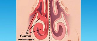
Morphological changes in chronic catarrhal rhinitis are less pronounced compared to other forms of rhinitis and are localized in the superficial layers of the mucous membrane. The vessels of the mucous membrane of the nasal concha are dilated, their walls may be thinned. With a long course of chronic rhinitis, sclerosis develops in the submucosal layer.
Clinic and symptoms of chronic catarrhal rhinitis
The symptoms of chronic catarrhal rhinitis generally correspond to the symptoms of acute rhinitis, but are much less intense. The patient complains of nasal discharge of a mucous or mucopurulent nature. Difficulty in nasal breathing is not permanent. It gets worse (like nasal discharge) in the cold. Alternating congestion of one half of the nose is often noted. It usually appears when lying on your back or side. In these cases, there is a rush of blood to the underlying parts of the nose. The vessels of the cavernous tissue of the turbinates, which are in a relaxed state due to loss of tone, become overfilled with blood, which causes nasal congestion. When you change your body position, the congestion moves to the other side.
Chronic catarrhal rhinitis may be accompanied by a violation of the sense of smell in the form of its weakening (hyposmia). Complete loss of smell (anosmia) is rare. The transition of catarrhal inflammation from the nasal cavity to the mucous membrane of the auditory tube with the subsequent development of tubo-otitis is possible.
Treatment of chronic catarrhal rhinitis
The success of treatment depends on the ability to eliminate unfavorable factors that cause the development of chronic rhinitis. Staying in a dry, warm climate, hydrotherapy and spa therapy are beneficial. It is necessary to treat common diseases accompanying chronic rhinitis, as well as eliminate intranasal pathology (deformation, sinusitis, adenoid vegetations).
Local treatment consists of the use of antibacterial and astringent drugs in the form of 3 - 5% solution of protargol (collargol), 0.25-0.5% solution of zinc sulfate, 2% salicylic ointment, etc. Prescribed to the nasal area UHF, endonasally UFO (tube -quartz). The prognosis is usually favorable.
Prevention
Prevention of the development of rhinitis, its transition to a chronic form or the development of exacerbations of the disease includes the following points:
- prevention of acute viral infections, flu vaccination;
- refusal of long-term use of decongestants;
- use of respiratory protection equipment in hazardous industries;
- eliminating contact with allergens;
- treatment of sinusitis, tonsillitis, pharyngitis;
- therapy for obesity, heart, kidney and endocrine system diseases;
- creation of favorable microclimatic conditions in everyday life (humidification and air purification);
- treatment at sea resorts or in sanatoriums of the Caucasian mineral waters;
- hardening, breathing exercises.
For timely diagnosis of chronic rhinitis, we suggest contacting the specialists of the NIKIO center. Experienced doctors will quickly make a diagnosis and select the most appropriate treatment tactics for each patient. This will help the patient restore nasal breathing and get rid of the unpleasant symptoms of the disease.
Chronic hypertrophic rhinitis
The causes of hypertrophic rhinitis are the same as catarrhal ones. The development of one form or another of chronic rhinitis is apparently associated not only with the influence of external unfavorable factors, but also with the individual reactivity of a person.
Pathomorphological changes
Pathomorphological changes in hypertrophic rhinitis differ from those in catarrh by the predominance of proliferative processes. The development of fibrous tissue is observed mainly in places where cavernous formations accumulate. Hypertrophy of the mucous membrane of the nasal concha often reaches significant sizes. There are three types of shell hypertrophy: smooth, tuberous and polypous. It can be diffuse and limited. The most typical sites of hypertrophy are the anterior and posterior ends of the inferior and anterior ends of the middle turbinate. Hypertrophy can also occur in other areas of the nose - in the anterior part of the nasal septum and at its posterior edge, on the vomer.
Swelling of the mucous membrane in the area of the nasal turbinates, especially the middle one, is possible, resembling nasal polyps. This swelling and polyp-like thickening, unlike polyps, has a wide base. In the future, polypous hypertrophy can gradually transform into polyps. This is facilitated by allergization of the body (auto- and exogenous). Occurring bony hypertrophy of individual turbinates is a variation of the anomaly of nasal development.
Clinic and symptoms of chronic hypertrophic rhinitis
Hypertrophic rhinitis is characterized by constant nasal congestion, which depends on excessive enlargement of the nasal turbinates, which practically do not contract under the influence of vasoconstrictors. Nasal breathing is complicated by copious mucous and mucopurulent discharge. Due to obstruction (narrowing) of the olfactory fissure, hyposmia occurs and then anosmia. In the future, as a result of atrophy of the olfactory marks, essential (irreversible) anosmia may occur. The timbre of the voice becomes nasal (rhynolaliaclausa).
As a result of compression of the lymphatic gaps by fibrous tissue, lymphatic drainage from the cranial cavity is disrupted, which causes a feeling of heaviness in the head, impaired ability to work, and sleep disturbances.
Shutting off nasal breathing leads to impaired ventilation of the paranasal sinuses, as well as disease of the underlying respiratory tract. Hypertrophy of the posterior ends of the inferior turbinates disrupts the function of the auditory tube and leads to tubo-otitis. Thickening of the anterior sections of the inferior turbinates can compress the nasolacrimal duct with subsequent development of dacryocystitis and conjunctivitis.
Treatment of chronic hypertrophic rhinitis
Endoscopic examination allows us to determine the nature of hypertrophy. Further treatment of hypertrophic rhinitis is predominantly surgical. Treatment methods for diffuse hypertrophy pursue the development in the postoperative period of a sclerosing scar process in the submucosal layer, which reduces the size of the nasal turbinates. For this purpose, various methods of intrashell cauterization of tissue are used (electricity - electrocautery, ultra-low temperatures - cryodestruction), as well as such effects as ultrasonic or mechanical disintegration. A laser beam is also used for this purpose.
Diagnostics
As part of the diagnosis of atrophic rhinitis, a histological examination is carried out, which shows metaplasia and thinning of the columnar epithelium, a decrease in the number of vessels and other characteristic changes in the tissues.
Anterior rhinoscopy is also performed. Due to pronounced atrophy of the turbinates, the posterior wall of the nasopharynx becomes visible.
A separated secretion is detected, which has a thick consistency and is colored yellowish-green. Sticky masses linger on the walls of the mucous membrane, forming crusts when dry.
Atrophic rhinitis
Simple atrophic rhinitis
This form of chronic rhinitis is based not on an inflammatory process, but on a dystrophic process, affecting mainly the mucous membrane. It may be a particular manifestation of a systemic disease, in which trophic disorders (atrophy) spread to the pharynx, larynx and other organs and systems. This is the so-called primary (genuine) atrophic rhinitis, the extreme degree of which is a fetid runny nose (ozena).
Secondary atrophic rhinitis
Secondary atrophic rhinitis is a consequence of exposure to various unfavorable environmental factors: hot, dry climate, dust and gas pollution in the atmosphere of industrial production (flour milling, woodworking, cement, silicate, chemical, etc.). Various injuries also play a role in the development of atrophic rhinitis - domestic, gunshot and surgical, causing damage to the tissues and blood supply of the nasal cavity.
Morphological changes
Morphological changes in simple atrophic rhinitis are most pronounced in the epithelial layer of the mucous membrane. The number of mucous goblet cells decreases, the ciliated epithelium loses cilia and degrades. The viscosity and pH of nasal secretions change, which leads to disruption of the functional activity of the ciliated epithelium.
Clinic and symptoms of atrophic rhinitis
Patients suffering from atrophic rhinitis are characterized by complaints of a feeling of dryness and itching in the nose, constant drying of yellowish-greenish crusts, the removal of which is accompanied by injury to the mucous membrane. In cases where the process affects the olfactory zone, hypo- and anosmia develops. In some cases, patients note an unpleasant odor that is not detected by others (kakosmiasubjectiva).
Treatment of atrophic rhinitis
When treating atrophic rhinitis, it is necessary, as with other forms of rhinitis, to eliminate or reduce the impact of harmful environmental factors. A course of treatment with ointments and iodine-glycerin is prescribed locally. In the morning and evening, the patient should inject Vojacek's diahilum ointment into the nose on a cotton swab for 10 minutes. Twice a week, the doctor or the patient himself lubricates the nasal mucosa with a solution of iodine-glycerin. This treatment is carried out for 2 months and is repeated 3 times a year. It is popular to insert cotton swabs into the nose with rosehip or sea buckthorn oil in olive or peach oil in a ratio of 1:3 - 1:4 or with the addition of an oil solution of vit. A (no more than 50,000 IU). Oral intake of Vit is also appropriate. And, and a complex of multivitamins with microelements (for example, Vitrum).
In the treatment of atrophic rhinitis, alkaline and oil inhalations through the nose are widely used. During spa treatment, irrigation of the nasal cavity and inhalation of mineral waters from local sources are useful. The most effective are inhalations and irrigation of the mucous membranes of the upper respiratory tract with 2 - 3% solutions of sea salt, as well as sea bathing. This is due to the fact that magnesium ions contained in sea water have a beneficial effect on the mucous membrane.
4.Treatment
Treatment usually begins with moisturizing rinses, tampons, ointments, inhalations and other physiotherapeutic procedures, including high-tech ones (for example, laser therapy has been widely practiced in recent years as a means of stimulating trophism). After determining the drug sensitivity of identified bacterial and/or fungal pathogens, appropriate antibiotics and antimycotics are prescribed in effective doses, often topically, less often in combination with systemic oral administration. According to indications, autohemotherapy, vitamin complexes, etc. can be used.
In case of advanced atrophy, significant destruction and deformation, they resort to plastic-surgical reconstruction using autografts or various artificial materials. The goal is to restore normal anatomy, ventilation and hydration of the nasal passages, but it should be understood that such intervention in all cases is forced and purely palliative.
Ozena
Ozena is a chronic atrophic fetid runny nose, which differs from a simple atrophic runny nose by deep atrophy of the entire mucous membrane, bone walls of the nasal cavity and nasal turbinates. Ozena is characterized by the secretion of a thick secretion that dries into foul-smelling crusts.
Despite the fact that the disease has been known since ancient times, the etiology and pathogenesis of ozena continue to remain insufficiently understood to this day. It is now recognized that in the pathogenesis of ozena, a significant role is played by hereditary-constitutional characteristics, transmitted by inheritance as a recessive trait, as well as environmental conditions, incl. nutritional and vegetative insufficiency. As a result, a patient with ozena develops various neuroendocrine, neurovegetative and neurotrophic disorders. The central links that form these disorders probably involve the hypothalamic-pituitary department of the central nervous system.
Clinic and symptoms of ozena
The disease usually begins at a young age, women are more often affected. After the onset of menopause, many clinical manifestations of ozena decrease, which is the basis for considering this disease as being associated with dysfunction of the endocrine glands.
Patients with ozena are characterized by severe atrophy of the nasal mucosa and a decrease in the size of the nasal turbinates, especially the lower ones. Sometimes the lower shells atrophy faster than the middle ones, and therefore the latter appear unusually large against the background of the small atrophied lower ones. As a result of atrophy of the mucous membrane and nasal turbinates, the nasal cavity becomes wide. Often during ozena, the entire nasal cavity is filled with crusts. They usually have a yellowish-greenish, brownish or dirty color and are easily removed in the form of casts. At the same time, the outer surface of the crusts is dry, and the lower surface is viscous, thick, and an extremely unpleasant fetid odor emanates from it, which is a mixture of indole, skatole, phenol, hydrogen sulfide and volatile fatty acids. Actually, of all the objective signs of ozena, stench (kakosmiaobjectiva) should be placed in first place, as the constant and most significant objective symptom. However, patients suffering from anosmia do not feel this smell. It is the stench emanating from ozena patients that makes it impossible for them to communicate closely with other people, determines the tragedy of their situation and puts ozena patients in the position of outcasts of society. All this allows us to consider ozena as a disease of social significance.
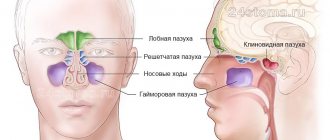
Atrophic phenomena during ozena affect not only the nasal cavity, but also the paranasal sinuses and underlying respiratory tract. The crusts filling the nasal cavity spread posteriorly into the nasopharynx and pharynx, closing the mouths of the auditory tubes. Unfavorable conditions are created for ventilation of the tympanic cavity, which contributes to its involvement in the inflammatory process. This is the reason for the often observed damage to the hearing aid in patients with ozena.
Treatment of ozena
Treatment of ozena, as well as other degenerative processes of the nose, is one of the most difficult problems of rhinology. In recent years, however, pathogenetically based conservative and surgical treatment methods have appeared, combined with the use of etiotropic drugs that directly affect Klebsiella ozena. However, symptomatic treatment methods aimed at removing fetid crusts and deodorizing the nasal cavity have not lost their importance and continue to remain in the arsenal of modern rhinologists.
Based on the infectious hypothesis of the etiology of ozena, antibiotics active against Klebsiella ozena are used in treatment. However, these treatment methods do not allow obtaining lasting clinical results.
In the treatment of ozena, surgical methods are mainly used. Thus, based on the idea that dystrophic (atrophic) processes in the nasal cavity develop as a result of a violation of the trophic function of the autonomic nervous system, a synthetic implant (polyurethane, ivalon, etc.) is implanted in the area where the diffuse autonomic ganglion of the nasal septum is located. Inserting a graft into the area of the autonomic ganglion aims to have a positive effect on the trophism of the nasal mucosa. The described surgical method allows for clinical recovery (i.e., cessation of drying of foul-smelling crusts and revitalization of the mucous membrane) in 75% of cases.
Also, at the discretion of the doctor, a conservative method can be used, based on the use of cholinomimetics, anticholinesterase drugs, vitamins E and D, long-acting nitrites, stimulants of tissue regeneration, in particular, metacil. This treatment is carried out for 1 month in a hospital setting.
2. Reasons
The causes and mechanisms of atrophic rhinitis today are not sufficiently clarified, so the results of new studies, hypotheses and statistical data on risk factors are constantly appearing. Thus, hypotheses about hereditary predisposition, the influence of endocrine imbalances and disorders, the role of psychogenic, autoimmune, hemodynamic, nutritional, environmental, and occupational factors are considered and discussed. Many authors see the influence of hypovitaminosis and iron deficiency. In some cases, there is a connection with previous medical interventions, injuries, infections, radiation and chemotherapy. A very serious factor in mucosal atrophy, the significance of which has been confirmed repeatedly, is the abuse of local vasoconstrictors (vasoconstrictor drops), which often turns into severe drug dependence.
In relation to ozena, the role of a specific pathogen has been proven - Klebsiella ozena, but fungal, Proteus, diphtheroid atrophic rhinitis also occurs; the possible role of viral infections as a trigger factor is assumed.
In general, the etiopathogenesis of chronic atrophic rhinitis needs further study.
Visit our Otolaryngology (ENT) page
Vasomotor rhinitis (“false rhinitis”)
A characteristic feature of vasomotor rhinitis is the manifestation of a runny nose symptom complex without pathological signs of inflammation of the mucous membrane. This is either a neurovegetative runny nose, which is a manifestation of autonomic neurosis, or an allergic one, which occurs in response to the action of allergens.
The basis of the neurovegetative form of vasomotor rhinitis is the inadequate “play” of the vessels that make up the main content of the cavernous bodies of the turbinates. These are cycles characterized by regular narrowing and expansion of the vessels of the nasal concha. Subjectively, no nasal breathing disturbances are felt. This alternating cyclical fluctuation of nasal congestion can be considered as a normal manifestation of physiological functional asymmetry. On the other hand, a more pronounced enlargement of the nasal turbinates, in which a clearly felt difficulty in nasal breathing appears, is a pathological condition, namely vasomotor rhinitis, in particular its neurovegetative form.
In healthy individuals, a change in the predominance of breathing through one or another half of the nose occurs according to a sinusoidal law with a period of 20 to 90 minutes, and the ratio of the volumes of exhaled air varies from 25% to 75%, i.e. complete congestion of one half of the nose is not observed. In patients with the neurocirculatory form of vasomotor rhinitis, there is a violation of the period and linearity of the law of change in the predominance of nasal breathing. Their exhaled air volume ratio ranges from 0% to 100%.
In the occurrence of the neurovegetative form of vasomotor rhinitis, the main role is played by functional changes in the central and autonomic nervous systems, as well as the endocrine system. The hypothalamus, the main integrating autonomic center in the regulation of nasal functions, plays an important role. The influence of dysfunction of the endocrine glands, primarily the thyroid gland, on the development of vasomotor rhinitis is also important. Vasomotor rhinitis is an endocrine-vegetative syndrome.
The development of vasomotor rhinitis is also facilitated by reflex effects, in particular: cooling, to which non-hardened people are especially susceptible; sedentary lifestyle; medications used for hypertension and coronary heart disease to dilate blood vessels, etc. Vasomotor rhinitis often occurs in people who have spines and ridges on the nasal septum.
Allergic form
The allergic form of vasomotor rhinitis occurs when exposed to various allergens, and depending on their name, seasonal and permanent (year-round) forms of allergic rhinitis are distinguished. Allergic rhinitis is one of the most common diseases. According to various studies, they occur in 10 - 40% of the population.
The cause of the seasonal form of allergic rhinitis (hay fever) can be pollen from various plants during their flowering period. Residents of many cities are especially concerned about the fluff and pollen of poplars, while rural residents are concerned about the cereals blooming in the fields (timothy, fescue, etc.). In recent years, ragweed, an overseas cereal that has penetrated into the southern regions of Russia from North America and has increased allergenic properties, has become relevant.
In the permanent (year-round) form of allergic rhinitis, the allergens are more diverse and can affect patients over a long period. These include: environmental allergens - book dust, relevant for library workers; house dust, bird feathers (feather pillows), hair, pet dander, daphnia (dry food for fish living in an aquarium); food products - citrus fruits, strawberries, honey, milk, fish, crayfish; medicines, perfumes, etc.
The pathogenesis of allergic rhinitis involves a specific reaction between the allergen and tissue antibodies, resulting in the release of chemically active substances (allergic reaction mediators) that contribute to the development of clinical manifestations of the disease. The possible mechanism of development of allergic rhinitis is described as follows.
An Ig E-dependent reaction occurs - activation of mast cells located in the nasal mucosa. The released mediators are contained in granules (for example, histamine and tryptase) or in the membrane of mast cells (leukotrienes and prostaglandins). Another mediator is platelet activating factor (PAF). Mediators have a vasodilating effect and increase vascular permeability, which leads to nasal congestion. Increased secretion is accompanied by the appearance of mucous discharge. Stimulation of afferent nerve fibers causes itching and sneezing. In addition, afferent stimulation (especially under the influence of histamine) can enhance the axonal reflex with local release of neuropeptides (substance P, tachykinins), which, in turn, cause further degranulation of mast cells, further enhancing the pathological response.
Neurovegetative form
In the neurovegetative form of vasomotor rhinitis, no specific changes in the mucous membrane are observed. The ciliated epithelium is thickened, the number of goblet cells is increased. The cavernous vessels are dilated. Microcirculation is disrupted. These disturbances are especially noticeable in the inferior turbinates. The movement of blood in the capillaries is uneven and often does not correspond to pulse impulses. In the allergic form of vasomotor rhinitis, stasis occurs. The epithelial cover of the mucous membrane thickens, in places metaplasizing into a multilayered flat layer. The number of goblet cells increases significantly. In the subepithelial layer, tissue infiltration with eosinophilic leukocytes takes place. They are also found in significant quantities in nasal mucus.
Clinic of the neurovegetative form of vasomotor rhinitis
As with all forms of vasomotor rhinitis, the neurovegetative form is characterized by the following symptoms: difficulty in nasal breathing, profuse serous or mucous discharge, attacks of paroxysmal sneezing, itching and burning sensation in the nasal cavity. These symptoms are often intermittent. They can occur after waking up from sleep (a change in the predominance of the parasympathetic over the sympathetic autonomic nervous system), with a change in ambient temperature, with overwork, emotions, stress, etc. Difficulty in nasal breathing is associated with the expansion of the cavernous spaces of the nasal mucosa, mainly the lower nasal shells The mucous membrane has a bluish color due to blood overflow (venous hyperemia). "Bluish or pale" spots are often visible. Lubricating the mucous membrane with vasoconstrictor drugs leads to rapid contraction of the conchae.
Clinic for seasonal allergic rhinitis (hay fever)
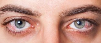
This form of vasomotor rhinitis is characterized by a clear seasonality of exacerbation, which occurs during the flowering period of plants to the pollen of which patients have increased sensitivity (sensitization). During this period, paroxysms of sneezing, itching and burning in the nasal cavity, eyes, and conjunctival hyperemia are noted. Almost complete nasal congestion and severe rhinorrhea occur, which leads to maceration of the skin in the vestibule of the nose. The mucous membrane in the initial period is sharply hyperemic, and there is a significant amount of clear fluid in the nose. Subsequently, the mucous membrane acquires a cyanotic appearance and then turns pale. Along with the noted rhinological symptoms, patients during this period often experience itching in the eyes, conjunctival hyperemia, a feeling of rawness in the pharynx, larynx and skin itching. In some cases, it is possible to develop Quincke's edema (enlargement of the face or part or limb) and larynx. Various discomfort manifestations are observed, incl. headache, increased fatigue, sleep disturbance, increased body temperature. The duration of the disease usually corresponds to the period of flowering of plants and ceases on its own after its end or after the patient changes the allergenic area.
An intermediate position between the neurovegetative form of vasomotor rhinitis and seasonal allergic rhinitis is occupied by the so-called. "neurosis of a reflected nature." Thus, in people suffering from hay fever, which occurs when smelling flowers, in particular roses, attacks of a runny nose are observed not only when inhaling the aroma of flowers, but also when just looking at an artificial rose, which the patient mistakes for a real one. In these cases, there is a combination of conditioned reflexes coming from the optic and olfactory nerves.
Clinic for permanent (year-round) form of allergic rhinitis
The disease is chronic from the very beginning. Severe swelling of the nasal turbinates is detected, associated with profuse sweating of transudate from the capillaries. The mucous membrane is pale; swelling affects not only the turbinates, but also the mucous membrane of the bottom of the nasal cavity and the nasal septum. The middle turbinates are also swollen, resembling polyps. Due to swelling of the mucous membrane, mucus is difficult to clear and can be discharged from the nose at random.
The allergic form of vasomotor rhinitis is characterized by the formation of mucous polyps. Gradually increasing in size and quantity, they can fill the entire nasal cavity, in some cases pushing apart the bone walls and deforming the external nose. Often there is a combination of allergic rhinitis with bronchopulmonary pathology - asthmatic bronchitis and bronchial asthma. An “asthmatic triad” may occur, including intolerance to acetylsalicylic acid, nasal polyposis and attacks of bronchial asthma.
Treatment of the neurovegetative form of vasomotor rhinitis
It consists of eliminating various causes, often of a reflex nature, that cause this disease. It is necessary to treat general diseases (neuroses, diseases of internal organs, endocrine dysfunction). If there are any local abnormalities in the nasal cavity (Conchabullosa, spines and ridges of the nasal septum), they should be eliminated. An active, mobile lifestyle and hardening procedures are recommended, in particular, effects on reflex zones - short-term dousing of cold water on the soles of the feet. Performing various gymnastic exercises.
Systematic use of vasoconstrictors is not recommended, because this gives only a short-term effect, and with prolonged use it damages the mucous membrane and is addictive. Acupuncture is also used, influencing the reflex zones of the nasal cavity (novocaine blockades in the inferior nasal turbinates, in the aggernasi, on the sphenopalatine node). Surgical interventions on the inferior turbinates (various options for intraturbinate disintegration) are more effective.
Treatment of allergic rhinitis
There are three main areas in the treatment of allergic rhinitis: elimination therapy, immunotherapy and drug therapy. The goal of elimination therapy is to eliminate allergens (pollen, dust, etc.) and control the state of the environment. Specific immunotherapy is very effective, but severe reactions are possible, especially in people suffering from bronchial asthma. It is not widespread in our country. In other countries (Great Britain, Scandinavian countries), the use of specific immunotherapy is sharply limited. To minimize the risk of immunotherapy, the question of its appropriateness should be decided by an allergist or clinical immunologist. Currently, drug therapy for allergic rhinitis has acquired the greatest importance.
Our medical center successfully treats various types of chronic rhinitis!
Cost of ENT services in our Medical Center
Rating 4.09 (11 Votes)
Symptoms characteristic of this disease
The symptomatic picture that occurs with this disease is as follows:
- Feeling of tightness in the nose;
- Decreased or complete loss of sense of smell;
- Viscous nasal compartments yellow-green in color;
- Dry mucous membranes;
- The appearance of crusts in the nose;
- Nosebleeds;
- Unpleasant smell from the nose;
- Acute headaches;
- Dizziness.
If such symptoms occur, you should contact an otolaryngologist for examination, diagnosis and treatment.