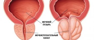
Diagnostics
The EXPERT Clinic has developed a clear algorithm for the primary diagnosis of glomerulonephritis. The necessary initial examination includes:
Laboratory methods
- general urine analysis
- clinical and biochemical blood tests are performed
- glomerular filtration rate is determined as a factor in assessing renal function
- immunological blood test.
Doctors at the EXPERT Clinic have compiled a list of necessary tests for a kidney screening examination.
Instrumental studies
Ultrasound of the kidneys with Doppler examination of the vessels of the kidneys, which evaluates ultrasound signs of changes in the blood flow of the kidneys.
The final diagnosis is made ONLY with a kidney biopsy in a hospital setting.
Important! The scope of the necessary examination can only be determined by a nephrologist.
Recommendations
Bacterial, viral infection, and the presence of foci of chronic infection can be considered as risk factors for the development of glomerulonephritis in patients with a genetic predisposition.
It is necessary to regularly monitor patients with systemic connective tissue diseases (systemic lupus erythematosus, scleroderma, rheumatoid arthritis, vasculitis, etc.), and women with kidney damage during pregnancy.
Basic preventive measures should be aimed at slowing the progression of glomerulonephritis. The diet should be monitored at every doctor's visit. The amount of salt, protein, and liquid varies depending on the clinical manifestations of the disease. Strong marinades, broths, and canned fish are prohibited.
Patients with glomerulonephritis are contraindicated in heavy physical activity, work at low temperatures, with increased dampness, and in the open air.
Patients who have suffered acute glomerulonephritis are observed for 2 years. For chronic glomerulonephritis, regular monitoring is recommended once a quarter.
Types and symptoms of the disease
Considering the clinical manifestations, glomerulonephritis occurs:
- Latent. This type is found in most cases. External signs are mild - the appearance of moderate swelling, and a slight increase in blood pressure. It can be determined by the results of laboratory tests, that is, by a general urine test it can be determined that protein, red blood cells, and white blood cells are increased.
- Hematuric. This form can be found in rare cases, no more than 4% of all cases. If we talk about external signs, then redness of the urine is observed. When taking a urine test, it can be revealed that the changed red blood cells have increased.
- Hypertensive. This type of glomerulonephritis occurs in approximately 15% of cases. The following external signs of the disease appear: blood pressure constantly rises, much more urine is released per day, and the urge to empty the bladder at night becomes more frequent. When submitting urine for a general analysis, protein and altered red blood cells can be detected. In addition, the density of urine decreases.
- Nephrotic. This type occurs in 30% of all cases. The following external signs appear: blood pressure rises, swelling appears. Also, much less urine begins to be released per day. Based on the results of laboratory tests, it can be revealed that there has been an increase in urine density and the amount of protein. A biochemical blood test can determine that total protein has decreased and cholesterol has increased.
- Mixed (nephrotic-hypertensive). With this form of glomerulonephritis, similar symptoms appear, which are inherent in the nephrotic and hypertensive types of the disease.
Complications of chronic glomerulonephritis
The most common complications are:
- left ventricular failure due to increased blood pressure
- renal failure
- intercurrent infections
- stroke
- nephrotic crisis
- thrombosis
Nephrotic crisis manifests itself as follows:
- stomach ache
- fever
- development of hypovolemic shock
- migrating erysipelas-like erythema
Complications associated with active immunosuppressive therapy may develop :
- infections
- cytopenia (including agranulocytosis)
- hyperglycemic conditions
- hemorrhagic cystitis
- osteoporosis
Acute glomerulonephritis
The classic picture includes a triad of symptom complexes: renal (renal) - urinary syndrome and extrarenal (extrarenal) - edematous and hypertensive syndromes. The pathology usually manifests itself 1-2 weeks after the etiological effect (infection, allergic reaction, etc.).
The appearance of edema is the earliest and most common sign of acute glomerulonephritis, occurring in 70-90% of patients, in half of them the edema is significant. Swelling is located mainly in the face: it is most pronounced in the morning and subsides during the day, giving way to swelling of the ankles and legs. In the future, the edematous syndrome can progress to anasarca, hydropericardium, hydrothorax, and ascites. In some cases, there may be no visible swelling, but the patient's daily weight gain indicates fluid retention in the tissues.
Arterial hypertension is usually moderate: in 60-70% of patients, blood pressure does not exceed 160/100 mm Hg. Art. However, persistent long-term hypertension has a poor prognosis. Acute glomerulonephritis is characterized by a combination of arterial hypertension with bradycardia less than 60 beats. per minute, which can last for 1-2 weeks. With acutely developing hypovolemia, phenomena of left ventricular failure, expressed by cardiac asthma and pulmonary edema, are possible.
Often there is the development of cerebral disorders caused by cerebral edema - headache, nausea and vomiting, decreased vision, “veils” before the eyes, decreased hearing, psychomotor excitability. An extreme manifestation of cerebral syndrome can be the development of angiospastic encephalopathy - eclampsia (tonic-clonic seizures, loss of consciousness, swelling of the jugular veins, cyanosis of the neck and face, decreased pulse, etc.).
The course of the pathology may be accompanied by pain of varying severity: lower back pain is often symmetrical and caused by stretching of the renal capsules and impaired urodynamics. Urinary syndrome is characterized by the early development of oliguria and even anuria in combination with severe thirst. In this case, there is an increase in the relative density of urine, the appearance in the urine of hyaline and granular casts, red blood cells, and a large amount of protein.
Erythrocyturia can occur in the form of microhematuria (Er-5 – 50 – 100 in the field of view) or macrohematuria, in which the urine becomes the color of “meat slop”. Proteinuria and hematuria are more pronounced on the first day of the disease. Less commonly, acute glomerulonephritis develops as a monosyndromic (urinary) form without edema and with normal blood pressure. Nephrotic syndrome may develop against the background of the disease.
Epidemiology
Mesangioproliferative glomerulonephritis is recorded in 5-10 cases out of 100 of idiopathic nephrotic syndrome in adults. Among patients with membranoproliferative glomerulonephritis, an equal proportion of women and men. Membranous nephropathy usually appears in people between 30 and 50 years of age. It is twice as common among men. It is found in 30-40% of cases of nephrotic syndrome in adults.
Minimal change nephropathy is found mainly in children aged 6 to 8 years. In 80% of cases, it is this form that causes nephrotic syndrome in children. Fibrillary immunotactoid glomerulonephritis accounts for less than 1% of all cases of glomerulonephritis in adults.
What processes occur in the paired organ?
The negative effects of glomerulonephritis mainly affect the glomeruli of the kidneys. In the walls of their vessels, the inflammatory process causes the following:
- The permeability of the walls of the renal glomerular vessels increases.
- Microthrombi form, resulting in blockage of the vascular lumen.
- Slowing down or completely stopping blood flow in blood clot-affected vessels.
- Penetration of blood cell elements into the Bowman's capsule, resulting in clogging of its lumen.
- The kidney tubules become clogged with blood cells.
- Blood and primary urine cease to be fully filtered.
- Impaired blood flow contributes to emptying of the lumen in the vessels of the renal glomeruli. The lumen is replaced by connective tissue.
- Since the renal tubules are clogged with blood cells, the lumen becomes empty, the walls stick together, and the entire nephron is replaced by connective tissue. The nephron consists of the renal glomerulus, Bowman's capsule and renal tubules.
- Nephrons gradually die, the volume of filtered blood decreases, as a result of which kidney failure develops.
- Due to the development of kidney failure, toxic substances accumulate in the blood.