Otitis externa is an inflammation of the outer ear, which includes the pinna and external auditory canal (EA).
Author:
- Filatova Evgenia Vladimirovna
otolaryngologist, rhinosurgeon
3.67 (Votes: 9)
- Causes of a boil in the external auditory canal
- Causes of development of bullous otitis media
- Treatment
Otitis externa is an inflammation of the outer ear, which includes the pinna and external auditory canal (EA). Like most inflammatory diseases of the ENT organs, otitis externa can be acute or chronic.
Features of the structure of the outer ear
The auricle and ear canal are covered with skin that extends to the eardrum. There are two sections in the ESP: cartilaginous and bone; at the border of these sections, a physiological narrowing is determined - the isthmus, which is why the three-dimensional model of the external auditory canal resembles an hourglass. Under the skin of the auricle and the cartilaginous part of the external auditory canal there is elastic cartilage, due to which this part of the auditory canal is mobile during chewing and articulation. In the bony section, under the skin, there is periosteum and bone. The skin of the external auditory canal differs from any other skin in that its thickness contains cerumenous glands that produce earwax (cerumen). The function of cerumen is to protect the skin from constant moisture and microorganisms, and the presence of a cerumen film is an important factor in the normal functioning of the outer ear. The dimensions of the external auditory canal may vary markedly depending on individual characteristics. Often in the area of the isthmus there are osteophytes - bony protrusions that reduce the lumen of the ESP, and sometimes completely block it. Anterior, posterior and inferior to the auricle in the subcutaneous tissue there are lymph nodes that can become inflamed with otitis externa.
Acute external otitis is an inflammation of the external ear that occurs within 1 month.
Forms of external otitis:
- diffuse external otitis;
- local external otitis or NSP boil;
- acute bullous (hemorrhagic) external otitis;
- myringitis;
- dermatitis of the auricle;
- erysipelas of the auricle;
- malignant (necrotizing) external otitis;
- chondroperichondritis of the auricle.
The most common is acute diffuse external otitis, in which bacterial inflammation of the skin of the entire ear canal occurs, often spreading to the eardrum or the skin of the auricle.
Risk factors
- Swimming or other exposure to water. Excess moisture leads to skin irritation and destruction of the skin-sulfur barrier.
- Injury. This includes excessive or aggressive ear cleaning. Not only leads to a decrease in sulfur, but can also lead to injury to the skin, allowing microorganisms to gain access to its deeper layers.
- Obstruction of the NSP. Earwax, foreign body, less often - devices that close the ear canal: hearing aids, in-ear headphones.
- Allergic contact dermatitis. Exposure to shampoo, cosmetics, allergic reaction to metal earrings.
- Dermatological diseases. Psoriasis, atopic dermatitis.
- Radiation therapy. It can lead to ischemia (local decrease in blood supply) of the NSP, alter the production of sulfur and its normal excretion.
Normally, NSP is populated by aerobic and anaerobic bacteria. Staphylococci are the most common bacteria (63%) found in the ear canal, most commonly Staphylococcus auricularis and Staphylococcus epidermidis.
The most common pathogenic organisms causing inflammation of the outer ear are Pseudomonas aeruginosa (38%). Less commonly - Staphylococcus epidermidis (9%) and Staphylococcus aureus (8%). Anaerobic bacteria are detected in 4-25% of cases. Fungal infections account for 2-10% of cases of ANO.
Acute diffuse otitis media
Provoking factors: excessive toilet of the ears, water getting into the ear, microtrauma of the skin of the NSP, factors of decreased immunity (hypothermia, excessive tanning, ARVI, chronic diseases). Pathogens: Staphylococcus aureus, Pseudomonas aeruginosa.
Symptoms
This is severe pain in the ear, ear congestion, liquid discharge from the ear canal in a small amount, there is also an increase in the behind-the-ear or parotid lymph nodes, inflammation of the skin of the auricle, there may be an increase in temperature, and symptoms of intoxication. On examination, hyperemia and swelling of the skin of the NSP are noted, the skin is uneven, with overlays of transparent yellow or purulent discharge, and desquamated epidermis is visible. The lumen of the ESP is often sharply narrowed and the eardrum may not be visible. If it is visible, it is usually also uneven and moderately hyperemic. Enlargement and tenderness of regional lymph nodes, thickening, tenderness and hyperemia of the auricle are often detected. Hearing, while maintaining the lumen of the lumen, is slightly reduced or normal.
Diagnostics
To make a diagnosis, in addition to examination, it is necessary to exclude middle ear disease, for which tuning fork tests (a hearing test using a set of tuning forks), tympano- and impedance measurements (measurement of the mobility of the eardrum), and a study of hearing thresholds (audiometry) are performed. These studies are carried out to exclude diseases of the middle ear. Laboratory diagnostics includes a general blood test, blood sugar, microflora culture from the diseased ear to prescribe antibacterial therapy.
Treatment
The main direction of treatment is antimicrobial and anti-inflammatory therapy. Antibiotics and anti-inflammatory drugs (non-steroidal anti-inflammatory drugs, corticosteroids) are prescribed locally or systemically. For severe pain, there is a good effect from behind-the-ear blockades with corticosteroids and local anesthetics. A gentle ear toilet and rinsing is carried out to free the ear from discharge and epidermal masses. Additionally, you can use physical treatment methods: quartz tube or laser therapy.
Limited (local) otitis externa or boil of the external auditory canal
A furuncle is an inflammation of the hair follicle that spreads to the subcutaneous tissue. Bristly hairs grow at the entrance to the NSP and around it, and inflammation develops in their follicles.
Causes of a boil in the external auditory canal
The most common pathogens are streptococci. Provoking factors and symptoms are similar to those of diffuse otitis media.
Diagnostics
Upon examination, a narrowing of the ESP is visible due to infiltration in the area of one of its walls. The boil goes through three stages in its development:
- Infiltration stage: at this stage, a painful infiltrate of the NSP skin is formed, local hyperemia and thickening of the skin about 0.5-1.5 cm in size appear.
- The abscess formation stage is the appearance of a purulent cavity and a necrotic core in the area of infiltration. At this stage, the presence of an infiltrate is also determined, in the center of which a yellow-green purulent core is visible, usually with a hair in the center; when pressing on the infiltrate, fluctuation (the presence of fluid in it) can be determined.
- The resolution stage is the release of pus from the cavity with gradual recovery.
Treatment
Treatment varies and depends on the stage. At the infiltration stage, systemic and local antibacterial and anti-inflammatory therapy is prescribed. In the stage of abscess formation, it is worth performing surgical treatment of the boil - opening and draining, cleaning the cavity from pus, dressings are performed within several days. Antibacterial and anti-inflammatory drugs are also prescribed. During the resolution stage, it is sufficient to use only local antibacterial drugs and ear toilet.
GBOU "NIKIO im. L.I. Sverzhevsky" of the Moscow Department of Health
Treatment of external otitis
Otitis externa is an inflammatory process that develops in the external auditory canal (the area that leads outward from the eardrum). Almost everyone is susceptible to this disease, but most often it occurs in preschool children. Regardless of the causes of inflammation, treatment of external otitis should be carried out in a timely manner and taking into account all the features of the clinical picture, since if left unattended, this disease can lead to serious complications.
Symptoms of external otitis
The symptoms of otitis externa are quite characteristic and “recognizable” - to identify inflammation of the ear canal, it is enough to know its key signs:
- Earache. In most cases, otitis externa develops on one side, and constant pain is felt in the inflamed ear canal, which patients describe as “pressing”, “bursting”.
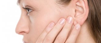
- Pain in the ear. When you touch the concha and, especially, the tragus (a small protrusion in front of the auditory opening, directed towards the cheekbone), the pain intensifies greatly and can take on a “shooting” character.
- Feeling hot. The auricle and surrounding tissues feel hotter than on the healthy side, and the feeling of heat persists regardless of the general body temperature.
- Discharge from the ear. Depending on the type of pathogen that caused the inflammation, discharge from the external auditory canal may be clear and watery, have a yellow tint, or be purulent.
- Decreased hearing acuity. On the inflamed side, there is a feeling of congestion, and sounds are heard muffled, as if through cotton wool. In severe cases of the disease, a “transfusion” of fluid may be felt in the ear, which causes serious discomfort.
- Increased body temperature. Depending on the severity of the disease, the pathogen that caused the inflammation and the age of the patient, the temperature can rise to 38-40°C. At the same time, lower values are more often observed in adults, and upper values in children, which is due to the immaturity of the child’s immune system, which reacts violently to the presence of infection.
In addition to the listed symptoms, which are directly related to otitis media, other signs are observed that indicate the negative impact of the inflammatory process on the entire body. Among them are increased fatigue, headache, irritability, sleep and appetite disturbances.
It should also be remembered that depending on the type of otitis externa, symptoms can be combined in various combinations, and also be supplemented by others (for example, the formation of an abscess in the external auditory canal).
Causes of otitis externa
In all cases of otitis, the disease is caused by pathogenic microorganisms that are literally present everywhere - in water, air, soil, food, etc.
But they cause inflammation in the external auditory canal only in the presence of predisposing factors - circumstances that facilitate the penetration of pathogens and create favorable conditions for their growth and reproduction.
These include:
- injuries to the external auditory canal, in which the integrity of the skin is compromised - most often this is caused by the use of unsuitable items for hygiene (matches, hair clips, etc.);
- insufficient hygiene of the external auditory canal, in which accumulations of sulfur and sebaceous gland secretions create a breeding ground for pathogens;
- recently suffered from acute respiratory viral infections, influenza, etc. - while the general state of the immune system worsens, which reduces the body’s resistance to infections;
- diseases of the ENT organs - rhinitis, tonsillitis, sinusitis, etc., left without treatment, can cause infection to spread to the middle ear cavity, from where pathogens move into the external auditory canal;
- hypothermia, temperature changes (for example, moving from a warm room to a cold one), which are also factors that reduce the body's defenses.
Treatment of external otitis necessarily involves identifying the reasons why the disease developed and eliminating them - otherwise, therapy may be ineffective.
Types of external otitis
Treatment of otitis media largely depends on the type of disease, which is classified according to the characteristics of the course and the extent of inflammation.
Thus, it is customary to distinguish between acute and chronic otitis media. In the first case, the disease manifests itself acutely, and the symptoms are pronounced. With chronic otitis media, the main symptoms (pain, fever, etc.) may be completely absent or appear very slightly. But with hypothermia, a cold and in the presence of other predisposing factors, the disease worsens, “activating” all the symptoms characteristic of otitis media.
According to the prevalence of the disease, limited and diffuse otitis media are distinguished.
Limited otitis is the introduction of infection into the lumen of the sebaceous ducts or hair follicle, after which a furuncle (painful swelling, which, as it matures, is filled with purulent contents) forms in the lesion.
With diffuse otitis, inflammation of the entire surface of the external auditory canal is observed, without clearly defined individual foci.
Diagnostics
When a patient consults a doctor with complaints of ear pain, decreased hearing acuity and other symptoms characteristic of otitis externa, a comprehensive examination is carried out.
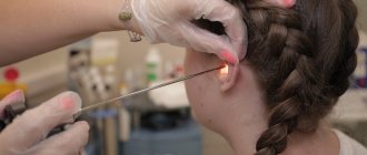
Among the routine methods (mandatory in all cases) is otoscopy, examination of the external auditory canal using a special device. This procedure is painless and does not require anesthesia, but it provides the doctor with enough information to confirm the diagnosis of otitis externa, determine the type of disease, identify complications if they have already developed and help set the direction for further diagnosis.
In addition to otoscopy, the following diagnostic methods may be recommended:
- laboratory tests of a sample of secretions taken from the ear canal, which make it possible to identify the causative agent of the disease and determine the group of drugs necessary for treatment;
- assessment of hearing acuity is necessary in cases where the inflammation is chronic and the doctor has reason to suspect hearing impairment as one of the complications of otitis media;
- Consultation with other specialists (infectious disease specialist, allergist-immunologist, etc.) may be recommended if the ENT sees this as necessary.
Comprehensive diagnostics is a necessary measure that allows you to accurately determine the cause of the disease, identify all possible complications or concomitant ailments, and prescribe treatment that best suits the given clinical situation.
Complications of otitis externa
Treatment of external otitis can rightfully be called not only a measure to eliminate the disease itself and its symptoms, but also the prevention of a number of complications.
The consequences of inflammation of the external auditory canal are so varied and severe that without adequate therapy, the development of complications is almost impossible to avoid. The most common of them are the following:
- Transformation of otitis media into a chronic form. The infectious and inflammatory process goes into a “dormant” state, only to be activated each time in the presence of unfavorable factors.
- Destruction of hard tissues of the external auditory canal. The bones and cartilage that form the ear canal can begin to break down and die when exposed to pathogens and the toxins they produce.
- Spread of infection to other tissues and organs. Treatment of external otitis can prevent infection of the eardrum and its perforation, inflammation of the middle and inner ear, orbital complications (developing in the soft and bone tissues of the orbit) and even brain damage. The most dangerous complication can be considered sepsis - the penetration of pathogens into the bloodstream and their spread throughout the body. Although such cases are rare, this does not diminish their severity - without providing the patient with emergency medical care, the prognosis is extremely unfavorable.
Drug treatment
Treatment of external otitis is carried out taking into account the results of the examination. So, when bacteria responsible for inflammation are identified, antibacterial drugs of the appropriate group are prescribed. For fungal infections of the external auditory canal, antimycotic agents are used.
In addition, the causes of the disease and the severity of the lesions determine the prescription of drugs with local or systemic action, or a combination thereof.
Physiotherapeutic treatment (UV therapy, electrophoresis, magnetic therapy and other methods) is also used.
The choice of drugs, the duration of the course of therapy and the combination of certain methods and means remains with the attending physician.
Treatment at the NIKIO clinic
At the Research Clinical Institute of Otorhinolaryngology named after. L.I. Sverzhevsky (NIKIO) treats external otitis using both classical regimens, which have proven to be highly effective, and with the help of the latest developments in the field of otolaryngology.
The diagnostic and treatment facilities of NIKIO are represented by the latest generation equipment, which allows one to quickly identify the causes and complications of the disease, as well as carry out therapy taking into account all aspects of the disease.
Outpatient and inpatient departments are presented to the attention of patients, as well as all currently existing treatment methods, as well as rehabilitation programs for people with hearing impairment and other pathologies.
To make an appointment, call 633-97-61 and remember: timely consultation with a doctor is the best guarantee of health and longevity.
Acute bullous or hemorrhagic otitis externa
This is a form of inflammation in which blisters (bullas) containing bloody fluid appear on the skin and on the outer surface of the eardrum. If such a bubble is opened, then bloody discharge flows out of the ear.
Causes of development of bullous otitis media
Most often, bullous otitis occurs against the background of acute respiratory viral infection or influenza, but a bacterial infection can also be the cause.
Treatment
An ear canal toilet is required. Antibacterial and anti-inflammatory drugs are prescribed locally, and drugs that strengthen the vascular wall and reduce vascular permeability should also be prescribed systemically. Opening bullae is not recommended.
Diagnosis and treatment.
Symptoms of otitis externa are usually intense and very disturbing.
Lack of proper treatment can lead to dangerous complications, so it is better not to delay visiting a doctor. It is necessary to conduct an ENT examination and determine the causative agent in order to begin appropriate treatment. For bacterial infections, topical antibiotics (eg in the form of ear drops) are used, and systemic antibiotics may be required for general symptoms. Fungal infections require the administration of antifungal agents, usually in the form of an ointment or suspension. Antiviral drugs are used to treat ear herpes. The goal of therapy is not only to eliminate the pathogen, but also to reduce unpleasant pain. As a rule, drugs based on paracetamol or ibuprofen are used, which can be purchased at the pharmacy without a prescription.
For otitis media, it is better to use a drug from the group of non-steroidal anti-inflammatory drugs based on ibuprofen or, in case of severe pain, metamizole sodium or ketoprofen. However, it is important to always follow the recommended dosage.
For topical use, you can use drops containing a local anesthetic - lidocaine) or an anti-inflammatory substance - choline salicylate.
Dermatitis of the ear
It can be separated into a separate form, since inflammation of the skin of the auricle often occurs, without involving the NSP. The causes of the disease are a bacterial infection plus a factor of sensitization of the body to bacterial toxins. With this type of inflammation, redness and thickening of the skin occurs, with the formation of small serous blisters, similar to those in eczema. When the bubbles open, yellow crusts form in their place. Diagnosis is made based on examination, and consultation with a dermatologist may be required. Treatment is also antibacterial and anti-inflammatory, and antihistamines may be prescribed.
Erysipelas of the auricle
Acute erysipelas is an infectious disease and is caused by streptococcus pyogenes; later, other flora may join. With such inflammation there are symptoms of skin damage and a general inflammatory syndrome - weakness, fever, chills. Factors provoking the disease: decreased immunity, abrasions and burns of the skin, swimming in polluted waters, contact with a carrier of pyogenic streptococcus or a sick person. Depending on the depth of damage to the skin, three forms are distinguished:
- Erythematous, when pain occurs, redness of the skin (it has a “varnished” appearance) slightly rises above the level of healthy skin and has a clear border with it.
- The bullous form, more severe, is characterized by the same skin lesions as in the erythematous form, but with the formation of large blisters above the surface of the skin with transparent or cloudy contents.
- Necrotic form. With this form, deep damage to the skin occurs, right down to the reticular and papillary layers, and deep ulcers with purulent plaques form. Due to the addition of various microflora, it is more severe than other forms. Typically, patients with this form require surgical debridement of areas of necrosis.
Erysipelas
This is a serious disease that often causes complications, and its treatment should take place in an infectious diseases hospital. If erysipelas is suspected, consultation with an infectious disease specialist is required. Isolated erysipelas of the auricle is rare and is usually combined with lesions of the skin of the face or neck.
Treatment
Massive antibacterial and anti-inflammatory therapy, detoxification therapy, and antistreptococcal gamma globulin are prescribed.
Our doctors
Gogolev Vasily Gennadievich
Doctor - otorhinolaryngologist
19 years of experience
Make an appointment
Zharova Galina Gennadievna
Doctor - otorhinolaryngologist, member of the European Society of Rhinologists, doctor of the highest category
Experience 39 years
Make an appointment
Debryansky Vladimir Alekseevich
Doctor - otorhinolaryngologist, doctor of the highest category
33 years of experience
Make an appointment
Malignant otitis externa (necrotizing otitis externa)
This form of external otitis is distinguished as a separate one, but in fact it is a negative course of ordinary diffuse external otitis. During the course of the disease, inflammation spreads to the structures of the temporal bone, damage to the cranial nerves and the development of intracranial complications may occur. The name of the disease is not associated with the development of a malignant tumor, it simply reflects the course of the disease. Fortunately, this disease is rare; it affects patients with severe immune disorders, such as AIDS, conditions after radiation and chemotherapy, decompensated diabetes mellitus, and patients receiving immunosuppressive and cytostatic therapy. The leading pathogen is Pseudomonas aeruginosa, but any infection is polymicrobial. During the process of inflammation, necrosis of the skin of the ear canal occurs and inflammation passes to the periosteum and bone, followed by its destruction. Complaints in this disease include severe pain, discharge from the ear of a purulent-inflammatory nature, with a putrid odor, and hearing loss quickly occurs. Later, symptoms of damage to the cranial nerves appear: difficulty swallowing, choking when eating, numbness of the face, paralysis of the facial muscles. When the process spreads to the middle and inner ear, systemic vertigo may develop. When inflammation spreads into the cranial cavity, cerebral and meningeal symptoms develop. On examination, one notices pronounced inflammation of the skin of the NSP, skin with foci of necrosis, a large amount of purulent-necrotic discharge, the eardrum and the structures of the middle ear are often indefinable. Symptoms of damage to the cranial nerves are determined - facial asymmetry, ptosis of the lower eyelid on the affected side, asymmetry of the soft palate, impaired mobility of the epiglottis. In diagnosis, culture for bacteriological examination and computed tomography of the temporal bones are important. Treatment is always carried out in a hospital setting. Massive antibacterial and detoxification therapy is carried out. Infusions of human gamma globulin, antipseudomonas serum or hyperimmune human plasma are prescribed. Surgical treatment is also performed - necrotomy (removal of necrotic areas of skin and bone), and subsequently, upon recovery, plastic surgery of the defect is performed using one’s own tissues.
Sources
- Kesser BW Assessment and management of chronic otitis externa. Current Opinion in Otolaryngology & Head and Neck Surgery. 2011, 19: 341–347.
- Roland PS, Stroman DW Microbiology of acute otitis externa // Laryngoscope. - 2002. - Vol. 112, No. 7, Pt. 1. - P. 1166-1177.
- Rosenfeld RM, Brown L, Cannon CR et al. Clinical practice guideline: acute otitis externa // Otolaryngology. Journal of Otolaryngology - Head & Neck Surgery. - 2006. - Vol. 134 (4 Suppl). - P. 4-23.
- Schaefer P., Baugh RF Acute otitis externa: an update. American Family Physician. 2012, Dec; 1-86 (11): 1055-61.
- Wiegand S., Berner R., Schneider A., Lundershausen E., Dietz A., Otitis Externa: Investigation and Evidence-Based Treatment. Archive of "Deutsches Ärzteblatt International". 2021, Mar; 116 (13): 224–234.
Gurov A.V., Yushkina M.A. Features of the clinical course and etiotropic therapy of external otitis. Russian medical journal. 2016. – T. 24. – No. 21. – P. 1426-1431.
Chondroperichondritis of the auricle
This disease is an inflammation of the perichondrium and cartilage of the auricle caused by bacterial flora. Chondroperichondritis can be caused by trauma to the auricle with damage to the skin, suppuration of an auricular hematoma, deep burns of the auricle, a boil or suppurating atheroma of the ear, and erysipelas. There are infiltrative and purulent forms. A few days after the described traumatic factor, infiltration and hyperemia of the skin of the auricle develops, accompanied by pain, with or without fever. An important diagnostic criterion is that the earlobe remains unaffected, since there is no cartilage in its thickness. Treatment: antibacterial and anti-inflammatory therapy. If there are purulent cavities, an opening with removal of pus and gentle necrotomy of the destroyed cartilage is performed. The disease is difficult to treat and can last for weeks or months. Often the consequence of chondroperichondritis is cicatricial deformation of the auricle; sometimes there may be complete destruction of the cartilage.
Treatment of otitis media during pregnancy
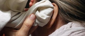
If you experience ear pain during pregnancy, you should urgently see an ENT doctor. Remember that in this case you cannot apply heating pads or warm compresses to the sore spot! This can be very dangerous if purulent inflammation begins in the ear.
If the pain increases and greatly bothers a pregnant woman, and there is no way to see a doctor in the near future, you can take several independent steps. For example, you should put vasoconstrictor drops into your nose.