Nowadays, fungal diseases, that is, diseases caused by fungal infections, have become widespread. The first thing that comes to mind when you hear the phrase “fungal disease” is nail fungus. But there are fungi that settle in the ears, causing the patient a lot of anxiety. Infection of the middle or outer ear with a fungal infection is called otomycosis.
Otomycosis is caused by mold and yeast-like fungi. Most often these are fungi of the Aspergillus and Candida species.
Another name for the diagnosis is fungal otitis media. If the inflammation affects the auricle and ear canal, the patient is diagnosed with external fungal otitis, or external otomycosis. If the middle ear is affected, fungal otitis media. When the eardrum becomes inflamed, a diagnosis of fungal myringitis is made. Otomycosis can also develop in postoperative cavities. ICD-10 codes - H62.2, H74.8. Otomycosis accounts for about 25% of cases among all ear infections.
In 90% of cases, the disease affects only one ear, and only in 10% the inflammatory process is bilateral. If the right ear is affected, it is a right-sided fungal otitis, if the ear is on the left, it is a left-sided one.
The disease can develop in both adults and children. Those at particular risk are residents of hot, humid climates, people with weak immune systems, swimmers and those who frequently use headphones. Only an ENT doctor can diagnose the disease.
What causes otomycosis?
Ear fungus belongs to the so-called opportunistic flora. Every person has fungi in the ear. They are harmless until favorable conditions appear for their activation and increased reproduction. Provoking factors include:
- Mechanical injuries and ear damage. This is a common cause of external otomycosis. Banal cleaning of the ear with cotton swabs can result in treatment in the ENT doctor’s office. When using cotton swabs, you can easily injure the skin of the ear canal. Infection is directed into microcracks and causes inflammation. Injuries can cause foreign objects entering the ear (children love to insert small parts of toys into the ear canal), as well as allergic reactions, accompanied by severe itching and scratching of the skin.
- Previous ear diseases (acute external otitis, acute otitis media, chronic purulent otitis, boils, etc.), due to which the composition of the microflora in the ear changes.
- Increased sweating in the ear area. This can be either a physiological feature of a person or a symptom of a metabolic disorder in the body.
- Taking medications (usually hormonal and antibacterial). More often, the disease occurs after uncontrolled use of medications among those who like to self-medicate.
- Chronic diseases that reduce immunity.
- Skin diseases such as dermatitis.
- Other diseases, such as diabetes, HIV, bronchial asthma, tuberculosis, etc.
- Swimming in ponds or pools.
- Unfavorable environmental conditions.
- Poor working conditions (work in dusty, cold, damp rooms).
- Frequent use of in-ear headphones.
- Wearing hearing aids.
- Allergic reactions, for example, to the use of shampoo.
As you can see, there is far more than one cause of fungal otitis media. Only an ENT doctor can determine the correct cause of the disease and carry out the correct diagnosis.
Causes
There are several groups of main causes of otomycosis:
- The presence of concomitant diseases that can provoke the development of a fungal infection. This can be eczema, atopic dermatitis, dermatoses accompanied by itching. When scratching certain areas of the skin, saprophytic fungi, which under normal conditions are asymptomatically located on the surface of the epidermis, can get onto the surface of the ear canal. Genital candidiasis present in humans also often causes otomycosis.
- The development of local dysbiosis, provoked by various inflammatory diseases of the ear. The use of antibiotics or corticosteroids included in antibacterial ear drops, as well as inflammatory mediators, make the skin of the ear canal susceptible to any fungal infection. A loose surface that does not have a natural protective film becomes an excellent breeding ground for fungi.
- External reasons, such as the constant presence of dust, high humidity or the presence of a large number of microorganisms in the environment. Otomycosis is often diagnosed in people of certain professions, such as ragpickers, workers at waste collection points, and cloakroom attendants. A certain percentage of patients report constant visits to the pool or frequent visits to public baths or saunas.
- Constant trauma to the ear canal with various foreign objects, including headphones and hearing aids for the hearing impaired.
- Postoperative periods.
Symptoms of otomycosis.
The main symptoms of the disease do not appear immediately. Otomycosis develops gradually as the fungus takes time to grow deeper into the skin.
Signs of the disease will differ depending on the affected part of the ear.
External fungal otitis begins with swelling of the ear canal. Its protective fatty film disappears. The ear “stuffs up” and itches inside. At this stage, few people go to the doctor. Many patients “blame” the cerumen plug for the itching and try to remove it themselves, which causes more injury to the skin. The fungus penetrates these microcracks, and the inflammatory process intensifies. A person's hearing deteriorates and ear pain appears, which intensifies while eating.
Fungal otitis media is an unpleasant consequence of purulent otitis media. This form of otomycosis is characterized by the following manifestations:
- severe pain in the ear cavity;
- discharge from the ear canal (curdled or dark);
- hearing loss;
- ringing in the ear.
When fungal flora gets on the eardrum, a person's hearing deteriorates, since the affected eardrum cannot vibrate normally. Fungal otitis may develop after ear surgery. In the postoperative period, antibacterial and antiseptic drugs are used for a long time to disinfect cavities, which changes the balance of microflora in the hearing organ. The ear begins to hurt, and discharge appears from it.
Risks of self-medication
Self-medication may not only not lead to recovery, but also cause irreparable harm to your body, so let’s immediately make a reservation about what is strictly prohibited and why this is so.
- Do not use alcohol-based solutions to instill or lubricate the ear. This will lead to a longer period of residence of the antifungal drug in the external auditory canal, causing irritation. And if it gets on the mucous membrane of the eardrum, it will further increase the swelling, cause severe pain and contribute to the development of granulations (they are a common factor in the chronicity of the process).
- Do not prescribe systemic medications for yourself, because... Each type of otomycosis requires a differentiated approach, which is based on the results of sensitivity to antimycotics. Otherwise, the treatment will be completely ineffective.
Otomycosis in children.
Illness in childhood is not uncommon. Most often, otomycosis occurs in children under the age of five. The causes of the development of the disease in childhood are previous inflammatory diseases, allergies, changes in the microflora in the ear, weakened immunity, and improper cleaning of the ears.
Children with ear fungus complain of itching, burning, and pain in the ear canal. In this case, parents need to examine the outer ear: it may be swollen and red. A cheesy or dark discharge may appear. Hearing may be impaired.
Mycotic otitis media
Average otomycosis is diagnosed half as often, it occurs in approximately twenty percent of patients, and most often is a complication of purulent otitis caused by the addition of fungal pathogenic flora to a bacterial infection. It is characterized by a sharp decrease in hearing, increased discharge, and sharp pain that does not subside during the daytime. Patients complain of congestion and tinnitus, frequent headaches and tenderness of regional lymph nodes. An increase in temperature and a general deterioration of condition accompany mycotic otitis media.
Diagnostics.
Diagnosis and treatment of otomycosis - the profile of an otorhinolaryngologist. Diagnostics includes a set of measures and studies. First, the ENT doctor interviews the patient about complaints and collects his life and health history. History helps determine the cause of the disease.
During diagnosis, the patient undergoes:
- otoscopy (examination of the ear cavity using a special funnel);
- endoscopic examination to examine distant areas not visible during otoscopy;
- laboratory testing of ear discharge (to determine the type of pathogen);
- hearing studies (audiometry, acoustic impedance measurement, otoacoustic emissions).
Based on the data obtained, the doctor selects effective treatment tactics.
Prevention
Otolaryngologists at the Nearmedic center warn that recurrent ear mycosis can be avoided. To do this, in addition to antifungal treatment, it is enough to follow the general rules:
- Avoid deep cleansing your ears using cotton swabs. They can only be used to remove wax outside the outer opening. In other parts of the ear, it performs a protective function, so it should not be removed.
- Do not self-medicate using antibiotics. These drugs should be prescribed only by a doctor, taking into account the indications (whether they are or not is also determined by a specialist).
- Promptly treat any concomitant diseases that can significantly undermine a person’s health.
- Regularly visit your treating otolaryngologist for 2 years, once every quarter, and if the condition is satisfactory - once every six months. If even minimal complaints from the ear appear, a visit to a specialist cannot be postponed until later.
- After the main stage of treatment, lubricate the skin with antifungal drugs for 1 - 1.5 months, and our doctors will tell you which one is best, based on the individual characteristics of your body.
Ear fungus can be defeated - turn to professionals! Make an appointment with Nearmedic for a consultation at a convenient time.
Treatment of otomycosis.
Many patients ask the question: “Is it possible to cure otomycosis yourself?” No. The treatment protocol should be drawn up by an ENT doctor depending on the type of fungal infection, the patient’s health condition and the cause of the disease. The treatment regimen is always selected individually.
An important element of therapy is rinsing the ear from pathological discharge. A high-quality toilet of the ear cavity is necessary, because if even a small amount of fungal waste products remains in the ear, the treatment may be very delayed. Only an otolaryngologist should rinse the ear using antifungal and anti-inflammatory agents.
Clinical recommendations for otomycosis may include:
- antifungal drugs (ointments, tablets, drops);
- vitamin complexes;
- antihistamines;
- physiotherapy (infrared laser therapy, vibroacoustic therapy, ultraviolet irradiation, ultrasound therapy).
You also need to adjust your diet during treatment and follow a diet. Diet is very important: sweets, flour, and foods that cause allergic reactions should be excluded from the diet.
Treatment in the network of Nearmedic clinics
When otomycosis is diagnosed, treatment is carried out only by conservative methods with the mandatory use of antifungal agents. At the first stage, doctors at the Nearmedic center give preference to local antimycotic drugs that do not have a systemic negative effect on the body. Usually, applications are used for this, which are performed professionally (painlessly and highly effectively) by doctors at our centers.
They select a unique combination of local antifungal agents for each patient based on the latest medical advances. It has been proven that the combined approach accelerates recovery and increases the percentage of destruction of pathogenic fungi, because Some antimycotics suppress reproduction, while others cause their immediate death.
But in order to prevent relapses, it is not always possible to cure otomycosis with the help of local therapy. Therefore, Nearmedic otolaryngologists initially identify patients who are candidates for systemic antifungal therapy. This helps not to waste precious time from the first minutes, but to immediately direct all efforts to destroy pathogenic fungi. Indications for systemic antifungal therapy are:
- Mycosis of the middle ear;
- Fungal postoperative infection.
Also, doctors face another main question - which antimycotic drug is best to prescribe. The answer to this depends on what type of fungus caused the mycosis. If it is candida, then Fluconazole is usually used, and if it is aspergillus, then Terbinafine or Itraconazole. This differentiated approach is determined by the different mechanisms of action (points of application) of these antimycotic drugs.
In some cases, surgical treatment may be required, but it is not aimed at curing mycosis, but at preventing its recurrence. Indications for surgery are:
- Ear polyps;
- Granulation.
They can be removed by excision or the use of silver nitrate, which causes necrosis (death) of the pathologically overgrown tissue.
Nearmedic doctors immediately warn patients about restrictive measures, non-compliance with which can slow down recovery or cause chronicity of the process. Therefore, the entire treatment period is necessary:
- Protect your ear from water getting into it;
- Avoid contact with gases and dust;
- Do not stay in rooms with high humidity.
Currently, the problem of mycotic diseases is acquiring important social significance. The increase in the incidence of mycoses is associated with many factors, primarily with the active use of antibacterial drugs in medical practice, which undoubtedly increases not only the incidence of candidiasis, but also mycoses caused by filamentous fungi. This is also facilitated by the use of cytostatics, corticosteroids and other drugs with immunosuppressive properties. The development of mycosis is promoted by a combination of many reasons associated with the urbanization of modern life of the population [1–4].
The wide distribution of fungi in nature, their constant presence both in the environment and in the body, makes contact and infection of humans inevitable. Particularly alarming is the increase in the number of patients with deep mycoses, which include some mycoses of the ENT organs.
Currently, approximately 100,000 species of microscopic fungi have been identified, of which more than 400 species are capable of causing infections in humans, but about 100 species are of real importance in clinical practice [5].
An analysis of clinical and laboratory studies conducted at the Moscow Scientific and Practical Center of Otorhinolaryngology showed the increasing importance of fungal pathology among general ENT morbidity. So, in 2010-2011. among patients with chronic inflammatory pathology of the ENT organs who applied to the mycology room of the Center’s advisory department, fungal infection was detected in 23% [6].
In this regard, the problem of mycoses of the ENT organs remains relevant, which determined the purpose of this work: to familiarize otorhinolaryngologists with modern principles of diagnosis and treatment of mycoses of the upper respiratory tract and ear, varying in localization and nature of clinical manifestations.
Nosological forms of fungal diseases of the ENT organs are otomycosis, pharyngomycosis, laryngomycosis, fungal infection of the nasal cavity and paranasal sinuses. There are superficial mycoses, which affect the skin, mucous membrane, external auditory canal, pharyngeal cavity, and deep mycoses affecting, for example, the SNP, the postoperative cavity of the middle ear, accompanied by invasion of the fungus not only into the epithelium, but also into surrounding tissues.
According to our data, in recent years, the frequency of detection of pharyngomycosis is 45%, otomycosis - 42%, mycosis of the nasal cavity and SNP - 8%, laryngomycosis -5% among all patients with mycosis of the ENT organs [3, 7].
The proportion of otomycosis among otitis of other etiologies reaches 25.3%, pharyngomycosis in chronic pharyngitis and tonsillitis - 26%. Fungal infection of the larynx in chronic laryngitis reaches 15%; in chronic inflammation of the nose and SNP, the share of the fungal process is 7%.
External fungal otitis, fungal otitis media and fungal infection of the postoperative middle ear cavity occur in 69, 18 and 13% of cases of otomycosis, respectively. The main causative agents of external fungal otitis are molds of the genus Aspergillus
(65%),
Penicillium
(5%), with mycosis of the postoperative cavity the proportion of mold fungi reaches 95%, while with fungal otitis media, yeast-like fungi of the genus
Candida
(79%).
In some cases, fungal diseases of the ears can be caused by fungi of the genera Mucor, Alternaria
, etc. In 17% of cases, we noted a combined infection with
Aspergillus
spp.
and Candida
spp.
in combination with representatives of Staph.
spp and
Pseudomonas
spp.
Infection with two or more species of fungi Aspergillus
spp.
+ Candida
spp.,
Aspergillus
spp.
+ Candida
spp.
+ Alternaria alternata
occurs in 1% of observations.
Candida are the leading pathogens
(97-99% of observations).
In mycosis of the nasal cavity and SNP, mold fungi account for up to 78% of the lesions. The main pathogen is a fungus of the genus Aspergillus
, species -
fumigatus
and
niger
, in some cases, in immunocompromised patients, fungal diseases of SNP can be caused by fungi of the genera
Mucor, Alternaria, Rhizopus, Rhizomucor
, etc.
Basically, all these types of fungi belong to the group of opportunistic fungi and cause disease only under certain conditions that predispose to the development and reproduction of fungi, ensuring their transition from saprophyte to the realization of pathogenic properties.
Among the risk factors for the development of mycoses of the ENT organs, the leading positions are occupied by iatrogenic immunodeficiency states that develop as a result of massive antibiotic therapy, long-term use of glucocorticoid and immunosuppressive drugs for oncological and autoimmune diseases. Mycoses of the ENT organs often accompany severe somatic pathologies, such as HIV infection and AIDS, diabetes mellitus, thyroid dysfunction, agranulocytosis, anemia, bronchial asthma, etc.
The pathogenesis of mycosis consists of adhesion (attachment of the fungus to the surface of the mucous membrane or to the skin), colonization of fungi and their invasive growth. In severe forms of ENT mycosis, the next stage is the generalization of the process: the penetration of fungi into the blood with dissemination and the appearance of secondary foci of mycosis in various organs and tissues. This is especially true for fungi of the genus Aspergillus
spp., the pathogenicity factor of which is their ability to grow at 37 °C, the presence of enzymes (protease, phospholipase), toxins (aflatoxin, fumagillin, etc.), pronounced angioinvasiveness and the ability to evade immune surveillance due to the production of inhibitors of the phagocytic function of macrophages and neutrophils, for example gliotoxin [1, 5].
A factor contributing to the development of otomycosis is, first of all, trauma. When the skin and mucous membrane are damaged, paths are opened for the fungus to penetrate deep into the tissues, and protective reactions at the site of damage are weakened. In addition, the secreted secretion of the injured integumentary epithelium is a good nutrient medium for the proliferation of fungi. Thus, a paradoxical phenomenon has been noted: fungal diseases of the outer ear more often occur in extremely careful people who intensively use cotton swabs and other objects to clean their ears, injuring the skin of the external auditory canal and often causing traumatic invasion of the pathogen. Speaking about the need for proper, gentle removal of excess sulfur, it should be remembered that its presence in a small amount in the ear canal is necessary, since it not only mechanically protects the skin, but also carries out bactericidal and fungicidal functions.
The occurrence of a fungal infection of the outer ear is often preceded by water entering the ear during bathing or removing wax plugs by washing. Long-term local use of glucocorticoid drugs, antibiotics, and antiseptics for otorrhea, which accompanies a purulent-inflammatory process in the middle ear, leads to the development of otomycosis in 20% of cases.
Surgical sanitation of the middle ear in the absence of a positive effect from previous courses of antibacterial therapy, local and physiotherapeutic conservative treatment often entails the development of fungal infection of the postoperative cavity. Perhaps in such cases there is undiagnosed fungal otitis media. Surgical treatment in this case can be performed only after negative repeated mycological studies.
Otomycosis can also develop when working in dusty conditions and in pressure complexes with high pressure and humidity.
Fungal infection of the pharynx and larynx often develops in patients after massive high-dose antibacterial therapy, long-term systemic use at a dose exceeding 0.3 mg/kg/day (in terms of prednisolone) for more than 3 weeks, and local use of inhaled forms of corticosteroids for hormone-dependent bronchial asthma. A stay in the intensive care unit for more than 10 days, accompanied by laryngeal intubation for more than 5 days, significantly increases the risk of developing mycoses. Smoking, the use of removable dentures and their improper treatment contribute to the development of mycosis of the pharynx and larynx.
Mycosis of the nasal cavity as an independent disease is extremely rare, usually due to trauma to the nasal mucosa in HIV-infected people. More often, the fungal process affects the maxillary sinuses after foreign bodies enter them (filling material when filling the canals of the 4th-7th teeth of the upper jaw).
Complaints and clinical manifestations of mycosis of the ENT organs
are determined by the characteristics of vegetation and parasitism of certain types of fungi and are largely determined by the localization of the pathological process.
In all forms of mycotic diseases of the upper respiratory tract and ear, there is a specific discharge in the form of crusts, plaques, thick caseous or liquid secretions, the color, quantity and consistency of which depend on the type of fungus and the phase of its development. The clinical picture, despite the diversity of pathogens, has a number of specific features. Main complaints with mycotic otitis media
- discharge from the ear, formation of “plugs” in the external auditory canal, itching, pain, ear congestion.
Thus, with otomycosis caused by dermatophytes, the skin of the auricle, postauricular area and external auditory canal is mainly affected. Patients are concerned about itching and peeling of the skin of the affected area. In the external auditory canal there is dry discharge in the form of plaque and films. Inflammatory phenomena are not expressed. Accelerated formation of sulfur plugs was noted.
For otomycosis caused by mold fungi ( Aspergillus, Penicillium, Mucor
etc.), the skin of the external auditory canal, the postoperative cavity, and less often the middle ear cavity and the skin of the auricle are affected. The patient experiences severe pain, severe hearing loss, and dizziness. The discharge looks like “wet newspaper.” The symptoms of inflammation are significantly pronounced. After removal of the pathological discharge, granulations are visualized, the skin is damaged and bleeds easily. The disease can occur as necrotizing external otitis with damage to the bones of the skull.
With candidal otomycosis, the middle ear cavity and the skin of the external auditory canal are affected. Upon examination, maceration and infiltration, reminiscent of weeping eczema, are determined. The process often spreads to the skin of the auricle and the area behind the ear. Pain and itching are moderate. Some patients in the acute stage may have complaints of increased body temperature, headache, increased sensitivity of the auricle and postauricular area.
Mycotic otitis media and mycosis of the postoperative cavity are characterized by the following clinical manifestations: hearing loss, noise in the ear, pain, discharge from the ear, periodic itching, congestion and dizziness.
Otomycosis, like other types of specific inflammation, has a chronic course with a subtle onset of the disease, its gradual development and significant duration. The development of a fungal disease is possible at any age; frequent exacerbations and relapses of the disease during treatment are typical.
For fungal infections of the pharynx
patients complain of discomfort in the throat, burning sensation, dryness, rawness, and soreness, which may be more pronounced than with bacterial pharyngitis. Painful symptoms are moderate; when swallowing and eating irritating foods, the pain intensifies. Sometimes patients note irradiation of pain in the submandibular region, on the anterior surface of the neck and in the ear. Depending on the degree of damage, pseudomembranous, hyperplastic, granulomatous and erosive-ulcerative clinical forms of pharyngomycosis are distinguished. Specific signs of the clinical picture of pharyngomycosis are the presence of plaque, swelling of the mucous membrane and pronounced general symptoms of intoxication; frequent (up to 10 times a year) exacerbations are also characteristic.
For fungal infection of the larynx
patients are worried about pain in the throat and on the front surface of the neck, a feeling of “rolling a lump” in the throat and neck, not associated with swallowing, constant hoarseness, rapid fatigue of the voice, and an unproductive cough. On examination, congestive hyperemia of the mucous membrane of the larynx, swelling of the mucous membrane of the vocal folds, and fibrin deposits are characteristic, when removed, the eroded surface is exposed. Often the fungal process is one-sided, which must be differentiated from cancer and tuberculosis.
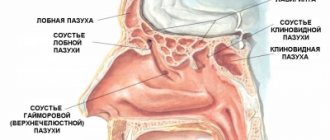
Fungal infection of the nose and SNP
divided into invasive and non-invasive forms. Invasive forms include acute (fulminant) and chronic forms. Non-invasive ones include fungal body and allergic fungal sinusitis.
Invasive forms of fungal infection of the SNP are extremely unfavorable in predicting the outcome of the disease. The fulminant form of SNP damage is most often caused by fungi belonging to the class Zygomycetes. This form of the disease occurs in patients with severe immunodeficiency, decompensated diabetic ketoacidosis, neutropenia, and burn disease. Zygomycosis is characterized by an extremely severe course and, without active antifungal therapy and early surgical treatment, usually ends in death.
The chronic form occurs as chronic sinusitis with reactive phenomena from surrounding tissues. Accompanied by copious purulent nasal discharge in the morning, severe pain in the projection of the affected SNP.
The fungal body is quite often a “diagnostic finding” when performing a CT scan of the ED and in some patients is asymptomatic.
With allergic fungal sinusitis, in addition to polyps in the nasal cavity and SNP, a thick, viscous secretion is detected in the lumen of the latter.
Lack of awareness among otorhinolaryngologists regarding the diagnosis of fungal infection of the ENT organs is often the reason for their late and/or incorrect diagnosis. This creates difficulties in treatment and leads to a chronic course of the disease.
Diagnosis of fungal disease of ENT organs
, like any infectious disease, is established only on the basis of complex laboratory mycological research methods. A pronounced clinical picture of a disease similar to a fungal infection, no matter how characteristic it may be, is not the basis for making a final diagnosis of mycosis.
Diagnosis of mycosis is carried out in the following areas:
1. Microscopy of pathological discharge.
2. Inoculation of pathological discharge on various elective nutrient media to isolate fungal cultures and their generic and species identification.
3. Determination of the sensitivity of isolated fungi to antifungal drugs.
4. Radiation diagnostics in cases of damage to the nose and special organs.
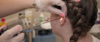
Sampling of biological material from the ear is carried out using an attic probe or a Volkmann spoon. Pathological discharge from the deep parts of the external auditory canal is placed between two fat-free sterile slides and microscoped at 100, 200 and 400 times magnification.
In the active stage of aspergillosis and mucorosis with abundant formation of mycotic masses with aerial mycelium, we recommend using an operating otorhinolaryngological microscope (otomicroscopic method of express diagnostics) for diagnosis.
If a fungal infection of the pharynx and larynx is suspected, microscopy of scrapings from the mucous membrane of the oral cavity, pharynx, tonsils, taken with a swab to detect elements of the fungus, and seeding of material from the affected areas of the mucous membranes are performed.
The main method for diagnosing mycosis of SNP is culture of aspirate or rinsing water (puncture using a Kulikovsky needle according to the usual method) with mandatory identification of the pathogen. A histological examination of the biopsy material is also carried out.
In addition to microscopy of native material, microscopic examination of preparations stained using the Romanowsky-Giemsa method is performed. The most informative method that reliably identifies the causative agent of the disease is staining the preparation with solutions of calcofluor white, followed by fluorescent microscopy. For mycological diagnosis, pathological material is inoculated on elective media (Saburo, Chapek, wort agar, etc.). Types of yeast-like fungi of the genus Candida
determined by morphological characteristics and the nature of fermentation of sugars. Inoculation of the material is carried out using the standard method [1, 2, 5]. To clarify the diagnosis, in some cases it is necessary to carry out serological, allergological and histological studies [2, 5].
Treatment
You should always start with eliminating the pathogen. Correction of general and local predisposing factors is carried out in parallel or in the second stage.
When treating a patient with mycosis of the ENT organs, it is necessary to take into account all possible conditions under which this disease arose specifically in this patient with a view to their possible elimination. It is necessary to identify and treat diabetes mellitus, blood diseases, gastrointestinal tract, and immune deficiency. The role of allergies in the pathogenesis of the disease should be taken into account, since mushrooms have pronounced allergenic properties, and hyposensitizing therapy should be carried out in parallel with etiotropic therapy.
Selection of rational antifungal therapy for mycosis of ENT organs
presents certain difficulties. On the one hand, a large number of antimycotic drugs expands the possibilities of treating fungal infections that differ in both localization and type of pathogen. On the other hand, it requires the clinician to have increased awareness of the spectrum and characteristics of their action (fungicidal or fungistatic), pharmacokinetics, compatibility with antibacterial drugs, side effects, etc. The ideal treatment is with an antimycotic that has the least toxic and greatest therapeutic effect, which is not always possible, especially in acute forms of fungal disease, when the choice of drug is made empirically, since the pathogenic fungus is not immediately identified and tests can take a long time. Determining the choice of drug are the results of laboratory mycological studies of the sensitivity of fungi isolated from a particular patient to known antifungal drugs.
Fungal disease of the oropharynx - pharyngomycosis is caused mainly by yeast-like fungi of the genus Candida
, which largely determines treatment tactics. Limited acute candidiasis of the tonsils, which developed as a result of antibacterial therapy, usually heals quickly after stopping the administration of antibiotics and performing only local antifungal treatment. However, widespread lesions of the mucous membrane of the oropharynx, with a tendency to chronicity and recurrence, require a long course of treatment using local and general antimycotic therapy. For systemic therapy, a drug from the azole group, fluconazole (mycoflucan), is used orally at a dose of 100-200 mg once a day. Treatment should be continued for another 14 days after clinical improvement. If such treatment is ineffective, itraconazole (200 mg per day orally) or amphotericin B (1 ml 4 times a day orally, or >0.3 mg/kg per day intravenously) is used [1, 8-10].
In the systemic treatment of pharyngomycosis caused by molds, it is advisable to use itraconazole and amphotericin B, which have a fungicidal effect not only on yeast-like fungi, but also on mold fungi of the genera Aspergillus, Mucor, Penicillium
[12].
In addition to systemic treatment, local treatment is also recommended. During treatment, the pharynx after meals should be lubricated with a solution of clotrimazole at a rate of 10 mg 5 times a day, or nystatin tablets should be dissolved/chewed after meals at a dose of 250-500 thousand units 4-5 times a day. For chronic tonsillitis of fungal etiology, courses of washing the lacunae of the tonsils every other day or 1-2 times a week at least 10 times with solutions of antiseptics with antifungal action (miramistin, quinozol, clotrimazole solution) [10, 11].
Drug therapy for mycosis of SNP always accompanies and/or precedes surgical treatment. In the treatment of fungal infections of the nose and SNP, both general and local measures are provided. For invasive forms of fungal infection of the maxillary sinuses, the most effective is a combination of drug antifungal and surgical treatment. To eliminate the Aspergillus infection at this location, surgical treatment is almost always necessary. Surgical intervention consists of performing radical surgery on the affected sinus using the extranasal method with complete removal of all pathologically altered tissues. In the postoperative period, daily rinsing with antiseptics and antimycotics (amphotericin B, miramistin, pimafucin suspension, clotrimazole) is carried out.
In acute toxic forms of invasive aspergillosis of SNP, antifungal therapy is mandatory. Voriconazole or lipid amphotericin B is used [1, 13]. It is possible to use caspofungin and amphotericin B. After stabilization of the process, treatment with itraconazole. The duration of use of antimycotics depends on the clinical picture of the disease and its course, but should continue for at least 14 days after clinical and laboratory cure [1, 2, 9].
For candidiasis of the nose and SNP, it is necessary to combine the use of systemic antifungals with local antifungal drugs. The drug of choice is fluconazole (mycoflucan), which is prescribed once a day at a dose of 50-200 mg. The course of therapy cannot be less than 14 days. If treatment with standard doses of fluconazole is ineffective, it is possible to increase the dosage to 400 mg/day, or prescribe itraconazole 200 mg/day [10].
Patients with allergic fungal sinusitis are advised to undergo surgery to completely remove the polypous mucous membrane and the viscous “rubber-like” contents of the sinus [1, 13]. In the early postoperative period, it is necessary to wash the SNP twice with an aqueous solution of amphotericin B. It is advisable to prescribe topical nasal corticosteroids to prevent relapse no earlier than 20 days after surgery.
Only in case of resistance to other antimycotics, amphotericin B is used for the treatment of candidal lesions of the nose and SNP (intravenous 0.3 mg/kg per day for 3-7 days). Treatment is carried out under the supervision of biochemical studies of liver and kidney function, since amphotericin B has a pronounced nephro- and hepatotoxic effect.
The specificity of the treatment of fungal infection of the larynx is the widespread use of the inhalation method for administering drugs. The effectiveness of the aerosol method for inflammatory diseases of the larynx is determined by the direct effect of the drug on the site of the disease and the higher concentration of the drug at the site of the lesion. With this method of administration, inhalations with the water-soluble sodium salt of nystatin or levorin are effective, especially for candidiasis. Treatment is carried out for 15-20 minutes 1-2 times a day. The initial course of treatment is 12-15 procedures. In case of resistance to other antimycotics, 12-15-day courses of aerosol therapy with amphotericin B are carried out. All patients with laryngomycosis, in addition to inhalation therapy, must undergo systemic therapy with antimycotics. The drug of choice is fluconazole (mycoflucan) once a day at a dose of 100-200 mg, in severe cases - 300 mg/day) [11]. For patients whose fungal disease in the larynx is accompanied by the formation of a large amount of viscous secretion, it is advisable to carry out inhalations with proteolytic enzymes that selectively break down necrotic masses and dilute the viscous exudate for their better removal. For this, inhalations with chemopsin and chemotrypsin (150 mg in 5 ml of saline) can be recommended.
When treating otomycosis, local and systemic drugs are used. For local treatment of mold lesions in the ear, the most effective are naftifine, nitrofungin, terbinafine (exifin), and for candidiasis - clotrimazole, terbinafine (exifin), naftifine. An indispensable condition for local therapy is preliminary thorough cleaning of the ear using endoscopic equipment or an operating microscope. Ear toileting is performed only by a doctor using an attic probe and a padded cotton pad moistened with an antimycotic drug. Thorough toileting of the ear is given special importance, since even a small amount of mycotic masses significantly lengthens the course of treatment until complete recovery.
In case of external fungal otitis, it is necessary to thoroughly clean the anterior inferior part of the external auditory canal. In case of fungal otitis media, mycotic masses are completely removed from the area of perforation of the eardrum. The postoperative cavity of the middle ear is cleaned in the same way - the entire cavity is thoroughly cleaned, especially in the posterior section, behind the spur. Before treatment, if polyps and granulations are present, they are removed or extinguished with a 20% solution of silver nitrate.
Local treatment with antifungal drugs must be carried out for at least 3-4 weeks under mandatory laboratory control (cultures before, during and after the end of the course of treatment). Local treatment is carried out by placing cotton wool moistened with a fungicidal preparation into the ear, which is left for 5-10 minutes 2-4 times a day, depending on the activity of the fungal process.
For systemic therapy of candidal lesions, the most effective is fluconazole (mycoflucan) - 50-200 mg per day for 14 days. For mold mycoses - itraconazole 200 mg/day for 14 days, terbinafine (exifin) - 250 mg/day for 16 days. If necessary, the course of treatment is repeated after 7 days.
The criterion for the effectiveness of treatment is complete clinical cure within a month, confirmed by both the clinical picture and negative results of mycological research [1, 9].
Quite often, with otitis externa, only a local effect on the mycotic focus of infection is carried out, while with fungal otitis media and mycosis of the postoperative cavity, systemic therapy is mandatory.
Since fungal diseases of the upper respiratory tract and ear are prone to recurrence, dynamic follow-up of the patient is necessary for timely implementation of preventive courses of antifungal treatment. Analysis of our own results of treating patients with ENT mycoses using the above drugs allows us to conclude that they are highly effective, accompanied by the elimination of fungi that cause the disease and normalization of the clinical picture [7, 8, 10].
In general, with proper treatment of mycoses of the ENT organs, therapy is quite effective. Unsatisfactory results are largely explained by the lack of awareness of doctors about the clinical manifestations of mycoses, incorrect diagnostic tactics, as a result of which adequate etiotropic treatment is delayed and carried out at a later stage, when the disease has already spread and become chronic.
1.General information
Otomycosis (from the ancient Greek “ear” and “mushroom”) is an infectious and inflammatory process in the external and, in severe cases, middle parts of the hearing organs, caused by pathogenic or opportunistic fungal cultures.
In the general etiological structure of otitis (ear inflammation), otomycosis accounts for about 20% in adult samples, and over a quarter in children’s samples, but among the primary cases there are more middle-aged people. Since a humid, hot climate generally favors the development of mold fungi, otomycosis is much more common in the subtropical and tropical zones, however, in northern latitudes they are not sporadic: according to modern epidemiological data, every tenth otitis has a fungal etiology. The incidence does not depend on gender.
A must read! Help with treatment and hospitalization!
What happens if otomycosis is not treated?
See also Treatment of ENT diseases Treatment of otomycosis
Fungal infection of the ear poses a significant danger due to the close location of the source of direct infection to the membranes of the brain. Separately, it is worth mentioning the likelihood of fungal infection of the bones of the base of the skull, especially if the patient has low immunity and the disease is not treated for a long time.
The main sign of an unfavorable course of fungal infection of the ear or the ineffectiveness of treatment is an increase in regional lymph nodes, which indicates the presence of a pronounced infectious process.