Diagnosisforeign body of the eye"is well known to almost everyone. It is unlikely that there is at least one person who has never in his life experienced unpleasant sensations caused by a small insect, a speck of dust, an eyelash getting into the eye, that is, what is called a “foreign body”.
Foreign bodies can be superficial, i.e. located on the surface of the eye or intraocular - penetrating into the eye cavity and damaging its membranes.
Most superficial foreign bodies are removed from the eyes as a result of intense blinking and increased tear production. If this does not happen, then it is necessary to seek specialized medical help as soon as possible.
Metal particles getting into the eye are especially dangerous. Often they are so small that they do not cause significant discomfort and the victim does not seek medical help.
However, after a few days, he begins to notice that his vision has noticeably deteriorated. This is due to the oxidation of the metal. The most dangerous in this regard are copper particles, the oxides of which have a toxic effect on the cornea, lens and retina. Therefore, it is so important to promptly contact an ophthalmologist in case of a foreign body in the eyes.
Types of foreign bodies
Foreign bodies entering the eye can be divided into:
- superficial - remain on the outer surface of the eyeball and can be detected during examination with the naked eye;
- intraocular - through the outer membranes they penetrate into the internal structures of the eye.
Superficially located foreign bodies in most cases are removed from the eyes as a result of frequent blinking and reflex lacrimation. If this does not happen, you should contact an eye clinic as soon as possible for specialized help.
According to their properties, foreign bodies can be divided into:
- magnetic (iron-containing) - particles of steel and other alloys containing iron;
- non-magnetic - fragments of glass, wood, stone, metals that do not have magnetic properties.
Metal foreign bodies are especially dangerous because they can be small in size and can oxidize, causing a toxic effect on eye tissue. Whatever the object that gets into the eye, it must be promptly identified by an ophthalmologist and removed.
Actions after removal
Even a small speck can cause serious harm. Therefore, after removing it, it is necessary to examine the eye in good daylight.
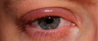
In case of redness or swelling, you should:
- Instill drops of antibacterial action: “Tobrex”, “Floxal”, “Tsiprolet”. They relieve inflammation and prevent the development of infectious eye diseases.
- Use eye drops such as Taufon or Albucid. They eliminate eye redness and promote rapid healing of the cornea.
In addition to drug therapy, traditional medicine methods can be used to relieve discomfort:
- The eyes are washed with strong black tea. A cotton pad is generously moistened with warm tea leaves and wiped over the eyes. A separate disk is used for each.
- Three times a day, one drop of infusion of sage, chamomile or calendula is instilled.
- Place a freshly cut cucumber or potato slice on closed eyelids and leave for twenty minutes.
- Dissolve a little honey in a tablespoon of boiled water. Moisten a cotton pad with the resulting solution and wipe your eyes.
Traditional medicine methods are applicable only if the foreign body is removed without complications.
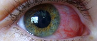
Signs of foreign body entry
Symptoms caused by foreign objects entering the eye can vary widely:
- mild discomfort or intense pain;
- redness of the eye;
- burning sensation;
- increased lacrimation;
- photophobia;
- reflex closing of the eyelids, difficulty when trying to open the affected eye;
- blurred vision.
The severity of the clinical picture depends on the type of foreign body, its size and location.
Injuries to the cornea of the eye
Most often, corneal injuries are caused by scratching with a fingernail or other foreign body, but more serious injuries, such as chemical burns, can also occur.
All corneal wounds are divided into linear and patchwork. They can be of various sizes and shapes. Clinical manifestations: lacrimation, photophobia, eye pain, blepharospasm. If an infection occurs, inflammatory infiltration of the wound edges can be detected.
To diagnose a non-perforated corneal wound, in addition to anamnestic and clinical data, the following method is used: drops of a fluorescein solution (1%) are instilled into the conjunctival sac, followed by rinsing with a sodium chloride solution (isotonic). The injured area of the cornea will turn yellow-green.
Treatment
Analgesics are not used for injury to the cornea of the eye, because this delays the healing process. Epithelization occurs within a few days without leaving a trace. If the damage was deep, it is possible that after healing there will be an area of cloudiness, which (sometimes) reduces visual acuity. In general, the prognosis for corneal injuries is favorable.
If a foreign body gets into the eye
If a foreign body gets into the eye, you must adhere to the following rules:
- do not rub the affected eye - or better yet, do not touch it with your hands at all, so as not to dislodge the foreign body and cause an infection;
- If you use contact lenses, you do not need to remove them after contact with a foreign body.
- try to keep the affected eye closed;
- do not try to remove a foreign body yourself or with the help of friends and family;
- consult an ophthalmologist as soon as possible;
- remember the circumstances of your injury and tell the doctor what materials and substances you worked with.
When can only a doctor help?
If you are unable to remove debris from your eye, then you should consult a doctor. Specialized medical care is also needed when:
- the eye is inflamed
- discomfort does not go away for a long time
- the eye is red and the redness does not disappear (it’s common for the eye to turn red when you rub it or try to get a speck out; you should sound the alarm if the redness lasts for a long time)
- you feel constantly watery or feel like you have a scratch on your eye
- you experience blurred vision
- the eye hurts and the pain intensifies.
In all of the above cases, you should run to the doctor. An ophthalmologist will give advice and help get rid of discomfort and, if necessary, prescribe treatment.
Diagnosis of a foreign body
A patient admitted with complaints of a foreign body in the eye is referred to:
- consultation with an ophthalmologist with a thorough eye examination;
- biomicroscopy, that is, examination of all eye structures using a slit lamp;
- ophthalmoscopy (examination of the fundus of the eye);
- gonioscopy (examination of the anterior chamber of the eye).
According to indications, the following instrumental studies are also prescribed:
- ultrasound examination of the eye;
- radiography of the eye sockets and skull;
- computer (or magnetic resonance) tomography (if there is a reasonable suspicion of a magnetic foreign body, MRI is contraindicated).
Hemorrhage into the vitreous body of the eye: symptoms and treatment
A blow to the vitreous area results in hemorrhage. Blood in the retrolental space of the vitreous expands it, and blood in the orbicular space leads to the formation of a specific rim (stripe) that surrounds the periphery of the lens behind.
Retrolental hemorrhage takes longer to resolve than orbicular hemorrhage. Sometimes minor hemorrhages may be invisible and are discovered later, when they “descend” to the lower part of the anterior chamber.
Hemophthalmos is understood as massive hemorrhage into the vitreous body, occupying a significant part of the latter.
Approximately on the third day after injury, the blood in the vitreous body undergoes a process of hemolysis with the loss of hemoglobin by red blood cells, as a result of which they become colorless and later disappear. And the hemoglobin of erythrocytes takes on the form of grains, which are subsequently absorbed by phagocytes. Hemosiderin is formed, which has a toxic effect on the retina. Sometimes the blood does not completely resolve and a blood clot is formed and replaced by connective tissue moorings. The clinical picture of hemophthalmos is dominated by loss of visual acuity from the state of light perception to complete blindness. Focal illumination and biomicroscopy make it possible to see behind the lens a dark brown granular, sometimes with a reddish tint, mass of blood that permeates the vitreous body. Ophthalmoscopy shows the absence of a fundus reflex. Later, when the blood clot dissolves, one can observe deformation of the vitreous with its liquefaction. Hemophthalmos must be distinguished from partial hemorrhage into the vitreous body, which quickly and completely resolves.
Hemophthalmos leads to the development of degenerative processes in the vitreous body.
Treatment
In case of vitreous hemorrhage, bed rest and a cold bandage are prescribed on the affected eye. Calcium preparations (tablets, eye drops and intramuscular injections), hemostatic agents (Vicasol) are used. To speed up the resorption of hemorrhage, heparin is used (subconjunctivally on days 1–2) and enzyme preparations and potassium iodide. Treatment of hemophthalmia: bed rest with the head end elevated. Use a binocular bandage for 2–3 days. Calcium chloride, pilocarpine 1% 2 times a day, glucose with ascorbic acid are used, and a solution of dicinone (12.5%) is injected subconjunctivally. Then, after 2-3 days, absorbable drugs are used: dionin, potassium iodide, lidase. Corticosteroids (under the conjunctiva) and fibrinolysin are also indicated. In the late period of treatment, ultrasound and physiotherapy help a lot. If there is no positive effect from therapy, then it is necessary to suction the vitreous, and sometimes excise part of it. The removed vitreous body is replaced with luronite (a hyaluronic acid preparation).
We must not forget that with somatic diseases (cardiovascular diseases, atherosclerosis, hypertension, blood diseases, endocrine pathology), the development of hemophthalmos is possible. But in these diseases, hemophthalmos occupies a small part of the vitreous body.
The prognosis depends on the area of hemorrhage. If the hemorrhage covers 1/8 of the vitreous body, it often resolves. With an area of 1/8-1/4 of the vitreous, moorings are formed, which leads to retinal detachment. In terms of restoring visual functions and preserving the organ in total hemophthalmia, when a blood clot occupies more than ¾ of the vitreous, the prognosis is unfavorable. Irreversible destructive changes occur in the vitreous body (organization of a blood clot, formation of adhesions). Tractional retinal detachment and atrophy of the eyeball may develop.
Treatment of a foreign body in the eye
Let us note once again that the removal of foreign bodies from the eyes, even those located on the surface of the eye membranes, should be performed by an ophthalmologist. Any attempts to cope with the problem on your own can worsen the situation.
Superficial foreign bodies are removed on an outpatient basis, and local anesthesia is used if necessary. Most often, this procedure is performed under a microscope (slit lamp). After removal, antibacterial and anti-inflammatory therapy in drops or ointments is prescribed.
Removal of intraocular foreign bodies is carried out in the operating room using a microscope and surgical instruments. Since penetrating eye injuries can lead to loss of vision, surgery is performed immediately.
To prevent foreign bodies from getting into your eyes, when carrying out carpentry, plumbing, construction and other work that involves the risk of eye injury, you must use safety glasses.
Removing visible debris
It's easier to remove something from the eye that you see. First you need to find a well-lit place and take a mirror with you.
- Gently open the affected eye using your thumb and index finger.
- Place your index finger on the eyebrow growth line, and your thumb just below the lower eyelid. This way you can see what's going on in the eye and find out what kind of debris is bothering you on the cornea (located in front of the iris and pupil) or on the sclera (the white part of the eye).
- When you find a speck, take a damp swab or napkin and carefully remove the dirt. The eyes are a sensitive organ and you may instinctively close your eyes. If you are unable to perform the manipulation yourself, ask a family member to help you.
- Another option is to put special drops or saline solution into your eyes. The drops moisturize the surface of the eye and the water “washes away” the dirt.
If the method of simply dripping drops into your eyes does not help you, then use a damp cloth. Both methods can also be used when you do not see the speck, but only feel it.
Introduction
Eye injuries have firmly taken a leading place among the causes of visual disability in Russia, accounting for 22.8% of the number of persons recognized as disabled for the first time. The main task of healthcare is to prevent eye injuries and ensure optimal medical and social rehabilitation of victims. A retrospective analysis of the problem indicates the dependence of visual injuries on the social, domestic, military-political and criminal situation in the country and in the world. Analysis of the political and economic situation in the country allows us to predict the high relevance of scientific research in the field of ophthalmology in this direction (R.A. Gundorova, 1997). Injuries to the organ of vision can be divided into industrial, agricultural, household, and children's. Each type of injury has its own characteristics. Where to begin examining a patient with an eye injury?
First of all, it is very important to correctly and carefully collect an anamnesis of the injury: where, when and under what circumstances it occurred, what was the traumatic agent, and what is the mechanism of injury, the time elapsed from the moment of injury to the patient’s admission, vaccinations that were previously administered to the patient. Then they begin directly to study the ophthalmological status of the patient, which includes: 1. External examination of the eyelids and palpation of the bony edges of the orbit. 2. Inspection of the conjunctiva of the eyelids and transitional folds using the method of focal illumination of the eye and eversion of the eyelids. 3. Inspection and palpation of the lacrimal sac, determining the nature of its contents. 4. Detection of superficial and penetrating wounds of the cornea using fluorescein test and Seidel test. 5. Study of pupillary reactions. 6. Assessment of the color of the pupillary zone in transmitted color. 7. Checking visual acuity (at least approximate). 8. Approximate assessment of intraocular pressure. 9. Examination of the ocular media and fundus (if possible). The variety of injuries is difficult to fit into classification frameworks. There are injuries to the orbit, appendages of the eye and eyeball. It is rational to divide injuries into mechanical, thermal, chemical, radiant energy, vibration, toxic, etc. Mechanical injuries, in turn, are divided into blunt injuries (contusions) and wounds; the latter are penetrating and non-penetrating, and they separately distinguish through injuries. According to the severity of the injury, injuries are divided into mild, moderate and severe, but for the eyeball this classification is to a certain extent arbitrary, since it is difficult to predict the course of the wound process in the eye. With relatively mild injuries, especially penetrating ones, the course of the wound process in the eye can be severe.
Injury to the eyelids and conjunctiva
They look different and are often accompanied by associated injuries, which are not always determined by doctors at the place of residence. The size and appearance of the wound of the eyelid and conjunctiva may not correspond to the severity of the concomitant damage to the deeper parts. A mandatory study of visual acuity, media transparency and examination of the fundus is necessary. It is necessary to pay attention to the presence of subcutaneous emphysema, indicating damage to the paranasal sinuses and nasal bones. First aid is the introduction of PSS (1500-3000 units) according to Bezredka. Cleaning the wound with tweezers, a tupper, washing with peroxide and furacillin, treating the edges of the wound with alcohol (1% brilliant green solution). Applying an aseptic dressing. Then urgent surgical treatment of the wound is indicated, but in principle, sutures can be applied at any time after the injury (if there is no infection). A wound to the conjunctiva requires surgical debridement if there is divergence of the edges of the wound or the sclera is exposed, which indicates damage to Tenon's capsule. Diagnosis of subconjunctival scleral tears
The first symptom of subconjunctival rupture of the sclera, which ophthalmologists paid attention to, was prolapse of the lens under the conjunctiva (Mandelstamm, 1888).
In addition to prolapse of the lens, prolapse of the iris, ciliary body, choroid and vitreous body was often observed (Shereshevskaya, 1959, Zolotareva, 1961, etc.). These symptoms should be considered characteristic of subconjunctival scleral tears. More often, we encounter not the classic picture, but cases without the listed symptoms. When can a subconjunctival scleral rupture be suspected?
- chemosis of the conjunctiva, - extensive or limited subconjunctival hemorrhage, - the appearance of iris coloboma and displacement of the pupil, - sudden massive hemorrhage in the anterior chamber and vitreous body, - folds of Descemet's membrane, - decreased IOP, - transillumination of the choroid under the conjunctiva, - prolapse shells under the con-vu, - the glow of the rupture in the form of a red stripe during diaphanoscopy (Linnik, 1964). Diagnosis of scleral tears in the posterior pole of the eye is especially difficult. Observation of the patient and indirect symptoms help in diagnosis: prolonged hypotony of the eye and the presence of incorrect light projection in the patient, as well as ultrasound examination. There is no doubt that the listed symptoms are unequal in importance, however, it should be recognized that pronounced hypotension caused by injury almost always indicates a subconvalal rupture of the sclera. It should be said about the “pain point symptom”, which occurs when pressing with a glass rod after local anesthesia, in the area of a suspected rupture, causing severe pain. This symptom may indicate not only the presence of a rupture, but also its location. Subsequently, patients are subject to treatment in a specialized hospital.