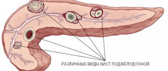
- Types of rhinosinusitis
- Factors predisposing to the development of rhinosinusitis
- Reasons for the development of rhinosinusitis
- Development mechanism
- Symptoms of rhinosinusitis
- Diagnosis and recommended clinical studies
- Laboratory research methods
- Instrumental research methods
- Differential diagnosis
- General principles of treatment
- Forecast
Rhinosinusitis is an inflammation of the mucous membrane of the paranasal sinuses.
Types of rhinosinusitis
- Acute (duration of illness less than 12 weeks and complete disappearance of symptoms after recovery).
- Recurrent (from 1 to 4 episodes of acute rhinosinusitis per year, periods between exacerbations (when there are no symptoms of the disease and no treatment is carried out) last at least 8 weeks).
- Chronic (presence of symptoms for more than 12 weeks).
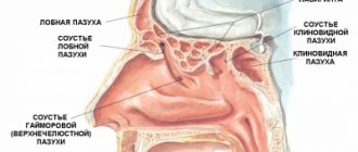
Localization
- maxillary sinus (sinusitis),
- sphenoid sinus (sphenoiditis),
- frontal sinus (frontitis),
- in the cells of the ethmoid bone (ethmoiditis).
Depending on the etiological factors, acute and recurrent rhinosinusitis is divided into:
- viral,
- bacterial,
- fungal.
Chronic are divided into:
- bacterial,
- fungal,
- mixed.
Taking into account the characteristics of pathogenesis:
- hospital,
- odontogenic,
- polyposis,
- developed against the background of immunodeficiency states of rhinosinusitis,
- acute (fulminant) form of mycosis of the paranasal sinuses.
Chronic fungal rhinosinusitis is divided into:
- allergic (eosinophilic) fungal sinusitis,
- mushroom ball,
- superficial sinonasal mycosis,
- chronic invasive form of mycosis.
Clinical features and diagnostic criteria for ARS
According to the definition of EPOS 2021 [5], ARS is an inflammation of the mucous membrane of the sinus and nasal cavity lasting <12 weeks, accompanied by two or more symptoms, which include:
difficulty breathing through the nose (nasal congestion) or nasal discharge;
pressure/pain in the face;
decreased or loss of sense of smell;
and:
rhinoscopic/endoscopic signs:
mucopurulent discharge, mainly in the middle nasal meatus
and/or
swelling/mucosal obstruction, predominantly in the middle meatus
and/or
changes during CT:
changes in the mucosa within the ostiomeatal complex and/or sinuses;
complete disappearance of symptoms no later than 12 weeks. from the onset of the disease.
EPOS 2021 also highlights recurrent ARS, which can occur once or several times over a period of time. It typically manifests as ≥4 episodes of ARS per year with complete resolution of symptoms between episodes [6, 7].
Depending on the location of the inflammatory process, maxillary sinusitis, sphenoiditis, frontal sinusitis or ethmoiditis develops. However, more often ARS occurs with simultaneous damage to several sinuses (polysinusitis). Involvement of all SNPs in the inflammatory process on one side is called hemisinusitis, and simultaneous bilateral involvement of all SNPs is called pansinusitis [1].
In children, the development of inflammation in the SNP depends on age-related anatomy. At birth, only the maxillary sinuses and the ethmoidal labyrinth sinuses are present, the size of which increases by age 14. The sphenoidal sinus begins to develop at the age of 2 years and reaches a constant size by 12 years. Frontal sinuses develop at 6–8 years of age and reach full size by 16 years of age [8]. Depending on the etiological factor, acute sinusitis is divided into viral, postviral and bacterial.
ARS only in 2–10% of cases has a bacterial etiology, and in 90–98% of cases it is caused by viruses [2]. Viral ARS often resolves in 5–7 days and requires only symptomatic treatment. If the disease lasts more than 10 days or the symptoms intensify after 5 days of the disease, then we can talk about the development of post-viral sinusitis. Patients with prolonged ARVI often see general practitioners, on whom further diagnosis and treatment tactics depend. It is important for these specialists to know the criteria for the development of ARS and to assume the possibility of developing ARS. If a patient suffers from ARS three or more times a year or has suffered ARS with complications, the general practitioner should refer such a patient for a consultation with an otolaryngologist.
The key points in choosing treatment tactics for ARS are timely diagnosis of ARS and deciding on the need to prescribe antibiotics [3].
According to the EPOS 2021 criteria [5], we can talk about the development of ABRS if at least 3 symptoms are present:
colorless discharge (more on one side) and/or purulent secretion in the nasal cavity;
severe pain in the face (more on one side);
fever (>38 oC);
increased ESR/C-reactive protein level;
“two waves”, i.e. deterioration after an initially milder phase of the disease.
In Russian clinical guidelines, the course of ARS is divided into degrees of severity, which determine the strategy and tactics of treatment and the need for antibacterial therapy [3]. Usually, with mild severity, the body temperature does not increase, headaches and the risk of developing intracranial complications are absent. Symptoms of ARS, such as difficulty in nasal breathing, rhinorrhea, nasal obstruction, cough, do not lead to a significant decrease in the patient’s quality of life and do not have a pronounced effect on the circadian rhythms of a person’s life, and do not distract him from everyday activities.
With moderate severity, symptoms of intoxication appear, hyperthermia up to 38 ° C, heaviness in the head and pain in the projection of the UL, especially when bending forward and sudden movements of the head. Symptoms of ARS are more severe and disrupt the patient's life and activities. In some cases, ARS is complicated by catarrhal or purulent otitis media and hearing loss, without the occurrence of intracranial and orbital complications.
A severe course is characterized by hyperthermia above 38 °C and a significant increase in symptoms of ARS, leading to a serious condition of the patient, against which intracranial or orbital complications arise.
When providing outpatient care in cases of mild to moderate ARS, without the threat of developing intracranial and orbital complications, patients with ARS can be treated by a general practitioner.
By decision of the council of experts, whose meeting took place in 2020 as part of an educational course conducted by the Russian Society of Rhinologists, a document was developed “Updating clinical guidelines for acute rhinosinusitis and adapting them to EPOS 2020”, which provides recommendations for the diagnosis and treatment of ARS, in including for general practitioners. It is recommended to conduct diagnostic studies to assess the severity of the inflammatory process, such as: general blood count, ESR, C-reactive protein level. It must be remembered that ARS can be a manifestation of blood diseases, systemic diseases, etc.
When making a diagnosis, general practitioners and pediatricians should assess the severity of the main symptoms, such as discharge and nasal congestion, as well as cough in children. Cases of increased body temperature over 38 °C, severe headache or predominantly unilateral facial pain, purulent discharge from one half of the nose, and the presence of “two waves” during the course of the disease should alert us to the development of bacterial ARS. The process and development of concomitant diseases, such as acute otitis media, bronchitis, pneumonia, are aggravated.
If, against the background of the therapy, there is no positive dynamics within 10 days or there is an increase in the symptoms of sinusitis, acute otitis media, severe headaches, neurological symptoms, swelling of the soft tissues of the orbit, a consultation with an otolaryngologist is necessary.
Symptoms such as unilateral periorbital changes (hyperemia, edema), meningeal symptoms, signs of sepsis, swelling of the skin in the projection of the frontal sinus, severe headaches that are not relieved by nonsteroidal anti-inflammatory drugs (NSAIDs), impaired mobility and position of the eyeball indicate the development of complications and require urgent referral of the patient to the otorhinolaryngology department of the hospital.
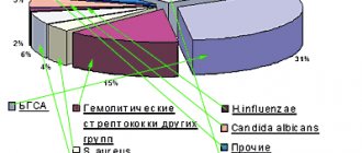
Streptococcus pneumoniae is most often cultured in smears of secretion from the middle nasal passage and discharge from the maxillary sinuses obtained during puncture.
and
Haemophilus influenzae
(total contamination - about 75%).
These data are confirmed by studies by JA Hadley and MA Pfaller, who noted that in ABRS, S. pneumoniae
was cultured in 20–43% of cases in adults and in 25–30% of cases in children.
The proportion of H. influenzae
was 22–35% in adults, which is 1.5 times more than in children (15–20%) [9].
According to the results of a multicenter microbiological study of SSSR microflora, conducted in patients with ABRS in Smolensk, Moscow and St. Petersburg, S. pneumoniae
was isolated in 42.0% of cases, and
H. influenzae
in 25.4%.
Streptococcus pyogenes
(6.9%),
Haemophilus parainfluenzae
(2.3%),
Staphylococcus aureus
(1.7%),
Moraxella catarrhalis
(1.1%)
were identified in smaller numbers 10]. A distinctive feature of this study was the identification of a low percentage of M. catarrhalis
in the studied material, which cannot be said about hemolytic streptococci (mainly group F), which were detected in severe patients with ABRS, which is presumably odontogenic in nature (Fig. 1) [10].
Despite the varying severity of ARS, antibiotic therapy is prescribed in 85–98% of cases. This leads to increased antibiotic resistance and reduces the effectiveness of treatment [7, 11].
According to the results of a multicenter prospective study of PeGAS conducted in Russia, pneumococci isolated from the maxillary sinuses were highly resistant only to co-trimoxazole (32.4% of resistant strains) and tetracycline (29.4% of resistant strains). Pneumococcal strains resistant to penicillin were identified in approximately 11% of cases, and to erythromycin - in 4.6%. The results obtained were encouraging, since there was no resistance of pneumococcus to protected penicillins, cephalosporins and fluoroquinolones [12].
However, repeated studies of antibiotic resistance in recent years have revealed clear negative dynamics and an increase in the resistance of pneumococcal strains to the group of macrolides (azithromycin) and cephalosporins, including the third generation. A characteristic feature was not only an increase in resistance, but also an increase in the bactericidal dose of cephalosporins to achieve a therapeutic effect even in sensitive strains of pneumococcus [13].
Factors predisposing to the development of rhinosinusitis
- Rhinitis.
- Intolerance to non-steroidal anti-inflammatory drugs.
- Anomalies in the structure of the nasal cavity and paranasal sinuses (deviated nasal septum; bulla of the middle turbinate; additional anastomosis of the maxillary sinus, etc.).
- Immunodeficiency states (X-linked agammaglobulinemia; common variable immunological deficiency; deficiency of IgG subclasses; selective IgA deficiency; hyper-IgM syndrome; HIV).
- Diseases accompanied by a slowdown in MCT (Kartagener's syndrome; Young's syndrome; cystic fibrosis).
- Wegener's granulomatosis.
- Hyperplasia of the pharyngeal tonsil, adenoiditis.
- Gastroesophageal reflux disease.
- Fistula between the oral cavity and the maxillary sinus.
Centrotherapy in combination with the “Light Breathing” irrigation technique for acute sinusitis
The combination of techniques gives a quick healing effect. As a rule, about 5 sinus lavage procedures and 10 centrotherapy sessions 2-3 times a week are required. Centrotherapy sessions are carried out from the first days of treatment, after the washing procedure, which enhances the therapeutic effect. In some cases, it is possible to shorten the course of antibiotic therapy.
*terminology of the founder of centrotherapy P. Bonnier
See the articles “Protracted runny nose in children and adults”, “Frequent colds in children and adults. Immune problems?
Reasons for the development of rhinosinusitis
The main causative agents of acute bacterial rhinosinusitis are Streptococcus pneumoniae and Haemophilus influenzae. Other pathogens include Moraxellacatarrhalis, Staphylococcusaureus, Streptococcuspyogenes, Streptococcusviridans, etc. The main anaerobic pathogens of rhinosinusitis are anaerobic streptococci. However, the spectrum of pathogens causing acute bacterial rhinosinusitis can vary significantly depending on geographic, socio-economic and other conditions.
The list of nosocomial pathogens that developed against the background of immunodeficiency conditions and odontogenic rhinosinusitis, along with the bacteria mentioned above, includes Staphylococcus epidermidis, Pseudomonasaeruginosa, Proteus spp., and in immunodeficient patients also saprophytic bacteria and fungal microflora. In recent years, the role of chlamydia and other atypical microflora in the etiology of rhinosinusitis has been discussed.
Fungal sinusitis is most often caused by Aspergillus fungi (in most cases A. fumigatus), less often by Candida, Alternaria, Bipolaris, etc.
The acute invasive form of mycosis of the paranasal sinuses is most often caused by fungi of the Mucoraceae family: Rhizopus, Mucor and Absida.
Development mechanism
Rhinosinusitis almost always develops when mucociliary clearance is impaired, when optimal conditions are created for the development of a bacterial infection.
The trigger point in the development of acute bacterial rhinosinusitis is usually ARVI. It was revealed that in almost 90% of patients with ARVI, changes in the paranasal sinuses are detected in the form of swelling of the mucous membrane and stagnation of secretions. However, only 1-2% of such patients develop acute bacterial rhinosinusitis.
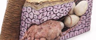
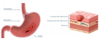
In the development of chronic rhinosinusitis, in addition to disturbances of mucociliary transport, abnormalities in the structure of the intranasal structures and ethmoidal labyrinth play an important role, blocking the patency of the natural openings of the paranasal sinuses and disrupting the mechanisms of sinus cleansing. The presence of two or more anastomoses of the maxillary sinuses also creates conditions for the throwing of infected mucus that has already been in the nasal cavity and back into the sinus. Under conditions of chronic inflammation in the mucous membrane, focal or diffuse metaplasia of multirow columnar epithelium occurs into multilayer epithelium, devoid of cilia and having lost the ability to remove bacteria and viruses from its surface through active mucociliary transport.
Nosocomial rhinosinusitis is most often caused by prolonged nasotracheal intubation.
Odontogenic sinusitis develops against the background of chronic foci of inflammation, cysts or granulomas in the roots of the teeth of the upper jaw, as a result of pieces of filling material, tooth roots entering the maxillary sinuses, or the formation of a fistula between the oral cavity and the maxillary sinus after tooth extraction.
A key role in the pathogenesis of polypous rhinosinusitis is played by eosinophils and IL-5, which causes their proliferation, migration into tissues and degranulation.
Mucopurulent discharge from the affected paranasal sinuses can be transported by the ciliated epithelium directly through the mouth of the auditory tube, which is the trigger point in the development of an exudative or chronic inflammatory process in the middle ear.
Superficial sinonasal mycosis is caused by the growth of fungal mycelium on crusts formed in the cavities of the operated paranasal sinuses, on the surface of neoplasms and on accumulations of antimicrobial drugs or glucocorticosteroids for topical use that remain in the nasal cavity for a long time.
Case from practice
Patient P., 15 years old. A course of nasal irrigation (washing) No. 5 and centrotherapy (10 sessions) was carried out as part of a complex treatment (systemic and local antibacterial therapy, antihistamines and vasoconstrictors) for bilateral acute purulent sinusitis. Before this, for several years he suffered from bilateral sinusitis twice a year and ARVI up to 4-5 times a year. Over the next 2.5 years, I only suffered from mild acute respiratory viral infection three times, using only vasoconstrictor drops for a runny nose.
Symptoms of rhinosinusitis
The main symptoms of rhinosinusitis are:
- difficulty in nasal breathing,
- headache,
- nasal discharge.
Variable symptoms:
- decreased sense of smell,
- stuffy ears,
- increase in body temperature,
- general malaise,
- cough (more common in children).
With inflammation in the maxillary and frontal sinuses, the pain is localized in the face, nose and eyebrow area. Sphenoiditis is characterized by pain in the center of the head and the back of the head. The discharge is mucous, purulent and can come away when you blow your nose or drain down the back wall of the throat. The latter is more typical for lesions of the sphenoid sinus and the posterior parts of the ethmoid labyrinth. Chronic rhinosinusitis is accompanied by the same symptoms as acute, but outside of exacerbation they are much less pronounced.
Clinical observation
Patient T., 18 years old. Complaints of difficulty in nasal breathing, congestion and purulent discharge from the nasal cavity. According to the patient, complaints appeared against the background of ARVI on the 6th day of illness. It is treated with vasoconstrictor drops and antipyretic drugs with a short-term positive effect. During endoscopy of the nasal cavity with a Karl Storz 0 g endoscope (Germany), inflamed and edematous mucosa, viscous mucopurulent discharge in moderate quantities, mainly in the area of the middle nasal passage on the right, were visualized, the nasal septum deviates to the right, in the nasopharynx and oropharynx, discharge of discharge into the oropharynx was noted with symptoms of pharyngitis.
It must be taken into account that endoscopic examination of the nasal cavity is carried out by an otolaryngologist. General practitioners and pediatricians should follow the diagnostic criteria for ARS.
The patient was prescribed complex topical therapy, which included rinsing the nasal cavity with a normalized solution of sea salt, Sinuforte, 1 injection 1 time per day into each half of the nose for 7 days. During the therapy, regression of symptoms and significant positive dynamics were noted when examined after 7 days (Fig. 2).
The patient's recovery occurred with monotherapy, without the use of antibacterial drugs.
Diagnosis and recommended clinical studies
The diagnosis of rhinosinusitis is established on the basis of:
- Anamnestic data.
- Clinical manifestations.
- Laboratory test results.
- Results of instrumental examination methods.
Acute bacterial rhinosinusitis is characterized by a connection with an episode of acute respiratory viral infection suffered 5-10 days ago.
Patients with a history of odontogenic and fungal sinusitis often have previous complex fillings of the teeth of the upper jaw, as well as a long history of repeated visits to an otolaryngologist and repeated diagnostic punctures of the maxillary sinuses, during which no contents were obtained.
Polypous rhinosinusitis is characterized by gradual progression of the main symptoms: difficulty in nasal breathing and decreased sense of smell. Often patients are bothered by the painful sensation of constant flow of a very viscous secretion down the back wall of the nasopharynx. In many cases, polypous rhinosinusitis is combined with bronchial asthma, intolerance to non-steroidal anti-inflammatory drugs, and cystic fibrosis.
Laboratory research methods
Bacteriological research
This is a study designed to isolate bacteria and study their properties in order to make a microbiological diagnosis. The material for the study can be obtained from the nasal cavity or from the affected sinus during puncture. When collecting material, there is a high probability of “traveling” microflora entering.
Study of mucociliary transport
Allows you to assess the condition of the mucociliary apparatus of the mucous membrane, that is, to identify one of the most important pathogenetic disorders in rhinosinusitis.
In clinical practice, the measurement of transport time is most widely used. One of the variations of this method is to measure the time during which a marker (charcoal, carmine, ink, foam, etc.) moves from the anterior parts of the nasal cavity to the nasopharynx. Due to its simplicity, the saccharin test has become more widespread.
Its principle is to measure the time it takes a particle to travel a conventional distance - from the anterior parts of the nasal cavity to the taste buds in the pharynx. Saccharin time in healthy people can range from 1 to 20 minutes, averaging 6 minutes. However, these indicators are very conditional.
Instrumental research methods
- Rhinoscopy. Anterior rhinoscopy against the background of diffuse congestive hyperemia and edema of the nasal mucosa reveals a typical sign of purulent rhinosinusitis - the presence of pathological discharge in the anastomosis area of the affected paranasal sinuses. With sinusitis and frontal sinusitis, the discharge can be seen in the middle nasal passage, and with sphenoiditis - in the upper.
- Endoscopic examination requires a minimum of time and is painlessly tolerated by the patient. The study includes three main points: sequential examination of the lower, middle and upper nasal passages. The method allows you to identify an additional anastomosis of the maxillary sinus. With a choanal polyp, a formation is detected, the stem of which comes from the anastomosis of the maxillary sinus.
- Diaphanoscopy. Illumination of pericutaneous formations or cysts with a narrow beam of light. Allows to identify a decrease in pneumatization of the maxillary and frontal sinuses.
- Ultrasound is a fast, non-invasive method that is used mainly for screening purposes, to diagnose inflammatory diseases and cysts of the maxillary and frontal sinuses. Both special devices for scanning the paranasal sinuses and standard equipment are used. The sensitivity of ultrasound in diagnosing sinusitis is lower than that of X-ray and CT.
- X-rays of the paranasal sinuses are usually performed in the nasomental projection. To clarify the condition of the frontal and sphenoid sinuses, an additional study can be carried out in the nasofrontal and lateral projections. X-ray of the ethmoid bone sinuses is not very informative. Poor quality radiography often leads to diagnostic errors.
- CT , which is performed in a coronal projection, is the most informative method and is gradually becoming the “gold standard” for studying the paranasal sinuses. CT not only makes it possible to establish the nature and extent of pathological changes in the paranasal sinuses, but also reveals the causes and individual features of the anatomical structure of the nasal cavity and sinuses, leading to the development and recurrence of rhinosinusitis. High-resolution CT allows you to visualize structures that are not visible with conventional radiography.
- MRI , although it provides better visualization of soft tissue structures, is not one of the main methods for diagnosing rhinosinusitis. This method gives virtually no idea of the patency of the air spaces connecting the paranasal sinuses with the nasal cavity. MRI is indicated only in certain situations - for example, if a fungal infection of the paranasal sinuses is suspected or the possible tumor nature of the disease, as well as with orbital and intracranial complications of rhinosinusitis. MRI is the most informative method for differential diagnosis between a cerebral hernia (meningoencephalocele) and a tumor or inflammatory process in the roof of the ethmoidal labyrinth.
- Diagnostic puncture and probing make it possible to assess the volume and nature of the contents of the affected sinus and indirectly obtain an idea of the patency of its natural opening.
To assess the patency of the anastomosis of the punctured sinus, a simple scheme is used, taking into account 3 degrees of violation of the patency of the anastomosis (see table). To do this, using a syringe connected to a needle or drainage tube, the contents are first aspirated, and then the sinus is washed.
Assessment of the patency of the natural anastomosis of the paranasal sinuses
| Normal patency of the anastomosis | During aspiration, air or liquid contents of the sinus enter the syringe; when rinsing, the liquid flows freely into the nasal cavity |
| Obstruction of the 1st degree | During aspiration, negative pressure is created; during rinsing, the liquid freely enters the nasal cavity (valve mechanism and negative pressure in the sinus) |
| Impairment of patency II degree | Aspiration from the sinus is impossible; rinsing is possible only by increasing pressure on the syringe plunger |
| Level III obstruction | Neither aspiration nor lavage of the sinus is possible: there is a complete blockage of the anastomosis |
Differential diagnosis
For viral and bacterial MS, simultaneous damage to several sinuses ( polysinusitis ) is more typical.
Isolated damage to one sinus ( monosinusitis ) is typical of fungal and odontogenic rhinosinusitis .
Signs of rhinosinusitis caused by typical pathogens (S. pneumoniae and H. influenzae) are a decrease in the sense of smell, the presence of a fluid level on the X-ray and the effectiveness of traditional therapy.
Distinctive features of rhinosinusitis caused by other microorganisms: foul-smelling nasal discharge, a total decrease in pneumatization of the paranasal sinuses on X-ray, slower positive dynamics of the X-ray picture during treatment.
Allergic (or eosinophilic) fungal rhinosinusitis is characterized by the detection of multiple polyps during endoscopy, as well as a very characteristic discharge of yellow, green or brown color with a very viscous rubber-like consistency. A similar discharge - allergic mucin - is found in all affected sinuses during surgery.
Odontogenic sinusitis usually acquires a primarily chronic course, accompanied by the formation of polyps, granulations or fungal stones in the sinus.
The chronic invasive form of mycosis is accompanied by the formation of fungal granulomas with their invasion into bone structures and soft tissues of the face.
Polypous rhinosinusitis is characterized by the formation and recurrent growth of polyps, consisting predominantly of edematous tissue infiltrated with eosinophils.
General principles of treatment
The main goals of treatment for rhinosinusitis are:
- Reducing the duration of the disease.
- Prevention of the development of orbital and intracranial complications.
- Eradication of the pathogen.
From this perspective, the basic method of treating acute bacterial rhinosinusitis (moderate and severe forms) and exacerbation of chronic rhinosinusitis is empirical antibacterial therapy.
The main indications for prescribing antimicrobial drugs include:
- Anamnesis characteristic of rhinosinusitis.
- Severity of clinical manifestations.
- The presence of purulent discharge in the nasal passages.
Antibacterial therapy, taking into account the type and sensitivity of a specific pathogen identified during a bacteriological study, does not at all guarantee success due to the high probability of “traveling” microflora entering the test material during sampling. In addition, the results of in vitro sensitivity studies of the identified microorganism do not always correlate with the clinical effectiveness of individual antibacterial drugs. The reasons for this may be a significant increase in antibacterial activity as a result of the unidirectional effect of the antibiotic and its metabolite and the ability to specifically achieve bactericidal concentrations precisely at the site of infection.
Antibacterial therapy for acute bacterial rhinosinusitis
On an outpatient basis, oral antimicrobial drugs are predominantly prescribed.
Taking into account the spectrum of typical pathogens and Russian data on their antibiotic resistance, the drug of first choice for acute bacterial rhinosinusitis is amoxicillin.
In the absence of a noticeable clinical effect, after three days the drug should be replaced with an antibiotic active against penicillin-resistant S. pneumonia and β-lactamase-producing H. influenzae. In this case, III-IV generation cephalosparins or new fluoroquinolones are prescribed.
If you are intolerant to penicillin drugs (and due to possible cross-allergic reactions, cephalosporins should also not be prescribed), the drugs of choice are macrolides.
If the patient is hospitalized, the parenteral route of administration of antimicrobial drugs is preferable.
Antibacterial therapy for exacerbations of chronic rhinosinusitis
When treating exacerbations of chronic rhinosinusitis, oral amoxicillin/clavulanate is considered the drug of choice.
Alternative drugs (prescribed in case of ineffectiveness of the antimicrobial drugs of choice) currently include fluoroquinolones of the III-IV generations. In patients under 16 years of age, alternative drugs include macrolides. Considering the significant role of obstruction of the natural openings of the paranasal sinuses in the pathogenesis of rhinosinusitis, vasoconstrictor drugs, which are prescribed either locally or orally, are of great importance in its treatment.
In the treatment of acute and chronic rhinosinusitis, herbal medicines that have anti-inflammatory and mucolytic effects are also used.
Puncture and probing of the paranasal sinuses
These methods allow you to rinse the affected sinus with an antiseptic solution, remove pathological secretions from it, and administer medications (antiseptics, proteolytic enzymes, glucocorticosteroids). In some cases, puncture and lavage of the paranasal sinuses can eliminate the blockade of its natural anastomosis. It is believed that regular removal of exudate during purulent rhinosinusitis protects local immune factors from proteolysis and increases the content of Ig and complement in the affected sinus by 2-3 times, stimulating the mechanisms of local antibacterial defense. The most common and easier to perform is puncture of the maxillary sinus. It is most often used in the treatment of rhinosinusitis.
Forced drainage method
The method has certain advantages over treatment with repeated punctures. The presence of a catheter creates an additional path for evacuating secretions from the affected sinus, increases air exchange, and eliminates negative pressure when the natural anastomosis is blocked or acts as a valve.
Other methods
Nasal showers, rinsing the nasal cavity with a warm isotonic solution and physiotherapy (ultrahigh-frequency currents, microwave therapy, ultrasound).
Surgery
Indications for surgical treatment for bacterial rhinosinusitis arise when antibiotic therapy is ineffective and orbital or intracranial complications develop.
In surgical treatment, the prevailing trend is towards minimal invasiveness. Less traumatic functional endoscopic interventions give better results, are accompanied by fewer complications, and are less likely to contribute to the progression of the disease and the development of bronchial asthma than classical operations with radical removal of the mucous membrane and nasal turbinates.
Therapy for fungal forms of rhinosinusitis
For a fungal ball, antifungal medications are not prescribed. Treatment is surgical (endoscopic). Complete removal of fungal masses from the paranasal sinuses guarantees recovery. Treatment of allergic fungal rhinosinusitis is surgical (in the presence of large polyps). Treatment of superficial sinonasal mycosis involves removing the substrate for the growth of fungal mycelium.
Safely boost immunity for rhinosinusitis (sinusitis)
Unfortunately, in some patients, despite all the efforts of conservative treatment, it is not always possible to achieve the desired effect and rhinosinusitis (sinusitis) continues to recur. It is well known that one of the factors of a protracted course is a decrease in immunity. Therefore, the body cannot properly cope with pathogenic flora that lives on the mucous membrane or is brought from the external environment. The slightest hypothermia or stressful situation (see the article “Immunity and stress. What is the connection?”) lead to a relapse. Trying to solve this problem, modern pharmacology offers many immune stimulants, which have sometimes significant side effects. Meanwhile, immunologists themselves are in no hurry to actively invade the immune system. Everything in it is not very simple and has not been fully studied. Today they are the last to prescribe immunotropic therapy. The leading positions are occupied by pediatricians, therapists, and gynecologists. And patients themselves often self-medicate and rush to buy something for immunity.
An alternative to medications are non-drug methods of increasing immunity, among which centrotherapy deserves special attention. This method is distinguished by high efficiency - up to 85% recovery and long-term disease-free course of the disease; absence of side effects, which is contained in an environmentally friendly mechanism of therapeutic action.
Illustrating the effectiveness of centrotherapy in increasing immunity, it is appropriate to mention the facts of treatment in France more than a hundred years ago of open forms of tuberculosis. After all, at that time anti-tuberculosis drugs had not yet been synthesized and the diagnosis of consumption sounded like a sad sentence. By means of minimal thermal effects applied to the “immune zones”* in the nose, it was possible to raise immunity and cure patients: fever and cough stopped, the general condition became normal, and there were no tuberculosis bacilli in the scanty sputum coughed up at the end of treatment.
Similarly, when treating a persistent runny nose, the effect of increasing immunity is used. Activation of the “immune center”* leads to the restoration of immune defense mechanisms to the proper level. This approach leads to the elimination of inflammatory phenomena and recovery.