Lymphoma is a malignant disease that affects the lymphatic system. More precisely, we are talking about a group of diseases, because there are several of them - and they differ in different features.
The lymphatic system itself is a complex system whose vessels cover all internal organs. It also includes lymph nodes, which form lymphocytes, which are very important for our immunity. This system has several key functions. The barrier cleanses the lymph and traps particles undesirable for the body (for example, dead cells). The transport function delivers nutrients to the organs, and the immune function helps fight viruses, bacteria, and any harmful cells.
If lymphoma develops, lymph cells begin to divide uncontrollably, tumors appear, which, without timely treatment, threaten the patient with death.
General information
Malignant lymphomas (cancer of the lymphatic system) are a large group of tumor diseases that originate from lymphoid tissue and belong to hematological malignancies .
Malignant lymphomas are more common in young people. Lymphoid organs, which include the lymph nodes, spleen, appendix, Peyer's patches of the intestine, tonsils and thymus, contain cells of lymphoid tissue. If lymphoma develops in lymphatic structures, then this is a nodal manifestation of the disease . If lymphomas originate from the lymphoid tissue of other organs (liver, stomach, brain), skin, then these are extranodal tumors .
The lymphatic system consists of lymph capillaries, lymph vessels and lymph nodes. Lymphatic capillaries collect lymph (interstitial fluid) with microorganisms, various cells, electrolytes and carry it to the lymph nodes for filtration. This is how the drainage function of this system occurs - the lymph nodes neutralize microorganisms, providing protection against infection. Lymphatic vessels have valves that prevent the reverse flow of lymph, so it flows in one direction into the venous bed. A primary tumor can develop in the lymph nodes (in the case of hemoblastoses, which will be discussed today), or there can be a secondary metastatic lesion when malignant cells spread from tumors, when there is oncology of various organs. Most often, malignant lymphomas affect the lymph nodes, which is why in everyday vocabulary it is called lymph node cancer.
Malignant lesions of the lymph nodes occur when:
- Hodgkin's lymphoma is a common variant that affects the lymph nodes. In terms of cure, it is a more favorable disease.
- Non-Hodgkin's lymphomas (the term lymphosarcoma , which was used earlier, is considered obsolete). This is a large group of malignant lymphoproliferative tumors. The identification of various variants is based on the morphological and immunophenotypic characteristics of tumors. Depending on this, there are differences in the clinical course and prognosis.
The starting point for diagnosing all these diseases is enlarged lymph nodes, which is the first and main symptom. Most often, malignant lesions of the lymph nodes occur in the groin area, neck and axillary region. An enlarged lymph node of more than 1 cm and lasting more than a month is an indication for a biopsy .
Diagnostic measures
Diagnosis of non-Hodgkin lymphoma begins with the first medical examination and medical history. Depending on the complaints, the doctor builds the entire strategy for examining the patient. For example, indolent non-Hodgkin's lymphoma is often diagnosed during routine examinations, since it does not produce clear symptoms.
During the examination, the doctor first of all pays attention to all groups of lymph nodes. He palpates and evaluates their size, consistency, sensitivity and shape.
In order to see deep lymph nodes, you can use: radiography, computed tomography , MRI .
If at least one of the nodes is suspicious, he directs the patient to puncture the contents of the node.
If a tumor is detected, it is necessary to perform a targeted tissue biopsy. Next, it will be assessed by a laboratory technician who will accurately determine NHL and its subtype.
Additionally, a biopsy and laboratory tests of the bone marrow may be prescribed, since this is where immune cells begin to form.
Of course, all these procedures are accompanied by a clinical blood test, where a laboratory technician can clearly count the number of pathological cells.
If an infection is present, screening tests are performed to identify the causative agent.
On each affected lymphocyte, there is a certain antigen that can be calculated and determine the specific type of NHL. To identify them, immunohistochemical analysis is used, which allows differentiation between different oncological diseases.
According to statistical studies, non-Hodgkin lymphoma is a fairly common disease. For example, in the USA, it is the most popular type of cancer. It accounts for 4% of all cases of malignant diseases. ( https://www.cancer.org/cancer/non-hodgkin-lymphoma/about/key-statistics.html )
Prognostic criteria
The prognosis of non-Hodgkin's lymphomas strictly depends on certain criteria
- Age;
- Stage;
- Lymphoma in or outside the lymphatic system;
- Patient activity according to the recommendations of the Eastern Cooperative Oncology Group (ECOG)
- Increased lactate dehydrogenase (LDH).
Depending on these criteria, the International Prognostic Index (IPI) for non-Hodgkin's lymphomas is calculated.
First of all, it is necessary to assess the patient's activity, according to ECOG recommendations.
0 - normal physical activity, without restrictions;
1 — minor limitation due to the onset of symptoms of the disease, but performs non-physical work without problems;
2 — the patient takes care of himself completely, but cannot perform tasks, requires a lot of rest, spends no more than half of the sunny day lying down;
3 — the patient spends more than half the day in bed and requires assistance with basic activities (eating, going to the toilet);
4- the patient is in bed all day, round-the-clock supervision and care is necessary;
5 — death.
(https://ecog-acrin.org/resources/ecog-performance-status)
To evaluate aggressive NHL, it is necessary to evaluate the patient.
| Criteria | Values |
| Age | More than 60 years |
| Patient activity according to ECOG | More than two points |
| Stage of development | III or IV |
| Lymphomas in or outside the lymphatic system | Yes |
| Increased LDH activity | High |
Each positive answer that matches the criterion in the criteria column means 1 point. This way you can get from 1 to 5 points.
Explanation:
* 1 or 0 points – the risk is very low, treatment prognosis is favorable.
* 2 points - average risk, favorable treatment prognosis;
* 3 points - High risk, unlikely cure;
* 4-5 points - very high risk, very unfavorable prognosis.
Indolent lymphomas
| Criteria | Meaning |
| Age | More than 60 years |
| Patient activity according to ECOG | More than two points |
| Stage of development | III or IV |
| Lymphomas in or outside the lymphatic system | Yes |
| Increased LDH activity | High |
Each positive answer that matches the criterion in the criteria column means 1 point. This way you can get from 1 to 5 points.
* 0-1 points – low risk
* 2 points – average risk
* 3 or more – high risk
(https://empendium.com/mcmtextbook/chapter/B31.II.15.13.)
Pathogenesis
Oncogenes are of primary importance in the pathogenesis of lymphomas. B-cell tumors are characterized by changes in the c-myc proto-oncogene in the cell, which promotes the transition of lymphocytes to the phase of division and constant reproduction. Chromosome abnormalities are also important in the development of lymphomas, as a result of which control over cell division is lost. Subsequently, the tumor tissue grows and has a separate metabolism. Tumor cells suppress the development of normal cells, often cause immunosuppression and susceptibility to various infections: pneumonia , urinary tract infections, herpesvirus infection . An increase in tumor mass causes exhaustion of the body.
Targeted therapy
A progressive technique that involves the use of genetically modified immune cells. Innovative targeted drugs act on specific molecules that are located directly in the cancer cell. They block the access of oxygen to tumor tissues, which is necessary for cell life and reproduction.
Unlike chemotherapy, targeted drugs have less effect on healthy cells in the body. They are often prescribed to elderly people and patients in serious condition when other treatment methods are contraindicated. Targeted therapy can also be used to prevent cancer recurrence, as well as to control the growth of cancer metastases.
Classification
All malignant lymphomas are usually divided into two large groups: Hodgkin and non-Hodgkin.
Classifications of lymphomas have constantly changed and been supplemented by histological and molecular genetic studies. Currently, neoplasms of lymphoid tissue include more than 20 types and are divided into 3 large groups:
- B cell.
- T cell.
- Hodgkin's disease.
- B-cell and T-cell lymphomas constitute “non-Hodgkin lymphomas.” In turn, they can originate from precursors of B and T lymphocytes and mature B and T lymphocytes.
Nodular lymphomas are divided into stages:
- Stage I - one lymphatic zone is affected.
- Stage II - two or more areas of lymph nodes are affected, but on one side of the diaphragm. One extranodular organ with regional lymph nodes may also be affected.
- Stage III - lymph nodes on both sides of the diaphragm and one extranodular organ are affected.
- Stage IV is widespread involvement of multiple extranodular organs with lymph node involvement.
Separately, it is worth mentioning secondary damage to the lymph nodes - these are metastases in the lymph nodes. In this case, tumor cells from the primary tumor travel through the lymph to the lymph nodes. Metastasis cells correspond to the cells of the primary tumor. The tumor embolus begins to grow in the node. In most cases, the lymph node cortex is involved in the metastatic process. As the tumor mass grows, the node's own tissue is replaced, the size of the metastasis increases and the unevenness of its contours increases. Subsequently, the tumor process progresses and extends beyond the capsule of the node. Very often, the affected lymph nodes are combined into a conglomerate. The growth of the metastasis beyond the node then involves surrounding tissue. Vessels are often involved in the tumor process - they are displaced and compressed. The less normal tissue of the lymph node and the greater the tumor mass, the more homogeneous the structure of the node and the less echogenic it is.
The main “target” is the lymph nodes of the neck, since lymph flow occurs here from the upper part of the body and from the lower half of the body. Metastases to the neck area occur in 40–70% of cases and are most often caused by oncology of the thyroid gland, nasopharynx, salivary glands, sweat glands, oral cavity and breast.
Types of lymphomas by aggressiveness
Since there are many diseases that are included in this group, they are usually divided into groups according to different criteria. One of them is the aggressiveness of the current.
So-called flaccid lymphomas (also known as indolent lymphomas) develop slowly and do not threaten the patient’s life for several years. With timely treatment, the chances of a long and lasting remission are high. This group includes lymphocytic as well as follicular lymphoma.
Aggressive forms, which include diffuse (mixed and large cell) forms, can kill the body within months, and highly aggressive forms - even within weeks. The latter, for example, include T-cell leukemia and Burkitt's lymphoma. In the treatment of any disease and in maintaining the adequate condition of the patient, timely professional assistance plays an important role.
Causes
Considering the causes of lymphomas, we can highlight the main ones:
- Infectious causes. Epstein-Barr virus causes Burkitt's lymphoma , Hodgkin's lymphoma , NK-T-cell lymphoma . T-cell leukemia virus has an association with T-cell lymphoma. Hepatitis C virus has been associated with splenic lymphoma and large cell lymphoma. The bacterium Helicobacter pylori has an association with MALT lymphoma.
- Exposure to carcinogens. The development of these diseases is facilitated by chemical carcinogens: polychlorinated biphenyls , phenytoin , dioxin , phenoxy herbicides . In addition, it has been noted that taking cytostatics (alkylating drugs cyclophosphamide, melphalan and others) also contributes to the development of lymphomas.
- Exposure to ionizing radiation (radiation therapy for medical purposes).
- Immunosuppression. This refers to long-term use of immunosuppressants ( cyclosporine , glucocorticoids ).
- Autoimmune diseases, among which are Sjögren's syndrome , systemic lupus erythematosus and rheumatoid arthritis .
- Some genetic diseases that predispose to the appearance of lymphomas are Chediak-Higashi and Klinefelter .
Radiation therapy
In most cases, radiation therapy is prescribed in conjunction with surgical treatment. It promotes the complete destruction of cancer cells that may remain after surgery. Also, this technique reduces the symptoms of the disease, for example, relieves pain, reduces the volume of the tumor mass under strong pressure on internal organs, etc. In this case, radiation therapy is carried out as preparation for surgery.
Symptoms of lymph node cancer
If we consider cancer of the lymphatic system using the example of lymphogranulomatosis, then patients may experience the following symptoms:
- enlarged lymph nodes above the neck and collarbone;
- enlargement of the mediastinal nodes, which is accompanied by cough and shortness of breath, and due to impaired venous outflow - swelling of the veins of the neck;
- lower back pain at night;
- pronounced weakness.
The first manifestations of lymphatic system tumors may vary, but most often the disease begins with enlarged lymph nodes that grow quickly. In addition, characteristic of malignant lymphomas are signs of intoxication, which can be used to suspect that there is oncology:
- heavy sweats at night;
- weight loss;
- temperature rise above 38;
- skin itching.
The appearance of one of these symptoms may precede the appearance of the primary tumor.
Symptoms of cancer of the lymph nodes in the neck
Almost half of lymphomas primarily develop in the lymph nodes. With local damage, the lymph nodes of the neck are most often involved in the process. They are dense, but painless, not fused with tissues and merge into conglomerates, forming a single tumor of the lymph nodes.
Photo of lymph node damage in Hodgkin's disease
A tumor in the neck (regardless of which side it is on the left or right) causes cosmetic discomfort, makes movement difficult, and, if large, makes breathing difficult.
Mediastinal lymphoma
This tumor is located in the anterior mediastinum and affects the intrathoracic lymph nodes and thymus nodes. At first, lymphoma does not manifest itself and is discovered accidentally during a chest x-ray. Then symptoms appear that are associated with compression of the mediastinal organs or tumor growth into these organs: hoarseness, cough, difficulty breathing, difficulty swallowing, heart pain, chest pain, shortness of breath, fainting, drooping eyelids and constriction of the pupil (this is associated with compression of the cervical sympathetic ganglion).
Lymphosarcoma
Non-Hodgkin's lymphoma begins with a single tumor node, which then spreads through metastasis. The tumor focus may be in the lymph node or in other organs. In non-Hodgkin's lymphomas, mediastinal lymph nodes are affected less frequently than in Hodgkin's disease, the spleen and bone marrow are affected in 30-40%, and the liver in 20-50% of cases, and damage to the gastrointestinal tract is also typical.
Just as with lymphogranulomatosis, patients are concerned about fever, night sweats and rapid weight loss. Moreover, elevated temperature can bother you for more than 2 months. Then, in half of the patients, the lymph nodes of the neck are affected one after another, merging into large groups. A primary tumor can also develop in the tonsils, which is accompanied by a sore throat and a change in voice. If the tumor develops in the chest cavity, which is much less common, coughing and shortness of breath appear. Possible damage to the gastrointestinal tract. B-large cell lymphoma is the most aggressive and accounts for 40% of all non-Hodgkin lymphomas.
Spleen cancer
Malignant tumors of the spleen are primary (marginal zone lymphoma) and secondary (metastatic spleen cancer). Splenic lymphoma is very rare and accounts for 1% of all non-Hodgkin lymphomas. The disease has a benign course and occurs in older people. Usually the disease is asymptomatic for a long time and the main manifestation is an enlargement of the organ, detected by chance, and mild itching. Sometimes the disease begins with a severe infection, for which patients are hospitalized and examined. With significant enlargement of the organ, patients experience abdominal pain on the left, weakness, sweating, and hemorrhages on the skin. Damage to the spleen develops in the third stage of Hodgkin's disease , when in addition to damage to the lymph nodes, organs are also affected - most often the liver and spleen.
Tests and diagnostics
Before a lymph node biopsy, a blood test is performed with a mandatory leukocyte count. The following blood test indicators for lymphosarcoma are characteristic:
- The number of red blood cells, and therefore hemoglobin .
- of platelets decreases .
- of leukocytes decreases . At the same time, in the leukocyte formula there is an increase/decrease in lymphocytes and a mandatory increase in neutrophils and eosinophils.
- Increased ESR.
Microscopy of a lymph node in Hodgkin's disease reveals characteristic Berezovsky-Sternberg cells. Currently, a morphological and immunohistochemical study of the material obtained from a biopsy is mandatory, and on the basis of this a diagnosis is established. A PET/CT or magnetic resonance imaging scan of three areas (chest, abdomen and pelvis) is performed.
An ultrasound of the lymph nodes is also indicated, which reveals a round shape and multiple nodes with a tendency to merge. They have an uneven contour, hypoechoic masses and the presence of blood vessels. A chest x-ray is also ordered.
Diagnosis of pulmonary lymphoma
Typically, medical specialists prefer MRI, since there is no radiation exposure; however, in the case of examining the air tissue of the lung parenchyma, which normally contains virtually no fluid, the most detailed examination results and detailed images can be obtained using a CT scan of the lungs. If lymphoma is detected on an MRI and the doctor suspects that cancer cells have migrated into the bone tissue, the patient will be advised to further examine the bones. During computed tomography, tissues of different morphologies that fall within the area of interest are examined: bones, internal organs, blood vessels. To diagnose the latter, additional contrast is required.
Read the article “CT with contrast”
In children
In children, any tumor of lymphoid tissue is manifested by general symptoms: fever, weakness, fatigue, pain in bones and joints. In the early stages, the malignant process is hidden under the “masks” of various diseases. Often, malignant tumors of the nose and nasopharynx occur under the guise of ARVI, and ENT doctors have to deal with this. Very often, when lymph nodes are enlarged, reactive lymphadenitis and a full examination is not carried out. And only if the treatment is ineffective, the patient is sent for examination to a specialized medical center. This approach leads to the fact that an accurate diagnosis is established late - 75-80% of patients with lymphosarcoma are detected at stage III-IV.
Among all diseases, Hodgkin's lymphoma in children accounts for 45%. Mostly boys under the age of 12 are affected. In most cases, the disease begins with an enlargement of the cervical-supraclavicular nodes, and it is not accompanied by a deterioration in well-being. Then from the lymph nodes the process spreads to all organs (most often the lungs and bones are affected). If we consider non-Hodgkin lymphomas, then in children the cervical nodes, intestines, nasopharyngeal ring and mediastinum are most often affected. Somewhat less frequently, the process involves the bone marrow, testicles, central nervous system, ovaries, soft tissues and skin. Of the large organs, the kidneys, liver, lungs and spleen are affected.
Before the age of 18, B-cell non-Hodgkin lymphomas and T-cell progenitor lymphomas are often detected. T-cell lymphosarcoma is found in the mediastinum, and B-cell lymphosarcoma is found in the nasopharynx and abdominal cavity.
When nodes in the abdominal cavity are affected, abdominal pain appears and intussusception often occurs. If the mediastinum is affected - cough, difficulty in breathing, stagnation of blood in the neck veins, compression of the trachea, pericardial tamponade . Damage to the central nervous system is manifested by cranial nerve palsy. Enlarged liver and spleen appear in the final stages, and in peripheral T-cell lymphomas - in the early stages.
Chemotherapy treatment
An effective treatment for non-Hodgkin's lymphoma is chemotherapy, which is performed in three stages: induction, consolidation and maintenance. For T-cell lymphomas, treatment is carried out as for acute lymphocytic leukemia: continuous courses of treatment for two years. For B-cell - short courses with a total duration of 6 months. Vincristine , Cyclophosphamide , Doxorubicin , Cytosar , Prednisolone , Methotrexate , Holoxan , Mesna , Rubomycin , L-asparaginase , interferon preparations , Etoposide are used in treatment .
Accompanying therapy
- A set of measures has been developed that can reduce the severity of adverse reactions during chemotherapy treatment.
- Prevention of toxic reactions associated with lysis of tumor mass. For this purpose, massive infusion therapy is carried out ( Glucose , Sodium chloride 0.9% , Sodium bicarbonate ).
- Diuresis is stimulated by Furosemide .
- To prevent uric acid nephropathy, Allopurinol for the first 3-5 days of treatment.
- When using Holoxan (ifosfamide), an antidote with a uroprotective effect, Mesna, .
- The greatest risk of relapse for B-lymphomas in children is 1.5 years from the start of treatment, and for lymphoblastic lymphomas - three years.
4. Treatment of non-Hodgkin's lymphoma
Treatment for lymphoma depends on the type of disease, its stage, and the symptoms of lymphoma in each patient. The treatment regimen for non-Hodgkin's lymphoma is individualized for each patient, and the goal of therapy is not only to eliminate the lymphoma, but also to minimize possible side effects of treatment and damage to normal cells.
The most common treatments for lymphoma
are chemotherapy, radiation therapy, immunotherapy, stem cell transplantation and, in rare cases, surgery. All these procedures can be used either independently or in combination with each other.
Prevention
- On the part of physicians, there should be oncological vigilance among general practitioners for the early detection of these diseases in adults and children.
- Eliminate/reduce environmental risk factors (solar radiation, exposure to chemical agents at work).
- Eliminate bad habits (smoking, drinking alcohol).
- Stick to a balanced diet.
- Have sufficient physical activity.
- Prevention of AIDS (protected sex, avoidance of contaminated syringes) is prevention of the development of NHL.
Consequences and complications
- Secondary malignancies - acute myeloid leukemia . This complication develops several years after the end of treatment.
- Disturbance of the hematopoietic germ.
- Infectious diseases (in particular, herpes infection and fungal infections ).
- Dermatitis , including allergic origin.
- Renal amyloidosis with renal failure.
- Intestinal amyloidosis.
- Blindness due to lymphomas of the anterior chamber of the eye.
- Damage to important organs (liver, spleen, kidneys, heart and lungs).
- With mediastinal tumors, compression of the superior vena cava, invasion into the pericardium, heart, esophagus and trachea. Compression of the trachea and cardiac tamponade due to the accumulation of fluid in the pericardial cavity.
Prognosis for cancer of the lymphatic system
Malignant lymphomas respond differently to treatment and will therefore have a different prognosis. The prognosis for Hodgkin's disease depends on the stage: in stages IA-IIA it is favorable, in stages IB-IIB, when there is damage not only to the lymph nodes, but also to organs (more than three areas) - the prognosis is intermediate, and in III IV (there is a massive mediastinum ) - unfavorable. However, any stage can be treated with a relatively good result: the five-year survival rate in the favorable prognosis group reaches 90%. Such results are achieved by chemotherapy (the higher the stage of the disease, the more courses are administered) and irradiation of the tumor mass.
How long do people live with cancer of the lymph nodes in the neck? The prognosis is relatively favorable, with a five-year survival rate ranging from 49 to 82% depending on the stage. High survival rate is observed at stages I-II and is 82% and 78%, respectively. Significantly lower at stage IV - 49%. The prognosis depends not only on the stage, but also on the age of the patient, the size of the tumor and damage to other groups of lymph nodes. Unfavorable factors regarding prognosis: systemic manifestations (especially itching), age over 50 years, tumors larger than 6 cm, stage II-IV, involvement of lymph nodes in 3 zones or more.
The prognosis for lymphosarcoma (meaning non-Hodgkin's lymphoma) is not so comforting. Five-year survival varies widely, depending on the morphological structure of the tumor. So, for marginal zone lymphomas, follicular and MALT lymphomas, it is more than 70%, and for T-lymphoblastic NHL, mantle zone lymphomas and peripheral T-cell lymphomas, it is below 30%. If we consider abdominal lymphomas, then gastric lesions are more common, and intestinal lesions occur only in 0.5% of cases. Non-Hodgkin's lymphoma of the stomach has varying degrees of malignancy and is most often a well-differentiated tumor.
The prognosis for gastric lymphoma is favorable, since it has limited growth for a long time. The five-year survival rate is 82%. If we consider non-Hodgkin's lymphomas by localization, then lymphomas of the gastrointestinal tract, tonsils, orbit, and lungs are considered favorable, and lymphomas of the bones, testicles and ovaries, breast, and central nervous system have a high malignancy. Low-grade tumors progress slowly, are moderately sensitive to standard chemotherapy, and therefore are often not cured with its use. Highly aggressive lymphomas are characterized by rapid progression, but are highly sensitive to standard chemotherapy, so they can be cured.
Splenic lymphoma is a sluggish lymphoma, and therefore does not require treatment in the early stages. If aggressiveness occurs, treatment is carried out. Splenectomy is the first line of treatment because it allows for stabilization of the disease. Chemotherapy regimens are also used, which achieve good results in 90% of patients.
The prognosis for metastases in the lymph nodes depends on the location and size of the primary tumor, as well as the stage of the underlying disease. In the third stage of many tumors, when metastases appear in regional lymph nodes, the five-year survival rate is about 50%. At the fourth stage and the appearance of distant metastases, the survival rate is 5-10%.
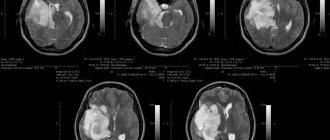
Lymphoma of the lungs on CT
Signs of pulmonary lymphoma are especially pronounced in the fourth stage of the disease, when the disease affects the respiratory organ. On a CT scan, enlarged lymph nodes will be visible, forming chains and conglomerates. In this case, the patient may also experience pulmonary edema. However, the high resolution of CT makes it possible to detect lymphoma at an early, first stage.
On CT scans, lymphomas, like any lumps, are visualized in a relatively lighter color. Normally, the airy pulmonary parenchyma is almost uniformly dark in color. Sometimes there are several such compactions and they are disseminated. The contours of the lymphoma are clear and even. Areas of “frosted glass” are found around pathological lesions.
List of sources
- Ponomareva O. V., Yurchenko O. V. Non-Hodgkin malignant lymphomas from marginal zone cells: diagnosis and treatment // Oncology - 2013. - vol. 15, no. 3. - p. 241-253.
- Klimenko A. A., Raksha A. P., Kopelev A. A. Hodgkin’s lymphoma // General Medicine. - 2007.- No. 2. - With. 15-20.
- Gluzman D.F., Sklyarenko L.M. Principles of modern diagnosis of lymphoid neoplasms // Oncohematology. - 2012. - No. 1. - pp. 44-46.
- Demina E. A., Tumyan G.S., Moiseeva T. N., Mikhailova N. B., Myakova N.V. Hodgkin's lymphoma // Modern oncology. - 2021. - volume 22, no. 2. pp. 6-33.
- Babicheva L.G., Tumyan G.S., Osmanov E.A. Follicular lymphoma // Modern oncology. — 2021.— volume 22, no. 2. pp. 34-51.